Aesthetic Rehabilitation of Maxillary Anterior Tooth Using Putty Index
Department-Conservative Dentistry and Endodontics, K.D. Dental College & Hospital, Mathura, UP, India, Department-Conservative Dentistry and Endodontics, P.D.M. University, Bahadurgarh
ABSTRACT
The adhesive dentistry allowed minimally invasive preparation through direct treatments with composite resin and indirect composite restoration. Various techniques and materials have been described to restore the traumatized tooth. This will allow a restoration of the original dental anatomy that maintains function and esthetics.
Keywords: composite restoration; teeth symmetry; central incisors; esthetic restorations; composite resin; thickness.
INTRODUCTION
A new procedure for symmetrical and consistent layer thickness control in esthetic direct replacement of anterior teeth is presented. To obtain a satisfactory result of an esthetic direct restoration. it is necessary to standardize either the design of the marginal preparation and the thickness of the dentin and enamel layers.1
Traumatic dental injuries (TDI) are the fifth most common disease worldwide.2
In a global systematic review, Petti et al. the estimated number of individuals aged 7 to 65 years with injured permanent teeth was approximately 900 million.3
Bilateral symmetry (also referred to as mirror symmetry, specular symmetry, and reflex symmetry) is found in the two maxillary central incisors in one of its highest expressions. Since these teeth are the most important part of the smile, every doctor should pay special attention to the following aspects related to symmetry:
- Symmetry and physical attractiveness are closely related.4
- Central incisors are often very symmetrical, while other teeth, such as the lateral incisors, can show huge asymmetries.5,6
- Central incisor asymmetry rarely exceeds 0.2 to 0.3 mm in one of three dimensions (length, width, or thickness)7,8
In an attractive smile, symmetry is very important near the midline. Some asymmetries may appear pleasing only if they are far from it.9
The main objective of this case report is to describe a new technique that can save chair time and dental laboratory costs in direct replacements of anterior teeth.
CASE REPORT
To fully evaluate the case a visual assessment was performed and the patient's occlusion was analyzed. (fig.1) Shade matching was done with the Vita Classic shade guide and A2 shade was selected for the case. The tooth was thoroughly cleaned and isolated with a cotton roll After that Alginate impression was made. (fig.2)
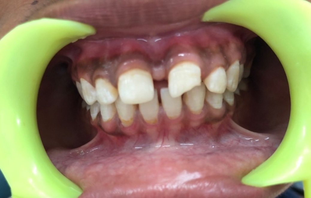
Figure 1. Pre-operative picture

Figure 2. Alginate impression
Study model was fabricated with dental stone and diagnostic wax-up performed on articulated casts. (fig.3)

Figure 3. (a),(b) & (c) wax mop-up
Vinyl polysiloxane (VPS) putty impression of the diagnostic cast was made and the excess material was removed. (fig.4)

Figure 4. Putty impression of diagnostic cast
LONG BEVEL AND SMOOTH MARGINS
A 40-micron diamond is used to smoothen out the jagged margins of the tooth and create a smooth bevel at least 2mm beyond the fracture line. This softens the edges and rounds off the fractured enamel prism. Sun burst appearance was made by beveling so that the light reflection in the finished restoration form a wavy line and not a straight line. (fig.5)

Figure 5. Beveling of the margin
Etch the tooth 2 mm beyond the beveled margins with 37% phosphoric acid cured for 15-20 seconds. The etchant is rinsed off. (fig.6)

Figure 6. Etchant Application
A bonding agent was applied to the etched surfaces and gently blown and cured for 20-30 secs using an LED curing light.
Multiple coats of bonding agent are applied. and after 30 seconds air is blown to let the solvent evaporate and for thinning the bonding agent. It is applied on all the area that have been etched and not only on the fractured margin. (fig.7)

Figure 7. Bonding agent application
A putty index was placed on the tooth. The first layer of enamel is placed and light-cured for 20 sec. The palatal wall was thus created. (fig.8) Placement of a thin layer of composite not more than 2mm in thickness, which was done in increments and cured every time. After removing the excess composite material and checking and adjusting the occlusion with articulating paper, the restoration was finished with diamond fine-coating burs and polished with a polishing system. (fig.9)
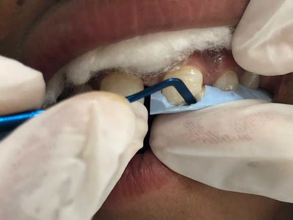
Figure 8. Placement of index on tooth

Figure 9. Post-operative picture
DISCUSSION
Traumatic dental injuries (TDI) have been proposed as the fifth most prevalent disease worldwide. In a worldwide systematic review, Petti et al. the estimated number of individuals between the ages of 7 and 65 with injured permanent teeth was approximately 900 million.10
Proper management for a predictable symmetrical result includes every aspect of the filling procedure. First, the margins should be prepared with the same design and geometry, while different materials, different thickness, and different substrates may provide different aesthetic results.11,12
One of the most common procedures is to use a silicone index created in the laboratory. With this technique.13 It is possible to shape both palatal and incisal edges, copying the planned anatomy. Several authors have attempted to shape the buccal surface, but this is very difficult to achieve predictably.14
Thus, the current approach to direct restorations of anterior teeth generally consists of three steps, as described by several authors:15-19
- Forming palatal wall and incisal edge;
- Building interproximal walls with matrices;
- Layering of the free buccal surface.
Vichi A. et al., who confirmed that layer thickness significantly affects the final aspect of a multilayer composite restoration. In fact, it is well known that exceeding the enamel thickness could cause a gray final result (low value).20
In the case presented, a step-by-step procedure was followed to secure the surface close to the adjacent teeth either for aesthetic reasons and to reduce biofilm formation that can cause discoloration and secondary caries.21-24
CONCLUSION
Direct bonded composite resin restorations are a conservative and valuable service that can be provided at a very reasonable cost. In this presented case, the dentist hid the fracture line and replicated the heavy surface texture.
This treatment option allows patients to enjoy an attractive smile and can delay more extensive removal of tooth structure, which could weaken the tooth and create a greater potential for pulpal necrosis.
REFERENCES
- Paolone, G.; Scolavino, S.; Gherlone, E.; Spagnuolo, G. Direct Esthetic Composite Restorations in Anterior Teeth: Managing Symmetry Strategies. Symmetry 2021, 13, 797.)
- Petti S, Andreasen JO, Glendor U, Andersson L. The fifth most prevalent disease is being neglected by public health organisations. Lancet Glob Health 2018;6:e1070?1.
- Petti S, Glendor U, Andersson L. World traumatic dental injury prevalence and incidence, A meta?analysis—One billion living people have had traumatic dental injuries. Dent Traumatol 2018;34:71?86
- Edler, R.J. Background Considerations to Facial Aesthetics. J. Orthod. 2001, 28, 159–168, doi:10.1093/ortho/28.2.159.
- Gallão, S.; Ortolani, C.L.F.; Santos-Pinto, A.; Santos-Pinto, L.; Faltin Júnior, K. Photographic analysis of symmetry and aesthetic proportion of the anterior teeth. Rev. Inst. Ciênc Saúde 2009, 27, 400–404.
- Wang, Y.; Song, Y.; Zhong, Q.; Xu, C. Evaluation of influence factors on the width, length, and width to length ratio of the maxillary central incisor: A systematic review and meta-analysis. J. Esthet. Restor. Dent. 2021, 33, 351–363, doi:10.1111/jerd.12606.
- Garn, S.M.; Lewis, A.B.; Walenga, A.J. Maximum-confidence values for the human mesiodistal crown dimension of human teeth. Arch. Oral Biol. 1968, 13, 841–844, doi:10.1016/0003-9969(68)90107-6.
- Mavroskoufis, F.; Ritchie, G. Variation in size and form between left and right maxillary central incisor teeth. J. Prosthet. Dent. 1980, 43, 254–257, doi:10.1016/0022-3913(80)90398-4.
- Rufenacht, C. Fundamentals of Esthetics; Quintessence Publishing: Chicago, IL, USA, 1990.
- Tewari N, Mathur VP, Siddiqui I, Morankar R, Verma AR, Pandey RM. Prevalence of traumatic dental injuries in India: A systematic review and meta-analysis. Indian J Dent Res 2020;31:601-14.
- Trifkovic, B.; Powers, J.M.; Paravina, R.D. Color adjustment potential of resin composites. Clin. Oral Investig. 2018, 22, 1601– 1607, doi:10.1007/s00784-017-2260-6.
- Pereira Sanchez, N.; Powers, J.M.; Paravina, R.D. Instrumental and visual evaluation of the color adjustment potential of resin composites. J. Esthet. Restor. Dent. 2019, 31, 465–470, doi:10.1111/jerd.12488.
- Vanini, L. Light and color in anterior composite restorations. Pract. Periodontics Aesthetic Dent. 1996, 8, 684.
- Ammannato, R.; Ferraris, F.; Allegri, M. The “index cutback technique”: A three-dimensional guided layering approach in direct class IV composite restorations. Int. J. Esthet. Dent. 2017, 12, 450–466
- Paolone, G. Direct composite restorations in anterior teeth. Managing symmetry in central incisors. Int. J. Esthet. Dent. 2014, 9, 12–25.
- Vanini, L. Light and color in anterior composite restorations. Pract. Periodontics Aesthetic Dent. 1996, 8, 684.
- Fahl, N., Jr. A polychromatic composite layering approach for solving a complex Class IV/direct veneer-diastema combination: Part I. Pract. Proced. Aesthet Dent. 2006, 18, 641–645.
- Fahl, N., Jr. A polychromatic composite layering approach for solving a complex Class IV/direct veneer/diastema combination: Part II. Pract. Proced. Aesthet Dent. 2007, 19, 17–22.
- Dietschi, D.; Ardu, S.; Krejci, I. A new shading concept based on natural tooth color applied to direct composite restorations. Quintessence Int. 2006, 37, 91–102.
- Vichi, A.; Fraioli, A.; Davidson, C.L.; Ferrari, M. Influence of thickness on color in multi-layering technique. Dent. Mater. 2007, 23, 1584–1589, doi:10.1016/j.dental.2007.06.026.
- Schroeder, T.; Da Silva, P.B.; Basso, G.R.; Franco, M.C.; Maske, T.T.; Cenci, M.S. Factors affecting the color stability and staining of esthetic restorations. Odontology 2019, 107, 507–512, doi:10.1007/s10266-019-00421-x.
- Marigo, L.; Nocca, G.; Fiorenzano, G.; Callà, C.; Castagnola, R.; Cordaro, M.; Paolone, G.; Sauro, S. Influences of Different AirInhibition Coatings on Monomer Release, Microhardness, and Color Stability of Two Composite Materials. BioMed Res. Int. 2019, 2019, 264, doi:10.1155/2019/4240264.
- Ionescu, A.C.; Cazzaniga, G.; Ottobelli, M.; Ferracane, J.L.; Paolone, G.; Brambilla, E. In vitro biofilm formation on resin-based composites cured under different surface conditions. J. Dent. 2018, 77, 78–86, doi:10.1016/j.jdent.2018.07.012.
- Cazzaniga, G.; Ottobelli, M.; Ionescu, A.C.; Paolone, G.; Gherlone, E.; Ferracane, J.L.; Brambilla, E. In vitro biofilm formation on resin-based composites after different finishing and polishing procedures. J. Dent. 2017, 67, 43–52, doi:10.1016/j.jdent.2017.07.012.
Disclaimer from Guident
The views and opinions expressed in articles published in Guident are solely those of the authors and do not necessarily reflect the views of the editors, the editorial board or the publisher, Ivory India. Guident does not endorse any product, technique or treatment described. Clinical content is intended for qualified dental and medical professionals and is not a substitute for individual clinical judgement. Guident, its editors and the publisher accept no liability for any consequence arising from the use of information published in the journal.
How to cite this article
Dr. Juhi Dubey, Dr. Rashmi Saini. Aesthetic Rehabilitation of Maxillary Anterior Tooth Using Putty Index. Guident. 2024 Jan;17(2). Available from: https://ivoryindia.in/articles/endodontics/aesthetic-rehabilitation-of-maxillary-anterior-tooth-using-putty-index.html