Endodontic Perforation Repair with MTA - Six Months Recall
Department of Conservative Dentistry & Endodontics D.J College Of Dental Sciences And Research, Modinagar, U.P
ABSTRACT
While undertaking endodontic procedures, perforations can occur.These accidents are defined as unfortunate events taking place while performing endodontic therapy. Some of these events are a result of poor attention to detail, others are totally unpredictable. Use of restorative materials such as Mineral Trioxide Aggregate (MTA) offer better prognosis in perforations treatments. A nonsurgical endodontic case was treated and repaired with MTA and recalls upto six months are presented. Itcan be concluded that if managed properly, perforation repairs can result in long-term clinical success.
Root canal treatment is associated with unforeseen and unwanted random circumstances, these are generally defined as procedure accidents or perforations.1 A perforation, or false connection
is the artificial communication between chamber or root canal system with support tissues of the tooth or to the oral cavity. Pathological causes like root resorption and caries can lead to a perforation. Nevertheless, the source is very often iatrogenic, as a result of an excessive use of the dental burr in the pulp chamber.3 In case of perforation a proper temporal seal should be established to avoid bacterial passage.5 The size of the defect also plays an important role since large perforations do not respond as well as the smaller ones.6 Large perforations can cause problems of incomplete sealing of the defect, thus allowing continual bacterial irritation at the perforation site.7. Smaller perforations are usually associated to lesser tissue damage and inflammation, and are easier to
repair. Healing is more predictable and has a better prognosis. Torabinejad informs that an immediate and typical sign is an abundant haemorrhaging emanating from the perforation site.
MTA has recently been proposed for repair of root perforations15. Several in vitro studies on
MTA have demonstrated its sealing ability.When used to repair perforations in animal models,minimal or no inflammation was presented and, in addition, cementum repair occurred at the material
interface (10, 11). It is reasonable to assume that the high surface pH of MTA supports repair and hard tissue formation in a similar fashion as calcium hydroxide. Thus, Holland et al.15 have proposed that calcium oxide in MTA reacts with tissue fluids to form calcium hydroxide, which in turn may encourage hard tissue deposition.
CASE REPORT
A 44 year old male patient was referred to the Department of conservative dentistry and endodontics at DJ College of dental sciences and research, modinagar, with a chief complaint of pain in left lower back tooth region since 2-3 months. . On clinical examination 37 had a carious lesion on the distal aspect of the tooth and a radiolucency was present in the furcation area.[Fig. 1 A]. On removal of the same, an ovoid shaped perforation was noticed in the furcation area and distal side of the canals. On probing the area, bleeding was noticed from the perforation site. Intra oral periapical radiographic analysis of 37 showed a radiolucency extending into the furcation area and distally. The root canals were negotiated, biomechanical preparation completed, calcium hydroxide saline paste was used as the intra canal medicament in between appointments and the root canal obturated using AH plus sealer (Dentsply DeTrey GmbH) and Guttapercha cones (DentsplyMaillefer), using a monocone technique. The site is cleansed with physiological solution. The perforation site was subsequently sealed using MTA and finally, radiographic evidence identifies the perforations’ sealing .[Fig. 1 B,C,D,E]. After one month, a control X-ray is taken.[Fig.2 A]. After three months the tooth is restored, with proper bone regeneration in the furcation zone.[Fig. 2 B]. After six months, radiographically, the repair of the defect is almost complete.[Fig. 2 C].
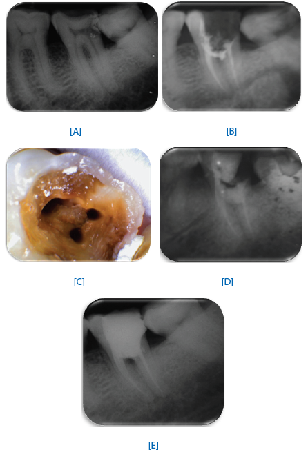
FIGURE 1: [A] Pre-opoerative Radiograph showing carious lesion on the distal aspect of the tooth and a radiolucency in the furcation area;[B]The perforation site was subsequently sealed using MTA showing the perforations’ sealing;;[C] Photographic view showing sealing of the perforation site;[D] The perforation site was subsequently sealed using MTA showing the perforations’ sealing;[E] Post endodontic restoration with perforation sealing


[C]
FIGURE 2. [A] One month, a control X-ray; [B] Three months X ray with proper bone regeneration in the furcation zone; [C] Six months, radiographically, the repair of the defect is almost complete
DISCUSSION
Perforation is a procedural accident that occurs during root canal treatment, creating an artificial communication between the root canal and the supporting periodontal apparatus.Currently
MTA has been the choice of material for perforation. For perforation repairs, it is important to also consider the periodontal and restorative prognosis of the tooth.
Application of MTA at the perforation site precluded, in this case. MTA provided good sealing of the defect, subsequently allowing a conventional root canal–filling technique. More importantly, the biologic response to this material was excellent, and complete resolution of the alveolar bone lesion.
Prognosis for a perforated tooth depends on the location of the perforation, the time in which the perforation allows entry of contamination, the possibility of sealing it and the accessibility of the main canal.15
Different materials have been used for endodontic perforation repair and the search for an ideal perforation repair material is a challenge. A repair material has to be placed in intimate contact with hard tissues of the tooth and soft tissues of the periodontium. These materials may pose a threat to endodontic treatment outcome by causing local or systemic adverse effects, either through direct contact with or leaching of chemical components into the periodontal tissues and alveolar bone (Breault et al, 2000). MTA may be the ideal material because it is the only material that consistently allows for the regeneration of the periodontal ligament, the apposition of a cementum like material and formation of bone.8
REFERENCES
- Walton RE, Torabinejad M. Endodoncia, principios y práctica; Accidentes de procedimiento. Ed. McGraw-Hill Interamericana, México. 2a edición, 1997: 329-336.
- Ingle J, Bakland L. Endodoncia; Percancesendodónticos: sudetección, corrección y prevención. 5a edición, Editorial: McGraw- Hill Interamericana. México, D.F. 2004: 856-868.
- Lasala A. Endodoncia; complicaciones y accidentesen el tratamientoy la obturación de conductos. Editorial Salvat. 1992: 407-
- Tsesis I, Fuss Z. Diagnosis and treatment of accidental root perforations. Endod Topics 2006; 13: 95-107.
- Lemon RR. Nonsurgical repair of perforation defects. Internal matrix concept. Dent Clin North Am 1992; 36: 439-457.
- Balla R, LoMonaco CJ, Skribner J, Lin LM. Histological study of furcation perforations treated with tricalcium phosphate, hydroxylapatite, mamalgam, and life. J Endod 1991; 17: 234-238.
- Fuss Z, Trope M. Root perforations: Classification and treatment choices based on prognosis factors. Endod Dent Traumatol1996; 12: 255-64.
- Kaufman AY, Fuss Z, Keila S, Waxenberg S. Reliability of different electronic apex locator to detect root perforations in vitro. IntEndod J 1997; 30: 403-407.
- Gutmann JL, Lovdahl PE. Problems encountered in tooth isolation and access to the pulp chamber space. In: Gutmann JL, Dumsha TC, Lovdahl PE, Hovland EJ et al. Problem solving in endodontics. Missouri. Mosby, 1997: 47-67.
- Kvinnsland I, Oswald RJ, Halse A, Gronningsaeter AG. A clinical and roentgenological study of 55 cases of root perforation. IntEndod J 1989; 22: 75-84.
- Rodríguez PA. Endodoncia, consider acion esactuales; accident esy compli caciones en enddoncia. Ed. Amolca. 1a edición, 2003; IX: 254-255.
- . Sinai IH. Endodontic perforations: their prognosis and treatment.J A6m Dent Assoc 1977; 95: 90-95.
- 13. De Moor RJ, De Boever JG. The sealing ability of an epoxy resin root canal sealer used withfive gutta-percha obturation techniques. Endod Dent Traumatol 2000;16:291–7.
- 14. Wedenberg C. Evidence for a dentin-derived inhibitor of macrophage spreading.Scand J Dent Res 1987;95:381– 8.
- 15. Frank AL. External-internal progressive resorption and its nonsurgical correction.J Endod 1981;7:473– 6.
Disclaimer from Guident
The views and opinions expressed in articles published in Guident are solely those of the authors and do not necessarily reflect the views of the editors, the editorial board or the publisher, Ivory India. Guident does not endorse any product, technique or treatment described. Clinical content is intended for qualified dental and medical professionals and is not a substitute for individual clinical judgement. Guident, its editors and the publisher accept no liability for any consequence arising from the use of information published in the journal.
How to cite this article
Dr. Charu Dayal, Dr. Saurabh, Dr. H.K. Bhullar. Endodontic Perforation Repair with MTA - Six Months Recall. Guident. 2014 Dec;8(1). Available from: https://ivoryindia.in/articles/endodontics/endodontic-perforation-repair-with-mta-six-months-recall.html