Corona Tree-Evolutionary Insights
Department of Prosthodontics & Implantology, K. D. Dental College & Hospital, Mathura (U.P) India
ABSTRACT
The coronavirus disease 19 (COVID-19) is a highly transmittable and pathogenic viral infection caused by severe acute respiratory syndrome coronavirus 2 (SARS-CoV-2), which emerged in Wuhan, China and spread around the world. Genomic analysis revealed that SARS-CoV-2 is phylogenetically related to severe acute respiratory syndrome-like (SARS-like) bat viruses, therefore bats could be the possible primary reservoir. The intermediate source of origin and transfer to humans is not known, however, the rapid human to human transfer has been confirmed widely. There is no clinically approved antiviral drug or vaccine available to be used against COVID-19. However, few broad-spectrum antiviral drugs have been evaluated against COVID-19 in clinical trials, resulted in clinical recovery. There is worldwide concern about the new coronavirus 2019-nCoV as a global public health threat. In this article, we provide a preliminary evolutionary and molecular epidemiological analysis of this new virus. Determining the origin and evolution of 2019-nCoV is important for the surveillance, drug discovery, and prevention of the epidemic. With more and more reported pathogenic 2019-nCoV isolates, it is necessary to re examne their origin and diversification patterns.
KEYWORDS: COVID-19, pathogenic coronaviruses, SARS-CoV, MERS-CoV, origin, evolution, Coronavirinae
INTRODUCTION
Since the first descriptions of AIDS in the early 1980s, much has been written about the causes and consequences of emerging viral diseases. Yet, despite an extensive research effort, viral infections continue to appear in human and wildlife populations, as demonstrated by the ‘new’ viruses identified since the rise of AIDS, such as HCV, Sin Nombre, Nipah, Hendra and most recently.SARS-CoV, and more venerable pathogens, including West Nile virus and dengue virus, that have recently expanded their global prevalence. Even less progress has been made in what is perhaps the ultimate goal of research into emerging viruses, predicting what viruses are likely to emerge in the future.1
Defining emerging viruses as those that are newly appeared or have recently increased in prevalence and/or geographical range reveals some important general patterns.
First, almost all emerging viruses have RNA rather than DNA genomes. Although RNA viruses are ordinarily more commonplace than DNA viruses, we will argue later that this difference primarily reflects the differing evolutionary rates of these two types of infectious agent.
Second, almost all emerging viruses have an animal reservoir, such that the process of viral emergence can usually be categorized as cross-species transmission (Cleaveland et al. 2001). For example, HIV type 1 (HIV-1), the major cause of AIDS, has its origins in the related SIV found in chimpanzees (Gao et al. 1999), while SARS-CoV has close relatives in Himalayan palm civets (Guan et al. 2003), although it is not yet established that these are the source population for the human form of the virus. The most significant exception to the rule that cross-species transmission is central to viral emergence is HCV, which was first identified in 1989 but which is likely to have a much longer history in human populations (Simmonds 1995).2
Despite surveying a number of animal species, the ultimate reservoir species for HCV remains a mystery, although this is more likely to reflect the fact that the wrong species have been surveyed or that the reservoir viruses are too divergent in sequence to be recognized, than the absence of an animal reservoir altogether.2,3
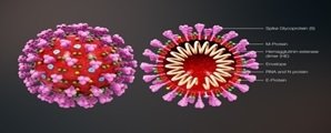
Coronaviruses belong to the Coronaviridae family in the Nidovirales order. Corona represents crown-like spikes on the outer surface of the virus; thus, it was named as a coronavirus. Coronaviruses are minute in size (65–125 nm in diameter) and contain a single-stranded RNA as a nucleic material, size ranging from 26 to 32kbs in length (Fig. 1).
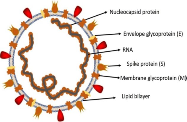
Fig 1
The subgroups of coronaviruses family are alpha (a), beta (b), gamma (c) and delta (d) coronavirus.2
The alphacoronaviruses and betacoronaviruses infect only mammals. The gammacoronaviruses and deltacoronaviruses infect birds, but some of them can also infect mammals. Alphacoronaviruses and betacoronaviruses usually cause respiratory illness in humans and gastroenteritis in animals. The two highly pathogenic viruses, SARS- CoV and MERS- CoV, cause severe respiratory syndrome in humans, and the other four human coronaviruses (HCoV- NL63, HCoV-229E, HCoV- OC43 and HKU1) induce only mild upper respiratory diseases in immunocompetent hosts, although some of them can cause severe infections in infants, young children and elderly individuals. Alphacoronaviruses and betacoronaviruses can pose a heavy disease burden on livestock; these viruses include porcine transmissible gastroenteritis virus, porcine enteric diarrhoea virus (PEDV) and the recently emerged swine acute diarrhoea syndrome coronavirus (SADS- CoV). On the basis of current sequence databases, all human coronaviruses have animal origins: SARS- CoV, MERS- CoV, HCoV- NL63 and HCoV-229E are considered to have originated in bats; HCoV- OC43 and HKU1 likely originated from rodents.
Primary reservoirs and hosts of coronaviruses
The source of origination and transmission are important to be determined in order to develop preventive strategies to contain the infection. In the case of SARS-CoV, the researchers initially focused on raccoon dogs and palm civets as a key reservoir of infection. However, only the samples isolated from the civets at the food market showed positive results for viral RNA detection, suggesting that the civet palm might be secondary hosts.4 In 2001 the samples were isolated from the healthy persons of Hongkong and the molecular assessment showed 2.5% frequency rate of antibodies against SARS-coronavirus. These indications suggested that SARS-coronavirus may be circulating in humans before causing the outbreak in 2003.5 Later on, Rhinolophus bats were also found to have anti-SARS-CoV antibodies suggesting the bats as a source of viral replication.4,5 The Middle East respiratory syndrome (MERS) coronavirus first emerged in 2012 in Saudi Arabia.9MERS-coronavirus also pertains to beta-coronavirus and having camels as a zoonotic source or primary host.6 In a recent study, MERS-coronavirus was also detected in Pipistrellus and Perimyotisbats, proffering that bats are the key host and transmitting medium of the virus . Initially, a group of researchers suggested snakes be the possible host, however, after genomic similarity findings of novel coronavirus with SARS-like bat viruses supported the statement that not snakes but only bats could be the key reservoirs .
Animal origins of human coronaviruses

Fig 2: Solid arrows indicate confirmed data. Broken arrows indicate potential interspecies transmission. Black arrows indicate infection in the intermediate animals, yellow arrows indicate a mild infection in humans, and red arrows indicate a severe infection in humans or animals.

Fig 3: The key reservoirs and mode of transmission of coronaviruses (suspected reservoirs of SARS-CoV-2 are red encircled); only a and b coronaviruses have the ability to infect humans, the consumption of infected animal as a source of food is the major cause of animal to human transmission of the virus and due to close contact with an infected person, the virus is further transmitted to healthy persons. Dotted black arrow shows the possibility of viral transfer from bat whereas the solid black arrow represent the confirmed transfer.
Phylogenetic relationships in the Coronavirinae subfamily.

Fig 4: The highly human- pathogenic coronaviruses belong to the subfamily Coronavirinae from the family Coronaviridae. The viruses in this subfamily group into four genera (prototype or representative strains shown): Alphacoronavirus (purple), Betacoronavirus (pink), Gammacoronavirus (green) and Deltacoronavirus (blue). Classic subgroup clusters are labelled 1a and 1b for the alphacoronaviruses and 2a–2d for the betacoronaviruses. The tree is based on published trees of Coronavirinae and reconstructed with sequences of the complete RNA- dependent RNA polymerase- coding region of the representative coronaviruses (maximum likelihood method under the GTR + I + Γ model of nucleotide substitution as implemented in PhyML , version 3.1)). Only nodes with bootstrap support above 70% are shown. IBV, infectious bronchitis virus; MERS- CoV, Middle East respiratory syndrome coronavirus; MHV, mouse hepatitis virus; PEDV, porcine enteric diarrhoea virus; SARS- CoV, severe acute respiratory syndrome coronavirus; SARSr- CoV, SARS- related coronavirus.
Key features and entry mechanism of human coronaviruses
All coronaviruses contain specific genes in ORF1 downstream regions that encode proteins for viral replication, nucleocapsid and spikes formation.4 The glycoprotein spikes on the outer surface of coronaviruses are responsible for the attachment and entry of the virus to host cells (Fig. 1). The receptor-binding domain (RBD) is loosely attached among virus, therefore, the virus may infect multiple hosts. Other coronaviruses mostly recognize aminopeptidases or carbohydrates as a key receptor for entry to human cells while SARS-CoV and MERS-CoV recognize exopeptidases.2 The entry mechanism of a coronavirus depends upon cellular proteases which include, human airway trypsin-like protease (HAT), cathepsins and transmembrane protease serine 2 (TMPRSS2) that split the spike protein and establish further penetration changes.8,9 MERS-coronavirus employs dipeptidyl peptidase 4 (DPP4), while HCoV-NL63 and SARS-coronavirus require angiotensin-converting enzyme 2 (ACE2) as a key receptor.2,11 SARS-CoV-2 possesses the typical coronavirus structure with spike protein and also expressed other polyproteins, nucleoproteins, and membrane proteins, such as RNA polymerase, 3-chymotrypsin-like protease, papain-like protease, helicase, glycoprotein, and accessory proteins.13,14 The spike protein of SARS-CoV-2 contains a 3-D structure in the RBD region to maintain the van der Waals forces.14,15 The 394 glutamine residue in the RBD region of SARS-CoV-2 is recognized by the critical lysine 31 residue on the human ACE2 receptor . The entire mechanism of pathogenicity of SARS-CoV-2, from attachment to replication is well mentioned in Fig. 5.
Fig 5: The life cycle of SARS-CoV-2 in host cells; begins its life cycle when S protein binds to the cellular receptor ACE2. After receptor binding, the conformation change in the S protein facilitates viral envelope fusion with the cell membrane through the endosomal pathway. Then SARS-CoV-2 releases RNA into the host cell. Genome RNA is translated into viral replicase polyproteins pp1a and 1ab, which are then cleaved into small products by viral proteinases. The polymerase produces a series of subgenomic.
mRNAs by discontinuous transcription and finally translated into relevant viral proteins. Viral proteins and genome RNA are subsequently assembled into virions in the ER and Golgi and then transported via vesicles and released out of the cell. ACE2, angiotensin-converting enzyme 2; ER, endoplasmic reticulum; ERGIC, ER–Golgi intermediate compartment.
Comparison of transmission of MERS-CoV and SARS-CoV
Both SARS-CoV and MERS-CoV are emerging zoonotic pathogens that crossed the species barriers to infect human.3,10 Evidence showed that SARS-CoV and MERS-CoV originated from bats, the nature reservoirs, then transmitted to human via intermediate hosts civets and camels, respectively Human SARS-CoV infection originated from the direct contact between humans and civets in markets or restaurants.Closing wet markets and cleaning civet cut off the spread chain of SARS-CoV and effectively ended the SARS epidemic In contrast, MERS-CoV is believed to have existed in camels for a very long time and camels are widely distributed in Middle East and African countries, serving as important transport vectors and sources of meat and milk for the local population.
Therefore, it is difficult to adopt the same strategy of SARS-CoV control in the prevention of future MERSCoV outbreaks. Until a comprehensive approach is found, which most likely will involve the effective vaccination of camels against MERS-CoV among other measures, it is envisaged that sporadic human infection will persist for some time in the future.
Bat coronaviruses and human coronavirus 229E (HCoV-229E) and NL63 (HCoV-NL63)HCoV-229E was found in the 1960s and causes comparatively mild common colds worldwide. A bat coronavirus detected in Hipposideros caffer ruber in Ghana termed Hipposideros/GhanaKwam/19/2008 was genetically related to HCoV-229E. Its RdRp fragment shared 92 % nucleotide sequence identity with HCoV- 229E and they were predicted to share a most recent common ancestor (MRCA) only 200 years ago.11 A recent study characterized more 229E-related coronaviruses discovered in hipposiderid bats from Ghana on full genome level. These bat coronaviruses were more diversified and formed a single viral species with HCoV- 229E. Interestingly, phylogenetic analysis revealed the intermediate position of a 229E-related alpaca virus between bat and human viruses. These findings suggested the ancestral origin of HCoV-229E in hipposiderid bats and the role of camelids as potential intermediate hosts was hypothesized.
Evolution of the 2019 Novel Coronavirus
Based on our phylogenomic analysis of the recently released genomic data of 2019-nCoV, we showed that the 2019- nCoV is most closely related to 2 severe acute respiratory syndrome (SARS)-like CoV sequences that were isolated in bats during 2015 to 20172, suggesting that the bats’ CoV and the human 2019-nCoV share a recent common ancestor(Figure 6A). Therefore, the 2019-nCoV can be considered as a SARS-like virus and named SARS-CoV-2.The 2 bat viruses were collected in Zhoushan, Zhejiang Province, China, from 2015 to 2017.2
There is speculation that the 2019-nCoV may have originated near Zhoushan or elsewhere. The new coronavirus was first isolated from stallholders who worked at the South China Seafood Market in Wuhan. This market also sells wild animals or mammals, which were likely intermediate hosts of 2019-nCoV, which originated from bat hosts (Figure 6B). It has been speculated that the intermediate hosts (wild mammals) may have been sold to the seafood market in Wuhan.
The 2019-nCoVs have long branches (0.09) for the 2 isolated in the phylogenomic tree (Figure 6A), indicating that the 2019- nCoVs likely share bat hosts. Similarly, the 2003 SARS-CoVs (human SARSCoVs) had short branches (0.03) for the bat hosts (Figure 6A). This indicates that there should be more bat viruses closer to 2019-nCoV.
According to their phylogenetic relationships, the 27 isolates of 2019-nCoV examined in this study can be divided into at least 6 genotypes (I-VI; Figure 6C).

Fig 6: The phylogenomic trees of the coronavirus and the genotypes of 2019-nCoV strains[4]. A, The maximum likelihood (ML) phylogenetic trees of the coronavirus with the approximate ML method by FastTree. This phylogenetic tree is a rooted tree with CoV BM48–31/BGR/2008 and BtKY72 as an outgroup. Coronaviruses from different sources are represented on the right side of the tree. The 2019-nCoV is labeled in red. The detailed sequence information used in this figure is shown in Supplementary Figure 6 B, A simple model of the 2019-nCoV origin inferred from the phylogenomic tree. C, The ML phylogenetic tree of 2019-nCoV with bat-SL-CoVZC45 as the root, with the approximate ML method by FastTree. The 27 isolates of 2019-nCoV can be divided into 6 genotypes. The 6 genotypes of 2019-nCoV strains are represented with different symbol shapes. Abbreviations: 2019-nCoV, 2019 novel coronavirus; SARS, severe acute respiratory syndrome.
Compared with the rapid reassortment and mutation of avian influenza (H7N9)3,4, the degree of diversification of 2019-nCoV is much smaller. But the 27 isolates can be divided into 6 genotypes, indicating that the 2019-nCoV has mutated in different patients. The magnitude of this variation is worthy of attention in the future, and it is necessary to be vigilant for any noticeable, rapid mutations. As of today, the intermediate host of 2019-nCoV has not been determined (Figure 6B). Considering that intermediate hosts are generally mammals,19 they are likely the living mammals sold in the South China seafood market. Therefore, strengthening the monitoring of wild mammals is an urgent measure needed to prevent similar viruses from infecting humans in the future. More than 1000 confirmed cases have been reported in China. The number of provinces and cities in China, as well as in other countries, with confirmed cases is steadily increasing. It is necessary to further strengthen monitoring to ensure that these virus strains will not cause diseases like the global outbreak of 2003 SARS.
Conclusion and future perspective
The collected data on genetic evolution, receptor binding and pathogenesis demonstrated that SARS- CoV most likely originated in bats through sequential recombination of bat SARSr- CoVs. Recombination likely occurred in bats before SARS- CoV was introduced into Guangdong province through infected civets or other infected mammals from Yunnan. The introduced SARS- CoV underwent rapid mutations in S and orf8 and successfully spread in market civets. After several independent spillovers to humans, some of the strains underwent further mutations in S and became epidemic during the SARS outbreak in 2002–2003. However, a recent serological investigation revealed the presence of antibodies against the SARSr- CoV nucleocapsid in humans living around a bat cave but who had not shown clinical signs of disease, suggesting that the virus can infect humans through frequent contact.4With human activity increasingly overlapping the habitats of bats, diseases outbreaks resulted from spillover of bat coronaviruses will continue to occur in the future despite the fact that direct transmission of bat coronaviruses to humans appears to be rare. To better prepare ourselves in predicting and preventing the next emergence of a coronavirus disease, it is necessary to maintain our vigilance in long-term coronavirus surveillance studies in bats as well as in other wildlife and livestock. Combined with other laboratory-based studies such as receptor specificity, pathogenesis and animal infection, a focus on continued surveillance will help us to improve risk assessment as well as to reveal the potential intermediate hosts that may play an important role in the interspecies transmission.2
According to phylogenetic trees, SARS-CoV is closer to SARS-like bat CoVs. Until now, no promising clinical treatments or prevention strategies have been developed against human coronaviruses. However, the researchers are working to develop efficient therapeutic strategies to cope with the novel coronaviruses. Various broad-spectrum antivirals previously used against influenza, SARS and MERS coronaviruses have been evaluated either alone or in combinations to treat COVID-19 patients, mice models, and clinical isolates. Remdesivir, Lopinavir, Ritonavir, and Oseltamivir significantly blocked the COVID-19 infection in infected patients. It can be cocluded that the homologus recombination event at the S protein of RBD region enhanced the transmission ability of the virus. While the decision of bring back the nationals from infected area by various countries and poor screening of passengers, become the leading cause of spreading virus in others countries.4
Most importantly, human coronaviruses targeting vaccines and antiviral drugs should be designed that could be used against the current as well as future epidemics. There are many companies working for the development of effective SARS-CoV-2 vaccines, such as Moderna Therapeutics, Inovio Pharmaceuticals, Novavax, Vir Biotechnology, Stermirna Therapeutics, Johnson & Johnson, VIDO-InterVac, GeoVax-BravoVax, Clover Biopharmaceuticals, CureVac, and Codagenix. But there is a need for rapid human and animal-based trails as these vaccines still require 3–10 months for commercialization. There must be a complete ban on utilizing wild animals and birds as a source of food. Beside the development of most efficient drug, a strategy to rapidly diagnose SARS-CoV-2 in suspected patient is also required.5,9,10
Furthermore, little is known about the replication and pathogenesis of these bat viruses. Thus, future work should be focused on the biological properties of these viruses using virus isolation, reverse genetics and in vitro and in vivo infection assays. The resulting data would help the prevention and control of emerging SARS- like or MERS- like diseases in the future. It is widely accepted that many viruses have existed in their natural reservoirs for a very long time. The constant spillover of viruses from natural hosts to humans and other animals is largely due to human activities, including modern agricultural practices and urbanization. Therefore, the most effective way to prevent viral zoonosis is to maintain the barriers between natural reservoirs and human society, in mind of the ‘one health’ concept.
REFERENCES
- Viral evolution and the emergence of SARS Coronavirus Edward C. Holmes* and Andrew Rambaut Department of Zoology, University of Oxford, South Parks Road, Oxford OX1 3PS, UK
- Origin and evolution of pathogenic coronaviruses Jie Cui 1, Fang Li2 and Zheng- Li Shi 1*
- Hu et al. Virology Journal (2015) 12:221 DOI 10.1186/s12985-015-0422-1Bat origin of human coronaviruses Ben Hu1, Xingyi Ge1, Lin-Fa Wang2 and Zhengli Shi1*
- Origin and Evolution of the 2019 Novel Coronavirus Article in Clinical Infectious Diseases • February 2020 DOI: 10.1093/cid/ciaa112
- Organization WH. Laboratory testing for coronavirus disease 2019 (COVID-19) in suspected human cases: interim guidance, 2 March 2020. World Health Organization, 2020.
- Peiris J, Guan Y, Yuen K. Severe acute respiratory syndrome. Nat Med 2004;10 (12):S88–97.
- Pyrc K, Berkhout B, Van Der Hoek L. Identification of new human coronaviruses. Expert Review of Anti-infective Therapy 2007;5(2):245–53.
- Rahman A, Sarkar A. Risk factors for fatal middle east respiratory syndrome coronavirus infections in Saudi Arabia: analysis of the WHO Line List, 2013– 2018. Am J Public Health 2019;109(9):1288–93.
- Memish ZA, Zumla AI, Al-Hakeem RF, Al-Rabeeah AA, Stephens GM. Family cluster of Middle East respiratory syndrome coronavirus infections. N Engl J Med 2013;368(26):2487–94.
- Wang C, Horby PW, Hayden FG, Gao GF. A novel coronavirus outbreak of global health concern. The Lancet 2020.
- Phan LT, Nguyen TV, Luong QC, Nguyen TV, Nguyen HT, Le HQ, et al. Importation and human-to-human transmission of a novel coronavirus in Vietnam. N Engl J Med 2020.
- Guan, Y. et al. Isolation and characterization of viruses related to the SARS coronavirus from animals in southern China. Science 302, 276–278 (2003). This paper is the first demonstration of SARS- CoV transmission from animals.
- Alagaili, A. N. et al. Middle East respiratory syndrome coronavirus infection in dromedary camels in Saudi Arabia. MBio 5, e00884–14 (2014).
- Hemida, M. G. et al. Middle East Respiratory Syndrome (MERS) coronavirus seroprevalence in domestic livestock in Saudi Arabia, 2010 to 2013. Euro. Surveill. 18, 21–27 (2013).
- Lau, S. K. et al. Severe acute respiratory syndrome coronavirus- like virus in Chinese horseshoe bats. Proc. Natl Acad. Sci. USA 102, 14040–14045 (2005).
- Woo PC, Lau SK, Li KS, Tsang AK, Yuen KY. Genetic relatedness of the novel human group C betacoronavirus to Tylonycteris bat coronavirus HKU4 and Pipistrellus bat coronavirus HKU5. Emerg Microbes Infect. 2012;1(11):e35. doi:10.1038/emi.2012.45.
- van Boheemen S, de Graaf M, Lauber C, Bestebroer TM, Raj VS, Zaki AM et al. Genomic characterization of a newly discovered coronavirus associated with acute respiratory distress syndrome in humans. MBio. 2012;3(6). doi:10. 1128/mBio.00473-12.
- de Groot RJ, Baker SC, Baric RS, Brown CS, Drosten C, Enjuanes L, et al. Middle East respiratory syndrome coronavirus (MERS-CoV): Announcement of the Coronavirus Study Group. J Virol. 2013;87(14): 7790–2. doi:10.1128/JVI.01244-13.
- Zaki AM. Novel coronavirus—Saudi Arabia: Human isolate. Int Soc Infect Dis. 2012;2012-09-20.(ProMED-mail.).
- Masters PS. The molecular biology of coronaviruses. Adv Virus Res. 2006;66: 193–292. doi:10.1016/S0065-3527(06)66005-3.
Disclaimer from Guident
The views and opinions expressed in articles published in Guident are solely those of the authors and do not necessarily reflect the views of the editors, the editorial board or the publisher, Ivory India. Guident does not endorse any product, technique or treatment described. Clinical content is intended for qualified dental and medical professionals and is not a substitute for individual clinical judgement. Guident, its editors and the publisher accept no liability for any consequence arising from the use of information published in the journal.
How to cite this article
Dr. C. Pradeep, Dr. Ruchi Kadian, Dr. Rahul Nagrath. Corona Tree-Evolutionary Insights. Guident. 2020 Jul;13(8). Available from: https://ivoryindia.in/articles/general/corona-tree-evolutionary-insights.html