Palatogingival Grooves: The Only Way Out Is Through....
Department of Periodontology, Department of Conservative and Endodontics, Department of Periodontology, K. D. Dental College & Hospital, Mathura
ABSTRACT
Palatogingival grooves are most common etiological factors affecting the maxillary anteriors and often lead to formation of Primary Periodontal lesion with secondary endodontic involvement. The only way out to salvage the affected tooth is to perform combined Periodontal and endodontic therapy. This case report discusses about a similar lesion and treatment plan for the same which improved the overall status of the affected tooth.
Keywords : Palatogingival grooves
INTRODUCTION
Combined Perio- endo lesions have been a nightmare for the endodontist and periodontist as clinicians. The small narrow grooves often lead to large mighty lesions, complicating the treatment plans as well as compromising the prognosis of the teeth involved. The preponderance of these lesions in maxillary anterior further complicates the treatment plan and also necessitates the complete resolution of the lesion to save the tooth, because a tooth loss in anterior region is esthetics lost.
Palatogingival grooves also known as radicular grooves, palato radicular groove, disto lingual groove, corono radicular groove or radiculo lingual groove are primarily formed as a result of infoldings in HERS (Hertwigs Epithelial Root Sheath) during root formation in teeth of maxillary anterior region primarily the Laterals followed by Centrals.1 (al, 2016). Several etiologies have been put forth for its development apart from this classical theory, out of which gained attention were :
(1) an aborted attempt towards formation of an additional root on the affected tooth and recently,
(2) alteration of genetic mechanisms and racial link has been proposed.2,3
Various classifications have been put forward to classify the Palatogingival grooves depending on their extent, location, etc. as summarized in table 1.4 Storrer et al. reported that 9.58% of maxillary lateral incisors showed PGG in an investigation of 73 extracted teeth, with the groove always located in the center of the lingual area.5

The radicular grooves are of clinical significance as they play a key role in initiating the pathogenesis of Periodontal disease by 1) site of harbor of plaque and calculus which initiate the Periodontal breakdown 2) grooves extending up till the Periapical regions can cause retrograde pulpitis thus also causing pulpal necrosis. An early diagnosis with efficient treatment planning aimed at obliterating the groove and promoting selective regeneration of periodontal tissue can completely obliterate the problem. This case report aims at showing the ruinous effect of Palatogingival groove present in association with lateral incisor which led to formation of Periapical granuloma and management of the same using amniotic membrane as a GTR membrane and biodentine as retrograde filling material
CASE REPORT
A 25year old male patient named Ram Rattan reported to the department of conservative and endodontics with the complain of dull pain in relation to left lateral incisor since 3 months. Dental history reveals no history of trauma and any previous dental treatment done for the same tooth. Upon examination of the tooth it was found a presence of deep Periodontal pocket on palatal aspect of the tooth. Electronic pulp testing showed the tooth was non vital. Patient was then referred to department of Periodontology for detailed analysis of the Pocket. Upon careful examination it was found that pocket of 10mm was associated with a groove on distopalatal aspect of the tooth with no mobility. Further a digital IOPAR revealed lateral periapical radiolucency associated with the groove. Based on these finding it was concluded that tooth#22 had palatogingival groove associated with it. For a better treatment planning to find the extent and depth of groove and periapical radiolucency patient was referred for CBCT of the same region.
CBCT reports confirmed the presence of radicular grooves which extended upto apical third of root (fig 1 ).Associated with a well-defined periapical pathology with irregular borders wrt 22 extending mesio-distally from the interdental bone between 21 and 22 to the mesial surface of root of 23 (measuring 8.9 mm); labio-lingually from the labial cortical plate to 5.0 mm anterior to the palatal cortical plate (measuring 6.7 mm); superio-inferiorly 5.0 mm above the crest of interdental bone to the floor of nasal 5.0 mm below the floor of nasal fossa (measuring 7.9 mm ) and Thinning and loss of labial cortical plate in relation to the periapical lesion. (fig 2)

Fig 1: Periapical lesion in relation with #22 and Palate gingival groove in relation to palatal aspect

Fig 2: Periapical pathology associated with the lesion.
A combined perio- endo treatment plan was chalked out for the same. First phase of therapy included conventional root canal treatment and scaling root planning of the same. The endodontic therapy involved determination of working length using electronic apex locator (Root ZX, J. Morita Mfg. Corporation, Kyoto, Japan) and CBCT slices. The biomechanical preparation was done using stainless steel K file upto 45 no k file in crown down technique. Irrigation was done using 3% NaOCL via manual syringe followed by intracanal medicament Ca(OH)2 for 2 consecutive weeks this resulted in decreased in Periapical radiolucency. Also After phase I periodontal therapy the pocket depth reduced upto 5mm.

FIG 3: Immediate Pre-op. (A)determining periodontal pocket depth in association with the groove (B) intrasulcular incision (c) Peri radicular defect with loss of buccal cortical plate
Phase II treatment involved periodontal surgery along with apicoectomy procedure to completely obliterate the groove and to remove the Periapical granuloma on the day of surgery the canal was sealed using ISO NO. 45 Gutta percha points and Zinc oxide eugenol sealer. Before Periodontal surgery anaesthesia was achieved after administering 2% lignocaine with 1:100,000 adrenaline following which a full thickness periosteal flap was raised on the palatal surface and buccal surface of tooth #22 using intra sulcular incision and two vertical releasing incision on the buccal aspect. On flap elevation a radicular groove extending upto apical third of root was seen on palatal aspect and on buccal aspect of an there was loss was buccal plate with Peri radicular lesion.

FIG 4: Surgical phase: (A) Debridement of peri-radicular lesion (B) apicoectomy done (C) biodentin as retrograde filling (D)sealing the groove with GIC
Meticulous scaling and root planning was performed using surgical columbia curette 2r/2l (Hu Friedy Manufacturing Co., Chicago, IL) on buccal and palatal aspect of tooth. This was followed by removal of periradicular lesion followed by apicoectomy in relation to #22. After the apicoectomy procedure the root canal was sealed/retrograde filling using Biodentine (Septodont, St. Maurdes Fosses, France).The material was allowed to initial set for about 9 min. The groove was sealed using GIC and an amniotic GTR membrane (tata memorial, tissue bank, Mumbai) was placed on the palatal aspect over the groove. The defect site was filled with alloplastic bone graft material (B-OSTIN 100% SYNTHETIC BONEGRAFT SUBSITITUTE). The flap was approximated and sutured using 4-0 silk suture.

FIG 5: (A) filling the defect with bone graft (B) B- ostin alloplastic graft (C,D)amniotic membrane used as a GTR membrane.
Analgesics and antibiotics were prescribed, and the patient was given regular oral hygiene instructions including chlorhexidine (0.12%) mouth rinse for 2 weeks.
The patient was recalled for checkup at immediate post op next day, 1week, 3months. At 3 months gingiva appeared healthy, and pocket depth was reduced upto 3mm. Radiograph also showed healing for the Periradicular lesion.
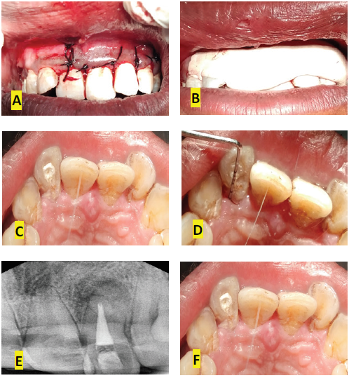
FIG 6: (A) Sutures placed (B) Periodontal Pack placed (C) Follow UP after 1 month (D) Reduced Probing Depth (1 month ) (E) IOPA RADIOGRAPH post op 1 MONTH
DISCUSSION
Palatogingival groove is the most common morphological and anatomical aberrancy affecting maxillary laterals followed by maxillary centrals and also most common etiological factor in developing the Perio-endo lesions. Present mostly on the palatal aspect of maxillary incisor it initiates the Periodontal breakdown by becoming an ideal site for harbouring Plaque and sometimes calculus both of which are detrimental for Periodontal health. When these grooves extend deep inside on the root surface they cause retrograde pulpitis. Amongst these grooves traversing the root, 43% have shown to extend apically <5 mm in distance, 47% between 6 and 10 mm and 10% have shown extension beyond 10 mm.Based on the invagination of the groove towards the pulp cavity, these have been termed as shallow/flat (<1 mm), deep (>1 mm) and a closed tube.
In this case report the groove present on the maxillary lateral belong to Type III as it was extending beyond the middle third of the root and also has a communication with the pulpal complex at the apex of the root , which become the supposed reason for large Periapical lesionas evident by CBCT. Also Probing depth of about 10 mm adjacent to the gingival margin is indicative of a deep isolated periodontal pocket which strongly suggested the presence of a long palatogingival groove.
The tooth in our case was nonvital with a Periapical lesion which initiated the need of starting with endodontic treatment before we go ahead with Periodontal surgical procedure aimed at GTR and GBR. After completion of endodontic treatment we moved ahead with the Periodontal therapy. After complete elevation of full thickness flap, followed by meticulous scaling and negotiating the depth of groove we chose to seal the groove with a suitable Restorative material that is GIC. On the buccal aspect after debridement of the defect, apicoectomy was performed and Biodentine was used as Retrograde filling material. The primary clinical advantage of Biodentine is its fast setting (between 12 and 15 min). This is an advantage when compared to the 170 min of MTA since a delayed setting time leads to an increased risk of partial material loss and alteration of the interface during the finishing phases of the procedure (cleaning and rinsing the crypt).7 Biodentine displayed bioactivity, i.e., activation of angiogenesis and activation of progenitor pulpal cells promoting healing and remineralization. From a biomechanical point of view, the sealing properties of Biodentine have been reported to be superior to MTA.8
Following this alloplatic bone graft (B-OSTIN 100% SYNTHETIC BONEGRAFT SUBSITITUTE) was placed in the defect. Alloplastic bone graft is the standard material as it is composed of natural form of Hydroxyapatite in its ceramic, crystalline form, essential for phase I osteogenesis, accounting for the proliferation of bone cells, especially from the osteoid. The rationale behind the filling of the surgical cavity with an alloplastic bone graft: is to be biocompatible, osteoconductive; to stimulate bone neoformation; to be hemostatic; to act as a barrier in guided tissue regeneration against the formation of both conjunctive and epithelial tissue; to be of easy handling; and to decrease the post-surgical healing time.9 After this we used amniotic membrane as a GTR membrane to enhance selective tissue proliferation. Amniotic membrane or amnion is the innermost layer of the placenta and consists of a thick basement membrane and an avascular stromal matrix. Human amniotic membrane (HAM) has been used successfully for over a decade for a wide range of surgical applications. Amniotic membrane is a gift of nature which not only protects the fetus inside the womb but also has several medicinal properties. It serves as a natural barricade to protect the fetus from bacterial infection and trauma. Amniotic membrane acts as a scaffold for proliferation and differentiation due to the presence of fibronectin, elastin, nidogen, collagen types I, III, IV, V, and VI, elastin, and hyaluronic acid. Another important advantage of using amniotic membrane in allotransplant or xenotransplant is lack of immunogenicity. Promotion of epithelialization, anti-inflammatory properties, antifibrotic properties, antibacterial properties, and antiangiogenic properties are confirmed by the presence of several related factors that makes amniotic membrane an ideal therapeutic for burns and wound healing.10 lastly Flap closure was done using 4-0 silk suture followed by Periodontal pack (Coe pack) placement.
CONCLUSION
Based on the literature review and case report presented it can be concluded that a well planned Perio-endo treatment plan can efficiently eliminate such lesion, also improve the overall status of the tooth by promoting regeneration of the lost tissues. The traditional root canal treatment with apicoectomy completely eliminate the periapical pathology and nectrotic pulp. Periodontal surgical procedure with careful selection and judicious use of regenerating materials for GTR and GBR further enhance the treatment plan.
REFERENCES
- Zahra Alizadeh Tabari et al,Treatment of a Developmental Groove and Supernumerary Root Using Guided Tissue Regeneration Technique, Hindawi Publishing Corporation, Pp1-5, 2016.
- Simon JH et al, Predictable endodontic and periodontic failures as a result of radicular anomalies. Oral Surg Oral Med Oral Pathol, vol 31(6),Pp 823-6, 1971.
- Ennes JP, Lara VS. Comparative morphological analysis of the root developmental groove with the palato-gingival groove. Oral Dis, vol 10(6), Pp378-82.2004.
- Hee-Jin kim et al, Recognition and management of palatogingival groove for tooth survival: a literature review, Restor Dent Endod,vol 42,issue 2,Pp 77–86,2017
- Storrer CM et al. Morphometric study of length and grooves of maxillary lateral incisor roots. Arch Oral Biol,vol 51,Pp649–654,2005.
- Sarang Sharma et al, Palatogingival groove: recognizing and managing the hidden tract in a maxillary incisor: a case report, Journal of International Oral Health ,vol 7(6),Pp :110-114, 2015.
- Gandolfi MG, Iacono F, Agee K et al. Setting time and expansion in different soaking media of experimental accelerated calcium-silicate cements and ProRoot MTA. Oral Surg Oral Med Oral Pathol Oral Radiol Endod ,vol 108(6), Pp e39–e45,2009.
- Gregory Caron et al,Use of a new retrograde filling material (Biodentine) for endodontic surgery: two case reports, International Journal of Oral Science ,vol 6, Pp 250–253,2014.
- Siddharth Shah et al, Surgical Management of a Periapical Cyst with an Alloplastic Bone Graft: A Case Report, IOSR Journal of Dental and Medical Sciences, .Vol 16, Issue 4 ,PP 116-119,2017.
- Ranjana Mohan et al, Human amnion membrane: Potential applications in oral and periodontal field, journal of international society of preventive and community dentistry ,vol 7 , issue 1, 2017.
Disclaimer from Guident
The views and opinions expressed in articles published in Guident are solely those of the authors and do not necessarily reflect the views of the editors, the editorial board or the publisher, Ivory India. Guident does not endorse any product, technique or treatment described. Clinical content is intended for qualified dental and medical professionals and is not a substitute for individual clinical judgement. Guident, its editors and the publisher accept no liability for any consequence arising from the use of information published in the journal.
How to cite this article
Dr. Pratishtha Khenwar, Dr. Jyoti Gupta, Dr. Jithendra K.D.. Palatogingival Grooves: The Only Way Out Is Through. Guident. 2019 May;12(6). Available from: https://ivoryindia.in/articles/general/palatogingival-grooves-the-only-way-out-is-through.html