
Dental Rehabilitation Using Alpha Dent Implant in Case of Reconstructed Mandible After Excision of Ossifying Fibroma
Indian Board Certified Oral and Maxillofacial Surgeon, Swiss Cleft Centre ( Cleft Children International, Zurich) BSES MG Hospital
CLINICAL CASE
18 year old male patient came with chief complain of Swelling over Rt side of lower jaw. He was diagnosed as Benign Ossifying fibroma. Surgery was made in Feb 2013 for excision of lesion and reconstruction using Iliac crest corticocancellous bone graft from right side. Bone graft fixed to the defect using a reconstruction plate. After 1 year we can see adequate bone formation at the defect side. He is planed for replacement of missing teeth using 3 Alpha Dent 5x13 mm classic implants.
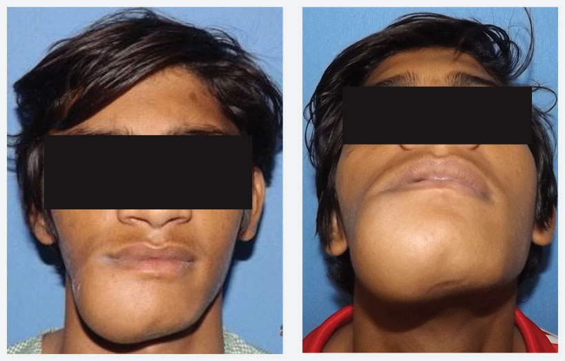
Fig 1: Pre operative photo of 18 year old boy Mr. V. N. showing hard, non fluctuant, non tender swelling over Right side of mandibular parasymphysis-body region extending from right corner of mouth to upper part of neck and midline to body of mandibular area.
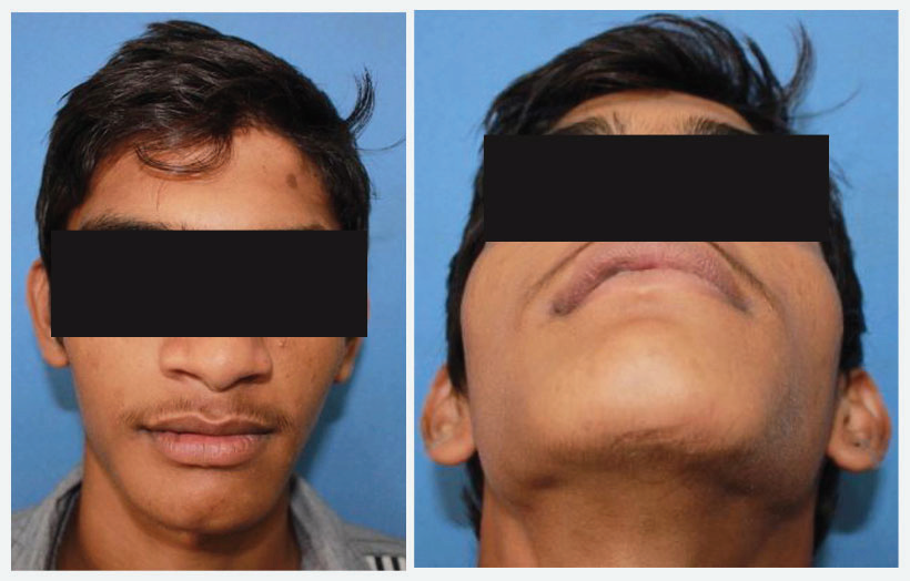
Fig 2: Pre operative Intra operative view showing hard bony swelling obliterating buccal vestibule from lower left central incisor to lower right first molar region.
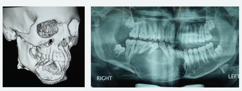
Fig 3: 3D CT of face and X-ray OPG show well defined, unilocular radiolucent area with cotton wool appearance and well demarcated radio-opaque border extending from lower left central incisor to lower right first molar area. There is expansion of both buccal and lingual plate.
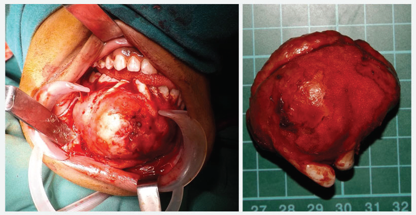
Fig 4: Show the lesion covered with thin shell of bone after mucoperiosteal flap reflection. Show 5x7 cm lesion after excision. Histopathology report suggests the lesion is Ossifying Fibroma.
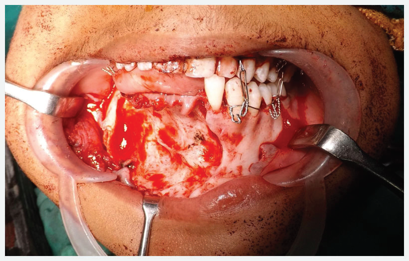
Fig 5: Shows the defect in the mandible after excision of lesion. Very thin shell of bone is left behind after excision. 2.5mm Reconstruction plate is fixed to the defect area to avoid pathologic fracture and Intermaxillary fixation done.
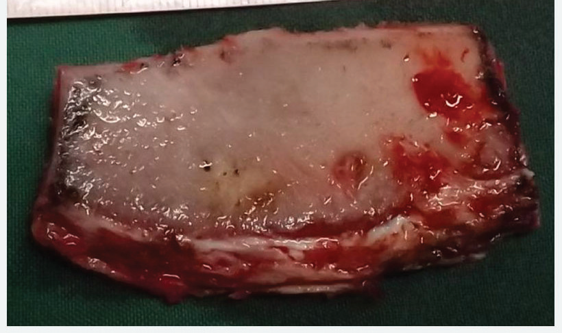
Fig 6: Iliac Crest Cortico-Cancellous Bone Graft Harvested From Right Iliac Crest Region Of About 7X5cm.
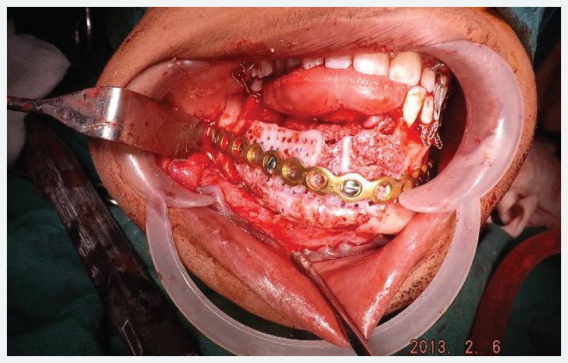
Fig 7: Graft Fixed To Mandibular Defect Using Reconstruction Plate And Screw.
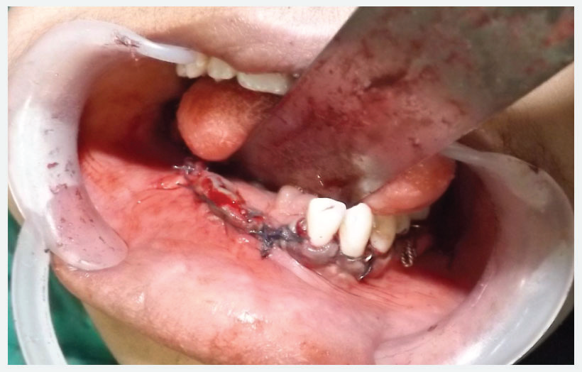
Fig 8: After defect closure.
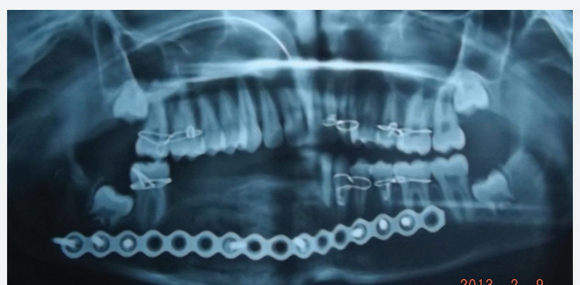
Fig 9: Immediate post op x-ray OPG.
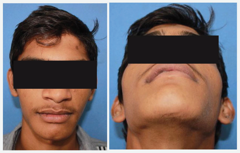
Fig 10: Eight Month Post Operative Photos Show Normal Face. No External Deformation On The Face.
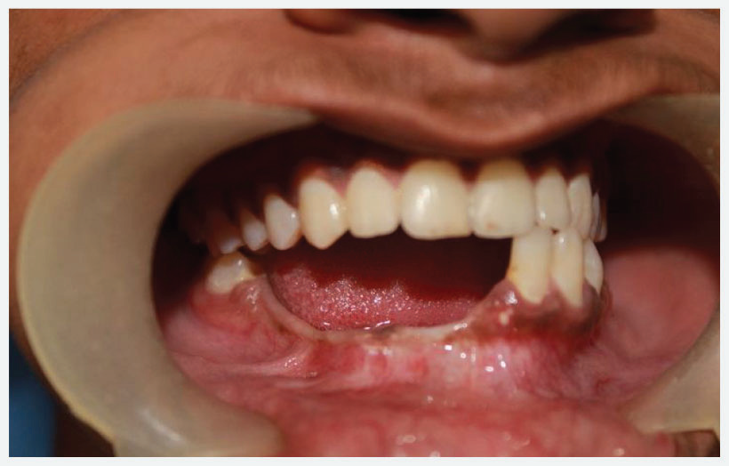
Fig 11: Show Healing Of Lower Ridge After 8 Months. Occlusion Is Normal.
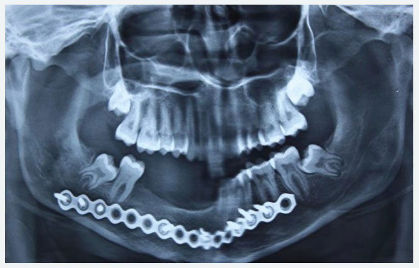
Fig 12: Eight Month Post Op Opg Shows New Bone Formation With Good Quality To Support The Implant.
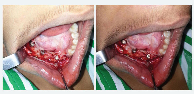
Fig 13: Shows Alpha Dent Classic 5X13 Mm Three Implants In Situ In New Bone.
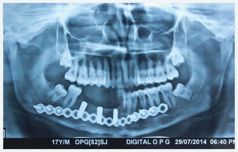
Fig 14: Shows 15 days post op x-ray showing Alpha Dent implant in situ.
Disclaimer from Guident
The views and opinions expressed in articles published in Guident are solely those of the authors and do not necessarily reflect the views of the editors, the editorial board or the publisher, Ivory India. Guident does not endorse any product, technique or treatment described. Clinical content is intended for qualified dental and medical professionals and is not a substitute for individual clinical judgement. Guident, its editors and the publisher accept no liability for any consequence arising from the use of information published in the journal.
How to cite this article
Dr. Parit Ladani. Dental Rehabilitation Using Alpha Dent Implant in Case of Reconstructed Mandible After Excision of Ossifying Fibroma. Guident. 2014 Dec;8(1). Available from: https://ivoryindia.in/articles/implantology/dental-rehabilitation-using-alpha-dent-implant-in-case-of-reconstructed-mandible-after-excision-of-ossifying.html


