Immediate Implantation and Provisionalization: An Approach Towards Preservation and Esthetics
Department of Prosthodontics K.D. Dental College & Hospital, Mathura, UP, India.
Dental implants present a valid treatment modality for completely and partially edentulous patients.Additionally,dental implants provide an excellent solution for patients who have lost a single tooth. The placement of dental implants immediately after tooth extraction is a popular treatment option, which has been reported to have high success rates.
Implant therapy success, especially in the anterior maxilla, is judged not only by osseointegration but also by the presence of sound and stable peri-implant tissues that appear identical to the gingiva surrounding the healthy adjacent teeth. Predictable peri-implant esthetics created immediately after tooth removal requires proper preservation, and often augmentation, of the osseous and soft tissues surrounding the failing tooth, proper three-dimensional implant placement, and proper planning and design of the prosthetic steps following the surgery. Extraction sites in the anterior maxilla present the greatest restorative challenges due to the need for high-quality esthetic results. Implant placement into fresh extraction sockets without using flap elevation has recently become more popular as a treatment modality.
Esthetics in implantology are becoming increasingly important in the contemporary reconstructive and surgical dental practice.Preservation and/or recreation of natural soft tissue emergence profiles lead to the foundation for esthetics in the final implant supported restoration .Provisionalization at implant placement supports the preservation of these natural tissue emergence profiles.
However, successful immediate implant placement depends on satisfactory patient selection, treatment planning, and atraumatic tooth extraction. This enables adequate primary stability and correct implant location in a site that has preserved the natural architecture of soft and hard tissues.
However, it is important to ensure that the interim crown does not contact the opposing dentition in maximal intercuspation or during excursive mandibular movements to allow non-functional loading for successful osseointegration.
CASE REPORT
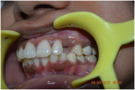
A 23 year old female patient reported with a chief complaint of unesthetic appearance and mobility in her upper front tooth and gave a history of root canal treatment in the same tooth restored with a metal ceramic crown 1.5 years back(Fig 1&2).

Fig 1: Pre-operative (Extra-oral)
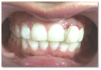
Fig 2: Pre-operative (Intra-oral)
The clinical examination revealed that the gingival margins were inflamed and the crown had grade 2 mobility i.r.t 22. The radiographic examination revealed that the tooth had undergone root canal treatment and horizontal fracture which needed to be extracted.
Different treatment plans were presented to the patient : conventional fixed partial denture, resin bonded prosthesis, placement of an immediate implant, with delayed or immediate loading. The patient consented to an implant-supported restoration with immediate loading of provisional crown replacing the failing tooth.
A CBCT of maxillary anterior region was prescribed for further treatment planning .
Preliminary impressions were made with irreversible hydrocolloid impression material and diagnostic casts for both the maxillary and mandibular arches were fabricated with dental stone.(Fig 3)

Fig 3: Diagnostic cast
Diagnostic waxup was done to ensure optimal esthetics. This cast was duplicated and a new cast was prepared. A clear thermoplastic template of 2-mm thickness was adapted on the new cast .This template served two important purposes:
Firstly as a surgical guide for optimal 3 D placement of the implant .
Secondly it was used to fabricate the provisional. (Fig 4)

Fig 4: New cast with thermoplastic template
On the day of the implant surgery, local anesthesia of 2% lignocaine with epinephrine (1:80,000) was administered and luxators were used for atraumatic extraction of the root (Fig 5& 6).

Fig 5: Removal of PFM crown
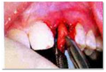
Fig 6 :Atraumatic extraction irt 22
Care was taken to preserve the buccal plate. A surgical curette was used to debride the socket of all granulation tissue.
An initial punch was made in the palatal wall .This was used for subsequent osteotomy for optimum positioning of the implant(Adin implant, 3mm*11.5mm) ensuring no contact with the buccal cortical plate. (Fig 7).
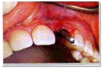
Fig 7: Osteotomy site irt 22
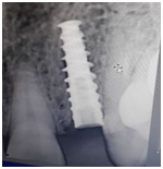
fig 8: Radiograph showing implant placed i.r.t 22
A radiograph was taken to verify the correct placement of implant.
The platform of the implant was placed 1.5 mm apically to the cementoenamel
junction of the left maxillary central incisor.(Fig 8)
A temporary abutment was placed on the implant .Provisional crown was fabricated in 2 steps. Initially the subgingival profile was fabricated followed by the coronal portion. (Fig 9& 10).

Fig 9: Temporary abutment

Fig 10: Modification of temporary abutment to get the subgingival profile
The access screw hole was blocked with PTFE Tape and putty .
For the fabrication of incisal/anterior of the temporary crown , the template was placed on the maxillary arch intra orally and hole was drilled to allow the proper flow of provisionalization material(Dmg Luxatemp-Fluorescence) followed by light curing for 20 seconds. After complete curing the template was removed and the provisional screw retained prosthesis was finished and polished.(Fig 11).
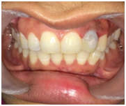
Fig 11: Finished crown on temporary implant abutment

Fig 12: Radiograph showing implant with provisional abutment and crown irt 22
A 2nd radiograph was taken with the temporary abutment and crown in place.(fig 12)
The occlusion was evaluated and any centric and eccentric contacts were eliminated to allow non functional occlusion.
Post surgical instructions were given to the patient for hygiene maintenance and medication was prescribed .Patient was recalled after day of surgery for post operative check up and final finishing of the prosthesis was done.
After 4 months ,radiographs were taken to verify optimum osseointegration .
For final impression ,provisional crown was removed and close tray transfer coping was modified with composite to transfer the subgingival profile and emergence profile and final impression was made with putty ( Dentsply Aquasil soft putty)and light body(Dentsply Aquasil ultra) impression material.(Fig 11)
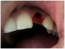
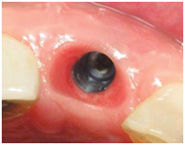
Fig 13: Soft tissues after 4 months of healing (after removal of provisional)
Shade selection was done in the day light and provisional crown was reseated in place.
The final PFM crown was placed along with abutment and x-ray was taken to verify the placement(fig 16) and occlusion was evaluated by doing centric and eccentric movements.(Fig 14 & 15)

Fig 14 : Cast with abutment and PFM crown

Fig 15: Abutment placed i.r.t 22

Fig 16: Radiograph showing implant with permanent
Patient was recalled for follow up after 15 days interval.(Fig 17 & 18)

Fig 17: PFM crown cemented on the abutment
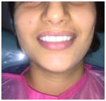
Fig 18: Post operative view
DISCUSSION
The extended treatment period and the need for a removable prosthesis during the healing phase of the traditional approach may be inconvenient to certain patients.The original approach has been modified by several authors to include 1-stage surgery, immediate implant placement, and immediate provisionalization. Immediate implant placement and provisionalization may be a good treatment option in the loss of anterior teeth, but this approach may be recommended
when there is no need for a bone augmentation procedure.
The surgery protocol should include the evaluation of bone quality and quantity as well as measures to ensure that the placement of prosthesis and other components is adequate.
A critical factor in implant rehabilitation of teeth in the anterior maxilla is the 3 D positioning of implants.Implant insertion at a palatal position in relation to the alveolar ridge preserves the buccal bone wall and provides better primary stability therefore it reduces the incidence of future gingival recession and implant exposure.
A surgical template is required to guide the optimal positioning of an implant immediately after tooth extraction and secure the immediate placement of an interim crown.
pre-existing gingival architecture surrounding the extracted tooth is to be preserved, the volume and shape of the prosthetic elements from the implant head up to the free gingival margin and papilla peaks should be designed and created to fill and support the pre-existing gingival cuff.
Support of the gingival margin with an interim restoration at the time of the tooth extraction and implant placement preserved 1 mm more facial gingival margin position than delayed implant placement.
An improved esthetics during provisionalization may sometimes be obtained if composite resins are used for the same purpose. Besides shade, other parameters such as surface texture and contour of crown are important for a successful result. Correct contour is also essential for proper support of the adjacent soft tissues.The mesial and distal papillae should be supported but not compressed with the crown form. In addition, the cervical third should be anatomically correct to avoid gingival margin recession.
CONCLUSION
Tooth extraction,immediate implant placement and provisionalization with immediate loading is a treatment option which offers several advantages compared to the conventional 2 stage protocol such as simplicity,superior esthetic outcome,immediate fixed prosthesis and less time required for the final restoration.
In this report, the harmony of soft and hard tissue was achieved by immediate implant placement with immediate loading of provisional crown in esthetically challenging region.
REFERENCES
- Volpato el al; A modified surgical template with dual function ;J Prosthet Dent 2013;110:232-233.
- Michalakis et al; Conversion of an existing metal ceramic crown to an interim restoration and nonfunctional loading of a single implant in the maxillary esthetic zone: A clinical report ;J Prosthet Dent 2014;111:6-10.
- Jun-Beom Park; Immediate Placement of Dental Implants Into Fresh Extraction Socket in the Maxillary Anterior Region: A Case Report; JOI 2010; vol 36:2 .
- M.F. Pasha et al;Immediate implant placement and provisionalization:optimizing esthetics and function ;International journal of oral implantology and clinical research;May –Aug 2014;5(2):72-78.
- Bo Rosenquist ; A comparison of various methods of soft tissue management following the immediate placement of implants into extraction sockets; INT JOMI 1997;12:43–51.
Disclaimer from Guident
The views and opinions expressed in articles published in Guident are solely those of the authors and do not necessarily reflect the views of the editors, the editorial board or the publisher, Ivory India. Guident does not endorse any product, technique or treatment described. Clinical content is intended for qualified dental and medical professionals and is not a substitute for individual clinical judgement. Guident, its editors and the publisher accept no liability for any consequence arising from the use of information published in the journal.
How to cite this article
Dr. Deeksha Agrawal, Dr. Rahul Nagrath. Immediate Implantation and Provisionalization: An Approach Towards Preservation and Esthetics. Guident. 2018 Jul;11(8). Available from: https://ivoryindia.in/articles/implantology/immediate-implantation-and-provisionalization-an-approach-towards-preservation-and-esthetics.html