Use of Platelet Rich Fibrin (PRF) and Bone Grafts as Adjuanct in Immediate Implant Placement
K.D Dental College & Hospital, Mathura, India
INTRODUCTION
The need and demand for increasing esthetic values and short duration treatment led to the evolution of immediate implants. Immediate implant is the placement of dental implant in a fresh extraction socket.1
The primary advantages of immediate implant placement are the reduction in time and cost of therapy, the reduction in surgical episodes, and preservation of the bone and gingival tissues.2
When a dental implant is placed into a fresh extraction socket, the space between the implant periphery and surrounding bone is called the gap or jumping distance. The gap consists of 2 dimensions: horizontal defect width and vertical defect height.(Fig 1) A gap can occur on any aspect of an immediately placed implant: buccal, lingual, or proximally. Bone fill in the gap between the implant and the peripheral bone is important. The dimensions of this gap will vary depending on a number of factors, such as tooth type, the particular morphology of the extraction socket and the diameter of the implant being placed. Gaps <2mm heal spontaneously, whereas larger gaps may require the use of bone grafts and barrier membranes better healing.
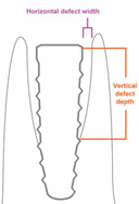
Fig 1: Jumping Gap
A number of bone grafting materials are available for use in site preservation. These fall into following four for categories.

PRF is an autologous grafting material . Besides, its jelly-like consistency favors stability of the clot and of the grafting material. This natural material seems to accelerate the physiological wound healing; besides, in association with bone grafts, it seems to accelerate new bone formation
There are certain clinical requirements which should be fulfilled for attaining high success rates for immediate implants such as absence of any frank active infection, good mechanical anchorage and primary stability of the implant fixture within the alveolar socket, atraumatic removal of unsalvageable tooth, preservation of labial plate of bone, use of the appropriate implant design that corresponds to the configuration and position of socket.3
INDICATIONS FOR IMMEDIATE IMPLANTS
Immediate dental implants may be considered the treatment of choice for an endodontically infected tooth, root fracture, root resorption, periapical pathology, root perforation, and unfavorable crown to root-ratio (not due to periodontal loss). However, site selection remains very controversial.
BONE GRAFT MATERIALS
Different types of bone grafting materials are available for preservation and ridge augmentation. They are:
Autografts:
These are also known as autogenous bone grafts. They are derived from the recipient and are traditionally in block or particulate form. Autogenous bone grafts can be harvested from extraoral or intraoral sites. Common intraoral sites include the tuberosity, the mandibular symphysis, and the palate, while common extraoral sites used for block bone grafts for ridge augmentation include the tibia and hip.
Xenografts:
Grafts may be osteoinductive. Xenogenous bone grafts, or xenografts, are derived from a different species, typically bovine or porcine grafts. The bone is treated during the manufacturing process to ensure biocompatibility and no risk of disease transmission. Xenografts are deproteinized bone and are always osteoconductive. A collagen-based grafting material is also available.
Alloplasts:
Alloplasts are derived from a number of synthetic sources, including a synthetic, resorbable sponge containing polylactide-polyglycolide acid, calcium phosphates, calcium sulfates, bioactive glasses, coral (which contains hydroxyapatite and TCP), hydroxylapatite, tricalcium phosphate and methylmethacrylate-based materials. Allografts are osteoconductive and can be resorbable or nonresorbable

Platlet Rich Fibrin (PRF)
Various pathological etiologies result in dysfunction of the oral tissues and creation of bony defects, thus affecting the quality of life in these patients. The pivotal goal in periodontal and maxillofacial tissue regeneration is to reconstruct these defects which led to the search of a biofuel.4
Various platelet-derived products or platelet concentrates have been introduced that act as biological mediators aiding the healing response. Platelet-rich fibrin (PRF) is one such product that has proved its worth and has edged past the others.5
Developed in France by Choukroun et al. in 2001.Unlike PRP, this technique does not require anticoagulants , bovine thrombin or any other gelifying agent. The PRF clot is yielded by a natural polymerization process during centrifugation, and its natural fibrin architecture seems responsible for a slow release of growth factors and matrix glycoproteins during ≥7 days.6; It consists of :
1.Transforming growth factor-𛽠(TGF-ð›½) Transforming growth factor-𛽠(TGF-ð›½):
It is released from ð›¼-granules of platelets and stimulates proliferation of osteoblasts. It has role in synthesis of collagen type I and fibronectin. It enhances woven bone formation and promotes chemotaxis of osteoblast cells formation. It also stimulates angiogenesis
2.Platelet-derived growth factor (PDGF) helps in migration and proliferation of mesenchymal lineage cells. It has an angiogenic effect on endothelial cells.
3.Vascular endothelial growth factor (VEGF)- initiates angiogenesis.
4.Insulin growth factor-1 (IGF-1)- It stimulates osteoblast proliferation and has chemotactic effects towards human osteoblasts. It Increases the expression of osteocalcin and enhances wound healing.
5.Fibroblast growth factor (FGF)- It stimulates osteoblast proliferation and has chemotactic effects towards human osteoblasts. It increases the expression of osteocalcin and enhances wound healing.
6.Epidermal growth factor (EGF) It stimulates cell proliferation and extracellular matrix turnover. It has chemotactic effect on periodontal fibroblast cells.
PREPARATION OF PRF
Preparation of PRF follows the protocol developed by Choukroun et al in 2001.
The steps involved are as follows:
- Blood specimen is collected from the patient,
- The blood specimen is placed in the centrifuge and is allowed to spin immediately for 10 minutes at 3000 rpm or 2700 rpm for 12 minutes.

Fig 2 Preparation of PRF
Its application and role:
1) PRF and PRF membrane have been used in combination with bone grafts to hasten the healing in lateral sinus floor elevation procedures.
(2) Protection and stabilization of graft materials during ridge augmentation procedures.
(3) Socket preservation after tooth extraction or avulsion
(4) PRF membrane has been used for root coverage with single and multiple teeth recession.
(5) Regenerative procedures in treatment of 3-walled osseous defect.
(6) In the treatment of combined periodontic endodontic lesion.
(7) Treatment of furcation defect.
(8) PRF enhances palatal wound healing after free gingival graft.
(9) Filling of cystic cavity.7
In the following case series, immediate implant placement along with the use of PRF (platelet rich fibrin) and various bone grafts have been discussed.
CASE REPORT 1
A 36 year old male patient reported to the Department of Prosthodontics with the chief complaint of mobility and discoloration in relation to 21. After the radiographic assessment the patient was diagnosed with internal resorption in relation to the 21. Various treatment options were discussed including fixed partial denture, resin bonded fixed partial denture, removable partial dentures and implant supported prosthesis. (Fig 3a,b) After discussion on the treatment options, immediate implant was chosen as the treatment of choice.

Fig 1a) Discoloration in relation to 21

Fig 1b) Radiographic assessment of 21
Technique:
A Diagnostic impression was made using irreversible hydrocolloid.A Stone (Type IV) cast was made and a surgical template using thermoplastic sheet was fabricated for accurate placement of implants in the surgical site.
Atraumatic Extraction
The tooth in question was extracted atraumatically using periotome and luxator. The surgical approach included intrasulcular incisions around the tooth to be extracted. The periotome was used to sever some of the periodontal ligament around the coronal aspect of the tooth. The luxator was used to widen the PDL membrane and tooth was extracted atraumatically. (Fig 4a,b)

Fig 2a) Extraction of 21

Fig 2b) Extracted Tooth (notice the resorbed root)
Socket Debridement
After extracting the tooth, socket walls were thoroughly debrided . This was done to induce fresh bleeding into the socket which promotes better healing. Failure to perform this step thoroughly can result in fibrous healing and poor graft consolidation within the socket, thereby potentially jeopardising subsequent dental implant healing.
An Implant of 4.25 mm in diameter and 13 mm in length was used (Adin) with the help of surgical stent. The osteotomy was initiated palatally to engage the palatal bone, to prevent the esthetic compromise because of the thin facial plate.

Fig 5 Palatal orientation of immediate implant
The graft material NovaBone (Bioactive synthetic bone graft) was lightly packed into the extraction socket in an incremental fashion.(Fig 6) Platelet rich fibrin was placed along with the graft.(Fig 7 a,b)

Placement of healing collar
A healing collar was placed after four months of implant placement. (Fig 8)

Fig 8: Placement of healing Collar
After two weeks of placement of the healing collar, a closed tray impression was made in Soft Putty (3M ESPE). Appropriate shade selection was done for the fabrication of final crown.
Final Restoration
The prepared abutment was torqued to the implant to restore the final crown. (Fig 9) Porcelain fused to metal crown was then cemented. (Fig 10).

CASE REPORT 2
A 21 year old female patient reported to the department of Prosthodontics with abnormal growth in the tooth in relation to 46. After the clinical and radiographic assessment the patient was diagnosed with pulp polyp in relation to the tooth in question. Due to the questionable prognosis of the tooth,it was advised for extraction and immediate implant placement.(Fig 11a,b)

Fig 11 a) Pulp polyp in relation to 46.

Fig 11 b) Radiographic assessment of 21
Technique:
A Diagnostic impression was made using irreversible hydrocolloid.A Stone (Type IV) cast was made and a surgical template using thermoplastic sheet was fabricated for accurate placement of implants in the surgical site.
Atraumatic Extraction
The tooth in question was extracted using atraumatically using periotome and luxator under local anesthesia. (Fig 12a,b).The socket was debrided and curetted in order to induce bleeding and promote better healing.

An Implant of 4.2 mm in diameter and 13 mm in length(Adin) was placed with the help of surgical template. BioGraft (synthetic bone graft) was used as a bone graft material. Platelet rich fibrin was placed along with the graft.The graft material was lightly packed into the extraction socket in an incremental fashion. (Fig 13-15)

Fig 13 a)Immediate Implant Placement irt 46

Fig 13 b) Radiograph Showing Immediate Implant irt 46

Fig 14 a)BioGraft Bone Graft

Fig 14 b)Platelet Rich Fibrin

Fig 15)Placement of platelet rich fibrin and bone graft
Placement of healing abutment
A healing abutment was placed after three months of implant placement. After 15 days healing abutment was removed and impression coping was placed and a closed tray impression with polyvinyl siloxane impression material was made(3M ESPE Soft Putty and Light body).
Final Restoration
The prepared abutment was torqued to the implant to restore the final crown. Porcelain fused to metal crown was then cemented.(Fig 16a,b)

Fig 16 a) Final prosthesis cemented

Fig 16 b) Radiographic view of final prosthesis
DISCUSSION
With the advent of immediate implant dentistry, implant fixtures are now regularly placed into fresh extraction sockets that, at their most coronal aspects, are sometimes much larger than the diameter of the implants being placed. Primary stability is generally achieved at the apical aspect of the implant fixture, but there is usually somewhat of a gap between the circumferential aspect of the immediate implant and the extraction socket wall. Management of this immediate peri-implant gap has become a novel challenge.8,9 This is particularly true of the buccal or labial gap because the appropriately heightened focus on both function and aesthetics in contemporary implant dentistry dictates that the facial profile of the implant restoration be developed in harmony with the surrounding tissues. The presence of a peri-implant gap affects both hard and soft tissue healing, and if not properly managed, may compromise function and jeopardize aesthetics.10
Preserving or reconstructing the extraction socket of a failed tooth according to the principles of guided bone regeneration enhances our ability to provide esthetically pleasing restorations to our patients without violating the predictability and function of those prostheses.
In this case report ,a PRF membrane was used along with bone grafts. Epithelial and connective tissue healing on PRF membranes is related both to the growth factors and fibrin matrix.Gingival fibroblasts easily migrate into this matrix and remodel it.PRF increases the cohesion between the graft materials as fibrin acts as a physiological glue between wounded tissues. Natural blood coagulation leads to the formation of a fibrin matrix that biologically links wounded tissue together allowing cell tissue proliferation,cell migration .neomatrix apposition and remodeling.Therefore combination of PRF with different kinds of filling materials improve the integration of grafted material,as PRF is an optimized blood clot.
CONCLUSION
Immediate implant placement is now accepted in clinical dentistry for reconstruction of partially or completely edentulous mandible or maxilla.8,9 This procedure has several advantages, such as prevention of bone resorption, reduced number of surgical visits, reduction of comprehensive treatment time, better esthetics and higher patient satisfaction compared with delayed placement of implants. However, because of the nature of this treatment method, a higher risk for complications and failures might be expected.10
Treatment protocols and guidelines should be followed to prevent complications. Case selection and evaluation of patient-related and implant-related factors are keys to the success of immediate implant placement. A thorough discussion between practitioner and patient is indispensable to discern patient’s desires. Practitioners need to consider these desires when planning immediate, early, or delayed implant placement.
REFERENCES
- Jordi Ortega-Martínez et al ( 2012) Mar 1 Immediate implants following tooth extraction. A systematic review, Med Oral Path Oral Cir Bucal.;17 (2):251-61.
- Abd El Salam El Askary ;Fundamentals of esthetic Implant dentistry;2007;2nd edition
- V. Gassling, T. Douglas, P. H. Warnke, Y. Ac¸il, J. Wiltfang, and S. T. Becker; Platelet-rich fibrin membranes as scaffolds for periosteal tissue engineering,Clinical Oral Implants Research;2010; 21; 543–549,
- V. Gupta, V. K. Bains, G. P. Singh, A. Mathur, and R. Bains, “Regenerative potential of platelet rich fibrin in dentistry: literature review,” Asian Journal of Oral Health and Allied Sciences, 2011; 1.;22–28.
- Choukroun J, Adda F,Schoeffer C,Vervelle A.PRF:an opportunity in perio implantology,Implantodontie 2000;55-62.
- Sujeet Vinayak Khiste and Ritam Naik Tari:Platelet Rich Fibrin as a Biofuel for tissue Regeneration.ISRN Biomaterials:2013;1-5
- Schwartz-Arad D, Chaushu G. Placement of implants into fresh extraction sites: 4 to 7 years retrospective evaluation of 95 immediate implants. J Periodontol 1997;68:1110-6.
- Schwartz-Arad D, Chaushu G. The ways and wherefores of immediate placement of implants into fresh extraction sites: A literature review. J Periodontol 1997;68:915-23.
- Schropp L, Isidor F. Timing of implant placement relative to tooth extraction. Journal of Oral Rehabilitation 2007;35;33-43.
- Araújo MG, Wennström JL, Lindhe J. Modeling of the buccal and lingual bone walls of fresh extraction sites following implant installation. Clin Oral Implants Res. 2006;17:606-614.
Disclaimer from Guident
The views and opinions expressed in articles published in Guident are solely those of the authors and do not necessarily reflect the views of the editors, the editorial board or the publisher, Ivory India. Guident does not endorse any product, technique or treatment described. Clinical content is intended for qualified dental and medical professionals and is not a substitute for individual clinical judgement. Guident, its editors and the publisher accept no liability for any consequence arising from the use of information published in the journal.
How to cite this article
Dr. Rahul Nagrath, Dr. Shipa Arora, Dr. Lijo Mathew Department of Prosthodontics. Use of Platelet Rich Fibrin (PRF) and Bone Grafts as Adjuanct in Immediate Implant Placement. Guident. 2016 Apr;9(5). Available from: https://ivoryindia.in/articles/implantology/use-of-platelet-rich-fibrin-prf-and-bone-grafts-as-adjuanct-in-immediate-implant-placement.html