
Traumatic Induced Mucocele of the Lower Lip
Department of Oral and Maxillofacial Surgery, Goa Dental College and Hospital Bambolim, Goa, Shetye Dental Clinic and Implant Centre, Vasco da Gama, Goa
ABSTRACT
Oral mucocele is an very common entity of the minor salivary gland usually caused due to trauma or obstruction of the minor salivary gland. They are classified either as retention or extravasation type based on their histopathological findings. Diagnosis is usually based on the clinical findings with lip being the most common site for occurrence. Surgical excision of the lesion is the choice of treatment.
Keywords: mucocele
INTRODUCTION
Oral mucocele is the seventeenth most common minor( accessory) salivary gland lesion of benign nature, caused by trauma to the excretory duct of the gland.1 It belongs to a category of reactive lesion being defined as mucous filled cavities which can appear in the oral cavity, appendix, gall bladder, paranasal sinuses and lacrimal sac.2,3
The term mucocele is derived from the Latin word mucous or mucus, and coele or a cavity.4 Usually two types are commonly seen based on their microscopic characteristics, mucus retention or mucus extravasations cyst.4,5,6 Mucus retention cyst is usually caused by obstruction of the duct of the minor salivary gland whereas mucus extravasation cyst is of traumatic origin like lip biting.
Mucocele are asymptomatic, appear well circumscribed, rounded, transparent and bluish color of variable size. They are found to be soft in consistency and fluctuant on palpation. They vary from 1mm to few centimeters in size, affecting both the genders equally with its peak in the 2nd and 3rd decade of life.6 Most common site of occurrence is lower lip where there is opening of accessory duct like buccal mucosa, anterior ventral tongue, floor of the mouth and rarely upper lip.
This case report is aimed to explain the history, clinical features and come to clinical diagnosis followed by removal of the mucocele by simple excision under local anesthesia which will help to enhance the knowledge of the clinician.
CASE REPORT
A 25 year old young man reported with swelling on the inner aspect of the lower lip on left side since 3 weeks. He gives history of swelling being small initially and increasing gradually to its current size. He gives history of sharp edge of upper left central incisor and lip sucking with no difficulty in chewing or speaking.
Intraoral examination shows a solitary, round well circumscribed swelling approximately 1cm in diameter with well defined margins. On palpation the swelling was noted to be soft in consistency, fluctuant, borders are well defined and surrounding mucosa appeared to be normal with no pulsation, not reducible or compressible.
This lesion was clinically diagnosed as a mucocele based on the history and clinical features. Complete hemogram was within normal limits.
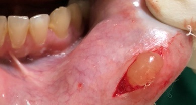
Figure: 1

Figure: 2

Figure: 3

Figure: 4
DISCUSSION
Mucocele is a self limiting mucoid containing cyst of the salivary gland with relatively rapid onset and fluctuating size. Decrease in size may be due to rupture of the lesion ,and subsequent mucin accumulation or reabsorption of the saliva deposits may cause the lesion to reform.1 Obstructive disorder are affecting the major and minor salivary gland, the causative mechanism being reduction or obstruction of salivary flow resulting in one of the two processes, destruction of the secretory elements of the gland or a mechanical blockage or disruption of the ductal system.2
Oral mucocele of salivary gland are rarely larger than 1.5cm in diameter and are always superficial. They rarely cause any problem. Discomfort, interference with speech, mastication swallowing and external swelling may occur depending on the location.1,2 Lower lip (90%) was the most frequent location followed by floor of the mouth (6% ) and central surface of the tongue( 4%).3,6 Ranula designates mucocele located on the floor of the mouth. Literatures showed oral habits such as lip biting/sucking is one of the etiologic factors for the oral lesion. Radiographic evaluation is needed to rule out sialolith. the fine needle aspiration cytology demonstrates the mucus retention phenomenon.2,5
The appearance of mucocele is pathognomonic. Location of lesion, history of trauma, rapid appearance, variation in size, bluish colour, and the consistency ,history and clinical findings lead to a diagnosis of superficial mucocele. This is an distinguishing feature from other lesion of the lip like fibroma, lipoma,sialolith,phlebolith arising from adipose tissue, connective tissue, blood vessels, nerves and salivary glands.4,6
The scalpel is one of the most commonly and simple method of excising a mucocele. Micro marsupialisation, cryosurgery, laser, electro cautery have also been tried. Scalpel is more popular as it does it need any extensive equipment, negligible cost and can be performed by trained dentist. With simple incision of the mucocele, the content would drain out but the lesion would reappear as soon as the wound heals. Total excision of the lesion is the choice of treatment. A semilunar incision is placed on the mucosa, the mucocele is seen popping out from the mucosa. Dissection is carried out all around the mucocele till the muscle layer underneath and it is dissected out for the base. Interrupted sutures are placed. To reduce recurrence the underlying cause must be addressed too.7,8
REFERENCES
- More CB,Bhavsar K, Varma S, Tailor M: J Oral Maxillofac Pathol 2017,2-8
- Anand S.,Wanjari Sonalika,Vanishree HS: Oral Mucocele: A clinicopathological analysis of 50 cases:Archives of Medicine and Health Sciences, Jan-Jun 2016,vol 4,40-44
- K.U.Nallasivam, B R Sudha: Oral Mucocele : Review of literature and a case report: J Pharm Bioallied Sci :2015 Aug: 7(Suppl 2) : S 731-733.
- P.Chaitanya,D. Praveen, Madhusudhan Reddy: Mucocele on lower lip: A case series, Indian Dermatology Online Journal 2017
- G.Giraddi, Aamir Malick Saifi: Micro-marsupialisation versus surgical excision for the treatment of mucoceles; Annals Of Maxillofacial Surgery,July-December 2016, Vol 6, Issue 2, 204-209
- Neville B,Damn DD,Allen CM, Bouquott JJ. Oran and maxillofacial pathology . 2nd ed. Philadelphia: W B saunders: 2002.pp 389-92
- Y.Gautam, M. Srivastava: Mucocele in pediatric patients: A case series with review; International Journal of Applied Dental Sciences 2018;4(2):100-103
- R.Badjatia,S.Badjatia, V. Kulkarni, D. Sharma; Oral Mucocle : a case report; NJDSR Number 2,vol1,January 2014 ,13-16
Disclaimer from Guident
The views and opinions expressed in articles published in Guident are solely those of the authors and do not necessarily reflect the views of the editors, the editorial board or the publisher, Ivory India. Guident does not endorse any product, technique or treatment described. Clinical content is intended for qualified dental and medical professionals and is not a substitute for individual clinical judgement. Guident, its editors and the publisher accept no liability for any consequence arising from the use of information published in the journal.
How to cite this article
Dr. Omkar Shetye, Dr. Nandita Keni Shetye. Traumatic Induced Mucocele of the Lower Lip. Guident. 2020 Jun;13(7). Available from: https://ivoryindia.in/articles/oral-surgery/traumatic-induced-mucocele-of-the-lower-lip.html


