Sublingual Ulcer Associated with Natal Teeth: Case Report and Review of Treatment
Department of Pediatric Dentistry, Sri Ramakrishna Dental College and Hospital, Coimbatore
ABSTRACT
Teeth present at birth or erupting shortly after birth is not very common. They are called the Natal and Neonatal teeth respectively. These teeth often present with numerous complications like difficulty in feeding, sublingual ulceration, risk of aspiration etc. Proper management of these teeth is important to prevent the complications and ensure a good oral health of an infant. In this manuscript, a case report of sublingual ulcer associated with natal teeth in a 20 day old child is presented and discussed. Extraction was chosen as the treatment option after considering the other treatment modalities.
Key words: Natal teeth, neonatal teeth, Sublingual ulcer
INTRODUCTION
Presence of teeth in newborns is uncommon.1 Normally, primary teeth begin to erupt at approximately 6 months of age and this is an important milestone in the child’s life. In rare cases, the first teeth are present at birth or will emerge shortly after birth and are called natal teeth and neonatal teeth respectively.2 This significant alteration in the chronology of tooth eruption brings about a chain of reactions due to the numerous problems associated with it like pain on suckling, refusal to feed, ulceration on tongue, fear of dislodgement and aspiration and so forth.
Sublingual ulcer, also known as Riga-Fede disease (RFD) is a rare traumatic ulceration of the ventral surface of the tongue and is most often associated with natal or neonatal teeth in newborns.3,4 It is also seen in older infants, in children with repetitive tongue thrusting habits5 , and in children with familial dysautonomia (insensitivity to pain).6 The problems associated with natal/ neonatal teeth makes early dental intervention mandatory.
CASE DESCRIPTION
A 20 day old child was brought to Sri Ramakrishna Dental College and Hospital with tooth in the lower jaw since birth. A history of subsequent development of an ulcer on the undersurface of the tongue was recorded. Since then, continuous crying of the child with much reduced feeding was noted by the parents.
No family history of any developmental disorder and congenital syndromes was obtained. On examination, there were two teeth in the mandibular anterior region which was milk white in color with sharp irregularities on the superior edge, erupted up to 1.5 to 2 mm into the oral cavity. The teeth showed Grade 2 mobility. The lesion on the ventral surface of the tongue was at the junction of attachment of lingual frenum. It was elliptical with erythematous, raised and indurated border with overlying whitish slough on the surface (Figure 1).
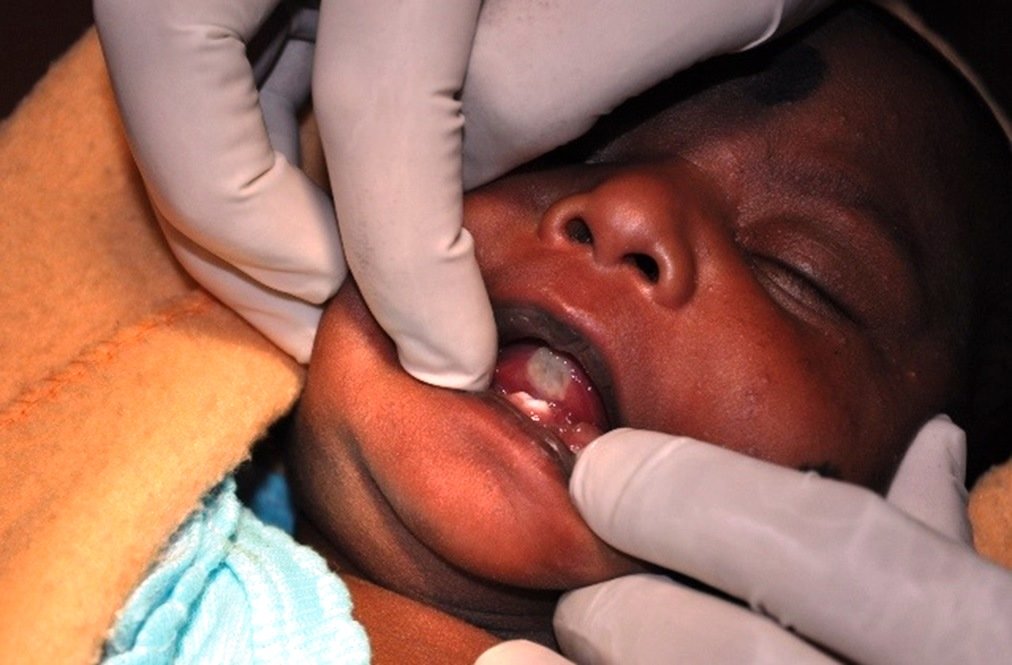
Figure 1 – Intraoral view showing the natal teeth with sublingual ulcer.
Based on the findings, the cause of the lesion on the ventral surface was associated to the tooth in the lower jaw and the lesion was diagnosed as Riga-Fede disease. As the size of the ulcer was medium, conservative treatment options like composite resin coverage and smoothening of the incisal edges were considered. But mobility and fear of aspiration and minimal tooth structure available in the oral cavity led to the selection of extraction as the treatment of choice over the above said treatment modalities. The child was administered Vit K (0.5mg) by the pediatrician. The extraction of the natal tooth was done under local anesthesia, followed by gentle curettage of the socket. The extracted tooth was found to be immature without any root formation. Ulcer healed completely in 2 weeks (Figure 2). The parents’ apprehensions were alleviated. The newborn’s parents were advised about the importance of future follow up.

Figure 2 – Regression of sublingual lesion observed after 2 weeks
DISCUSSION
Natal and neonatal teeth was first documented by Titus Livius, 59 BC, and Gaius Plinius Secundus, 23 BC,7 and since then many cases have been reported in the dental literature.
Though many terminologies were used previously the analogous terms of “natal” and “neonatal teeth” described by Massler and Savara are now the most accepted.2 Teeth erupting beyond the natal period of thirty days (within 1-3.5 months) are usually referred to as early infancy teeth.8,9
The reported prevalence of natal and neonatal teeth in a normal population has varied considerably from 1 in 11 to 1 in 30,000 births.10 Natal teeth are three times more common than neonatal teeth and they erupt 85% in mandibular incisor region, 11% in maxillary incisor region, 3% in mandibular canine region and 1% in maxillary canine and molar region.2,8,9 Most commonly, these teeth are precociously erupted from the normal complement of primary teeth (90%-99%). Only 1% to 10% of natal and neonatal teeth are supernumerary11,12,13 Spouge and Feasby 14 however, believe that such structures, present at birth, undoubtedly represent only the dental lamina cyst of the newborn.
Over the years there has been many postulations about the cause of premature eruption including endocrine disturbances, hypovitaminosis, syphilis, febrile status, environmental factors, familial trait (8-62%) and trauma but a cause and effect relationship has not yet been established. But the most accepted theory is based on superficial localization of dental follicles, probably related to a hereditary factor.1,11
Complications include discomfort during suckling, irritation and trauma to infants' tongue, sublingual ulceration (RFD), laceration of the mother's breast and risk of aspiration of the mobile teeth 10 Prolonged gingival irritation from natal or neonatal teeth may cause localized inflammation of the gingiva or fibrous hyperplasia.15
Sublingual ulceration
It is a reactive, traumatic mucosal disease characterized by persistent ulcerations of the oral mucosa. The lesion was first described by Antonio Riga, an Italian physician in 1881.4 Histological studies and additional cases were subsequently published by Fede and has since been known as “Riga-Fede disease"5 Few reports cite Carderelli (1857) first described the condition in a cachectic infant, which was later referred to by several Italian authors as ‘afta cachectica’.16 A variety of other terms have been applied to this lesion, including sublingual growth in infants, lingual traumatic ulceration, traumatic atrophic glossitis, and traumatic granuloma of the tongue. Elzay called it “Traumatic ulcerative granuloma with Stromal eosinophilia” (TUGSE) 16 and Goho3 described it as “Neonatal sublingual traumatic ulcer”.
The lesion usually begins as an ulcerated area on the ventral surface of the tongue. On repeated trauma it may progress to an enlarged, fibrous lesion with the appearance of an ulcerative granuloma (Elzay, 1983). Based on these characteristics the differential diagnosis of RFD should include neoplasms and infective conditions. However, once the clinician is familiar with the diagnosis RFD, the history and clinical features are most often so typical that there is seldom a need for additional histopathological examination.
Presentation appears to be bimodal. Early cases (before six months of age) are related to natal or neonatal teeth. Late cases (six months of age and older) occur with primary dentition, and are frequently habitual and may be related to neurological or developmental disorders.17 Domingues–Cruz et all 18 called them ‘precocious’ and ‘late Riga Fede disease’. A relation with chronic trauma due to the teeth has been proposed as the etiology by many authors.3,17,18
Treatment
Natal and neonatal teeth present a challenge when deciding on appropriate treatment. In the past, the natal teeth and the ulcer have been treated both radically as well as conservatively. Conservative approaches are generally used to deal with small ulcers 19 One such treatment option is Composite resin coverage of the incisal edges.3 Though conservative, there are practical difficulties associated with this procedure. Hypomineralisation of enamel and reduced tooth structure and moisture control make bonding and hence retention of the resin difficult. Other conservative procedures include smoothening of sharp incisal edges,3,7,12 Stomadhesive wafers4 and modifying the feeding behavior or using a feeding device.5 Some authors recommend “leaving them alone, unless they are causing difficulty to the infant and mother. Teeth that are not excessively mobile or not causing feeding difficulty are to be preserved and maintained with close monitoring at frequent intervals.2,5
Extraction is indicated if the tooth is supernumerary or if the tooth is excessively mobile and is associated with the risk of aspiration.4,15 Complications like RFD indicate extraction. However, according to Zhu & King [19] RFD by itself is not an indication for extraction. Size of the ulcer is an important consideration, and in case of large and denuded ulcers, the effectiveness of the conservative techniques is questionable and these are best treated by extraction.3 Potential for hemorrhage should be considered when planning extraction for newborns. Excision of the lesion, radiation and liquid nitrogen has been practiced earlier as treatment options.
CONCLUSION
Many cases of natal and neonatal teeth have been reported in medical and dental literature. Its prevalence, etiology, associated syndromes, complications and treatment modalities have been vastly discussed. Yet awareness and early management is not satisfactory. Failure to diagnose and treat the lesion in the early stages can lead to inadequate nutritional intake and dehydration in newborns.
Pediatricians are usually the first to come across these cases. Early referral to a Pedodontist will help in devising a proper treatment plan and minimizing the complications.
REFERENCES
- Goncalves FA, Birmani EG, Sugayai NN, Melo IS. Natal teeth: Review of literature and report of an unusual case. Braz Dent J 1998; 9:53-6
- Massler M, Savara BS. Natal and neonatal teeth: a review of twenty-four cases reported in the literature. J Pediatric 1950; 36:349–359
- Goho C. Neonatal sublingual traumatic ulceration (Riga-Fede disease): reports of cases. J Dent Child 1996; 63:362-4.
- Buchanan S, Jenkins CR. Riga-Fede syndrome: Natal or neonatal teeth associated with tongue ulceration, case report. Aust Dent J 1997; 42:225-7.
- Slayton RL. Treatment alternatives for sublingual traumatic ulceration (Riga-Fede Disease). Pediatr Dent 2000; 22:413-4.
- Rakocz M, Frand M, Brand N. Familial dysautonomia with Riga-Fede’s disease: Report of a case. ASDC J Dent Child 1987; 54:57-59
- Anegundi RT, Sudha P, Kaveri H, Sadanand K. Natal and neonatal teeth: a report of 4 cases. J Indian Soc Pedo Prev Dent 2002; 20(3):86–92.
- G. A. Kates, H. L. Needleman, and L. B. Holmes, “Natal and neonatal teeth: a clinical study,” The Journal of the American Dental Association 1984; 109(3):441–443.
- Kamboj M, Cougule R. Neonatal Tooth—how Dangerous Can it Be? J Clin Pediatr Dent 2009; 34(1):59-60.
- Rao RS, Mathad SV. Natal teeth: Case report and review of literature. J Oral Maxillofac Pathol 2009; 13:41-6.
- Available from: www.newdao.com/natal-teeth-baby-born.html. (Last updated on 2007 Nov.9)
- Delbem AC, Faraco Junior IM, Percinoto C, Delbem AC. Natal teeth: Case report. J Clin Pediatr Dent 1996; 20:325-7.
- El Khatib K, Abouchadi A, Nassih M, Rzin A, Jidal B, DaninoA, et al . Natal teeth: Study of five cases. Rev Stomatol Chir Maxillofac 2005; 106:325-7.
- Spouge JD, Feasby WH. Erupted teeth in new born. Oral Surg Oral Med Oral Pathol 1966; 22:198–208.
- Singh S, Subbba Reddy VV, Dhananjaya G, Patil R. Reactive fibrous hyperplasia associated with a natal tooth: A case report. J Indian Soc Pedo Prev Dent 2004; 22:183-6.
- Padmanabhan MY, Pandey RK, Aparna R,Radhakrishnan V. Neonatal sublingual traumatic ulceration - case report and review .Dent Traumatol 2010;26:490-95
- Hegde RJ. Sublingual traumatic ulceration due to neonatal teeth (Riga-Fede disease). J Indian Soc Pedo Prev Dent 2005; 23:51-2.
- Domingues-Cruz J, Herrera A, Fernandez-Crehuet P, Garcia-Bravo B, Camacho F: Riga-Fede disease associated with postanoxic encephalopathy and trisomy 21: a proposed classification. Pediatr Dermatol 2007, 24:663–5.
- J. Zhu and D. King, “Natal and neonatal teeth.” ASDC J Dent Child 1995; 62(2):123–128.
Disclaimer from Guident
The views and opinions expressed in articles published in Guident are solely those of the authors and do not necessarily reflect the views of the editors, the editorial board or the publisher, Ivory India. Guident does not endorse any product, technique or treatment described. Clinical content is intended for qualified dental and medical professionals and is not a substitute for individual clinical judgement. Guident, its editors and the publisher accept no liability for any consequence arising from the use of information published in the journal.
How to cite this article
Dr. S. KarthiKeyani, Dr. R. Bharathan, Dr. N. C. Srihari. Sublingual Ulcer Associated with Natal Teeth: Case Report and Review of Treatment. Guident. 2022 Aug;15(9-10). Available from: https://ivoryindia.in/articles/pedodontics/sublingual-ulcer-associated-with-natal-teeth-case-report-and-review-of-treatment.html