Management of Endodontically Treated Maxillary Molar Root Fracture
New Delhi, India, Department Of Periodontology, Yenepoya Dental College, Mangalore, India
INTRODUCTION
Root fractures may be horizontal, vertical or oblique in pattern. Horizontal root fracture is usually characterized by a fracture line perpendicular to the long axis of the root of a tooth. Horizontal or oblique root fractures usually occur in the anterior teeth of young adults because of traumatic injury. Other than as a result of trauma, horizontal root fractures are rare in the absence of previous endodontic treatment.1 A number of contributing factors have been implicated in root fractures, including traumatic injury, iatrogenic dental procedures (excessive compaction forces during root filling) and trauma from occlusion.2
Besides exogenous traumatic injury, understanding of the causes of horizontal/oblique root fracture is unclear. Yeh suggested that root fractures might be related to special dietary or chewing habits.3 On the contrary, Bader et al suggested that patient behaviors such as chewing hard food or clenching teeth, were not major risk indicators for posterior tooth fracture.4
Accurate diagnosis of root fractures is usually accomplished by detailed clinical and radiographic examinations. However in the early stages, root fractures might not show any specific signs and symptoms, making diagnosis difficult. Diagnosis of such cases, thus might only become possible when the patient develop secondary complications. Today, a number of treatment modalities are available for salvaging teeth with root fractures. However, various factors determine the predictability as well long-term survival of the treated teeth. Thus, accurate case selection for carrying out certain specific therapies become invincible.5
Root resection is the surgical procedure by which one or more of the roots of a multirooted tooth are removed at the level of the furcation whilst the crown and remaining roots are left in function .6 The procedure of root resection was first introduced by Farrar in 1884 .7
The indications for root resection include:
1. Advanced periodontitis affecting only one root of a multirooted tooth
2. Cervical enamel projections
3. Inoperable root canals of endodontically treated teeth
4. Unfavourable root proximities
5. Sub-alveolar root fractures
6. Advanced caries extending to or beyond the alveolar crest
7. Developmental grooves
8. Iatrogenic damage
Contraindications:
1. Poor oral hygiene
2. Fused roots
3. Unfavourable tissue architecture
4. Retained roots endodontically untreatable
Treatment planning for root resection encompasses a multidisciplinary approach that includes periodontic, endodontic, and restorative considerations for the tooth being treated. The following case report describes a conservative approach towards management of maxillary molar with root fracture of mesiobuccal root, comprising of root resection and prosthetic rehabilitation.
CASE REPORT
A 36 year old female patient reported to the Department of Periodontology, with the chief complaint of pain in relation to upper back left tooth since 1 month. Pain aggravated on chewing and was relieved on having pain killers. Patient reported history of root canal treatment with respect to upper back left tooth 1 year back, with no permanent crown placement. Patient also complained of food lodgement in the upper back left region since 6 months. Patient reported history of localized swelling in relation to the upper back left tooth 2 months back, with intermittent pain and pus discharge for which she received symptomatic treatment from a private clinic.
On clinical examination, a mesio-occlusal amalgam restoration with mesial over-hanging was observed in relation to 26. A 5 mm periodontal pocket with 2mm recession was seen with respect to the buccal aspect of 26. There was presence of a localized swelling of 2 mm diameter with regressed sinus opening with respect to buccal gingival margin of 26, which was also tender to vertical percussion. (Figure 1)

Figure 1 – Draining sinus
Radiographically, there was an oblique radiolucent line extending from the cervical one third to the middle one third of the mesio-buccal root of 26. There was widening of periodontal ligament space in relation to the same root was also observed. (Figure 2)

Figure 2 – Pre-operative radiograph
Clinical and radiographic findings indicated oblique fracture in relation to the mesio-buccal root of 26 and therefore, root resection of the same root was planned as a part of conservative periodontal therapy. Patient education and motivation was carried out followed by full mouth scaling and root planing. The patient was prescribed 0.2% Chlorhexidine mouthwash [Guard-OR® mouthwash] and inter-interdental brush [STIM-Proxa interdental brush]. The patient was reviewed after 1 week for surgical phase.
Patient was administered local anaesthesia (2% Lignocaine hydrochloride ; 1:80,000 Adrenaline ) for posterior superior alveolar nerve block . Two vertical incisions were placed in relation to the mesio-buccal and disto-buccal line angle of 26, followed by a crevicular incision buccally with No.15 BP blade. (Figure 3) A muco-periosteal flap was reflected beyond the mucogingival junction, providing accessability to the apical extent of bone overlying the mesio-buccal root. (Figure 4) A rotary cutting carbide round bur was used to carefully remove the thin plate of bone overlying the mesio-buccal root till it’s apical extent, under copious saline irrigation. (Figure 5)

Figure 3 – Horizontal and vertical Incisions

Figure 4 – Flap reflection
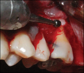
Figure 5- Exposure of fractured Root
The fracture line was observed clinically extending obliquely from the level of furcation on the mesial side to the middle third of the root on the distal side. (Figure 6) A long tapered diamond point was used to resect the exposed mesio-buccal root at the level of furcation in horizontal direction . (Figures 7 and 8) The resected fragment of root was disengaged from the socket using a periosteal elevator.(Figures 9 and 10) The socket was then thoroughly curetted to remove all granulation tissue and rough bony edges were filed and thoroughly irrigated. (Figures 11 and 12) The tooth crown height was reduced using diamond points minimally to relive 26 from occlusion. Flap was approximated using single interrupted sutures (3-0 Mersilk black braided non-resorbable suture) and periodontal dressing was placed. (Figures 13 and 14)

Figure 6- Oblique fracture line

Figure 7- Resection of fractured root

Figure 8- Exposure of resected root fragement

Figure 9 - Disengaging the resected root fragment

Figure 10- Resected root fragment

Figure 11-Degranulation of the socket

Figure 12 – Curetted socket

Figure 13- Sutures placed

Figure 14 – Dressing placed
Patient was prescribed Amoxicillin 500mg (t.i.d) and Diclofenac sodium 50mg (s.o.s) for five days. Oral hygiene instructions were reinforced. Patient was recalled after 10 days for suture removal. (Figure 15) Patient received permanent metal ceramic crown in relation to 26 , one month post-surgical healing.(Figures 17 and 18) 6 month post-operative radiograph revealed satisfactory bone fill.(Figure 16)
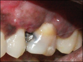
Figure 15 – 10 days post-operative

Figure 16- Post-operative radiograph

Figure 17- Post-operative restoration

Figure 18 - 6 months post-operative
DISCUSSION
Weine emphasized that a greater number of endodontically treated teeth are lost because of fracture due to improper restorations than of poor endodontic result. Comparing teeth with and without crowns demonstrated a significant difference in longevity. With permanent crown placement, the average time before extraction was found to be 87 months. Without a crown, the average prosthetic failure was found to occur at 50 months.8 Kern et al stated that specimens prepared by a specific restorative technique fractured in a consistent predictable pattern.9 In the current case, the failure to prosthetically restore the tooth after endodontic treatment as well the bulky overhanging amalgam MO restoration, would have caused undue occlusal forces to concentrate and fracture the mesio-buccal root.
Root fractures often present no specific signs and symptoms. After progression for several months or years, the symptoms and signs of root fractures may become more obvious with the development of pulpal and periodontal lesions. Finally, the fractured segments may separate from the rest of the tooth structure and be observed during periodontal or root canal treatment. In most cases, horizontal/oblique root fractures can be suspected when a combination of the following symptoms is observed: pain, local swelling, tooth mobility, periodontal pocket, radiographic fracture lines, sinus tract, abscess formation, sensitivity to percussion or palpation or detection of fracture lines over root surfaces with an explorer. As seen in the present case, the patient became symptomatic only after the development of abscess following the root fracture.10
The main goal of periodontal therapy is to eliminate the subgingival infection and through subsequent supragingival plaque control measures, prevent recolonization of the subgingival area. The posterior segments of the dentition present particular problems, due to the complex morphology of the molars.11 Hence, additional treatment means, varying from furcation plasty to root separation/resection, are commonly used in order to improve the accessibility for proper root debridement and to facilitate post-treatment plaque control.12
Treatment alternatives for teeth with horizontal/ oblique and vertical root fractures may depend upon the location and extent of the fracture, the length and bony support of the remaining roots. Additional factors include the extent and quality of remaining tooth structure, root morphology and distribution of occlusal forces for the final prosthesis .13 The 4-year survival rate of maxillary molars after root amputation is reported to be 93% and long term survival of teeth after root amputation ranges from 87% to 95% .14,15
Svardstrom and Wennstrom conducted study in first and second molars to analyze the factors influencing non-regenerative treatment moadalities for molars as well as the outcome of those treatment decisions after 8-12 year follow up. They concluded that, other than the degree of furcation involvement, tooth mobility, tooth position and lack of occlusal anatagonism markedly influenced the decision with respect to non-regenerative therapy. Among the factors evaluated with respect to root resection, degree of furcation involvement strongly influenced the treatment decision. Factors like endodontic conditions, root and root canal anatomy and overall treatment strategy, has also shown to have a strong influence on the treatment decision for molars.16
When performing root resection of maxillary molars, care should be taken not to overload the furcal concavities and provide a favourable environment for maintenance of adequate oral hygiene. After resection, regular maintenance treatment, consisting of sub-gingival instrumentation, might be required periodically to prevent periodontal disease.17
CONCLUSION
Root resection therapy is still a valid treatment option for molars with furcation involvement as well for teeth free of periodontal disease. However, root resection to treat periodontal problems showed a better prognosis than root resection performed for nonâ€periodontal purposes.18 The present study shows satisfactory results in short-term follow up period. Periodontal problems around resected molars have a tendency to recur and should be maintained through meticulous supportive periodontal treatment. In addition, a careful prosthetic plan should be designed to avoid a fracture of resected molars related to biomechanic impairment.
REFERENCES
- Caliskan MK, Pehlivan Y. Prognosis of root-fractured permanent incisors. Endod Dent Traumatol 1996;12:129-36.
- Bender IB, Freedland JB Adult root fracture. J American Dent Ass 1983; 107:413-419.
- Yeh CJ .Fatigue root fracture: a spontaneous root fracture in non-endodontically treated teeth. British Dent J 1997 ; 182 :261-266.
- Bader JD, Shugars DA, Martin JA (2004) Risk indicators for posterior tooth fracture. J of the American Dent Ass 2004; 135:883-892.
- Wang P, Lv H, Sun H, Lin Y, He W. Horizontal root fractures in posterior teeth: a case series. Dent Traumatology. 2011 ;27(2):152-155.
- American Academy of Periodontology. Glossary of Periodontal Terms. Chicago: American Academy of Periodontology ; 2001:45.
- Farrar JM. Radical treatment of alveolar abscess by amputation of roots of teeth. Dental Cosmos 1884 ;26:79-82.
- Weine FS. Endodontic therapy. 3rd ed. St. Louis: C. V. Mosby Co.,1982:4.
- Kern SB, VonFraunhofer JA, Mueinghoff LA. An in vitro comparison of two dowel and core techniques for endodontically treated molars. J Prosthet Dent 1984; 51:508-514.
- Lin. Horizontal/oblique root fractures the palatal root of maxillary molars with associated periodontal destruction: case reports .Int Endo J 2008 ; 41:442–447.
- Bower, RC. Furcation morphology relative to periodontal treatment. Furcation entrance architecture. J Periodontol 1979;50:23-27.
- Carnevale G, Pontoriero R, Lindhe J. Treatment of furcation- involved teeth. In: Lindhe J, Karring T, Lang NP, eds. Clinical Periodontology and Implant Dentistry, 3rd ed. Copenhagen: Munksgaard; 1997:682-710.
- Silverstein LH, Moskowitz ME, Kurtzman D, Faiella R, Shatz PC.Prosthetic considerations with periodontal root resective therapy, Part 1: Root amputations Dent Today 1999 ;18: 82-85.
- Babay NA, Almas K . A four-year clinical follow-up of nonvital root resection in maxillary molar teeth. Indian Journal of Dental Research 1996 ; 7:29-32.
- Hemptom T, Leone C . A review of root resective therapy as a treatment option for maxillary molars. Journal of American Dental Association 1997; 128: 449-455.
- Svärdström, Wennström Periodontal Treatment Decisions for Molars: An Analysis of Influencing Factors and Long-Term Outcome. J Periodontol 2000;71:579-585.
- Cercek JF, Kiger RD, Garrett S, Egelberg J. Relative effects of plaque control and instrumentation on the clinical parameters of human periodontal disease. J Clin Periodontol 1983;10(1):46-56
- Park SY, Shin SY, Yang SM, Kye SB. Factors influencing the outcome of root resection therapy in molars: A 10-year retrospective study. J Periodontol 2009 ; 80(1):32-40.
Disclaimer from Guident
The views and opinions expressed in articles published in Guident are solely those of the authors and do not necessarily reflect the views of the editors, the editorial board or the publisher, Ivory India. Guident does not endorse any product, technique or treatment described. Clinical content is intended for qualified dental and medical professionals and is not a substitute for individual clinical judgement. Guident, its editors and the publisher accept no liability for any consequence arising from the use of information published in the journal.
How to cite this article
Dr. Shashikanth Hegde, Dr. Onisha Vijay, Dr. Ashish Kakar. Management of Endodontically Treated Maxillary Molar Root Fracture. Guident. 2019 Feb;12(3). Available from: https://ivoryindia.in/articles/periodontics/management-of-endodontically-treated-maxillary-molar-root-fracture.html