Complete Dentures- the Unconventional One a Review Part- I
MP, India, Rama Dental College, Kanpur, U.P, India, Senior Lecturer, ITS Dental College, Muradnagar, Ghaziabad, UP, India, Depatment of Prosthodontics
Abstract:
During fabrication of complete dentures there are some routine complications faced by the dentist. These are atrophic ridge, microstomia, flabby tissue, xerostomia, bony exostosis, labially inclined premaxilla, aesthetic demand, bruxism, systemic disorders, patient’s demand for duplicating dentures, etc. Proper incorporation of suitable materials and advanced techniques helps in managing these complications. This article will be covered in two parts which describes the unconventional approaches to various modalities so as to provide ultimate satisfaction for the patient.
Keywords: complete dentures, unconventional.
INTRODUCTION
Conventional complete denture is a removable dental prosthesis that replaces the entire dentition and associated structures of maxillae and mandible. (GPT)1
Many edentulous patient regardless of the excellence of professional skills, techniques and humanitarian concern are maladaptive to complete dentures. Transforming conventional into unconventional approach is a peculiar feature of advancing prosthodontics. The increasing demand of patients and revolutionary thought of prosthodontists have led to the outcome of the special, i.e. the unconventional approach for fabricating complete dentures. The unconventional complete dentures is known as starring new techniques based on same old fundamentals of Prosthodontia. It has been prove to be a manifestation of new vision in prosthesis construction.
There are some routine complication faced by the prosthodontist include atrophic ridges, microstomia, flabby tissue, xerostomia, bony exostosis, labially inclined premaxilla, aesthetic demand, bruxism, systemic disorders, etc. Proper incorporation of suitable material and advancing techniques can be used to manage these difficulties.
Sometimes the conventional approach may not fulfil the five basic principles of complete denture like retention, stability, support, aesthetics and preservation of supporting structures which are of utmost importance for the complete satisfaction of the patient. When complete denture does not suit patient’s needs, certain modifications are made to meet his requirements. This article will be covered in two parts, reviewed some simple, effective and non-invasive alternative treatment to the classical conventional technique in a completely edentulous patient.
Commonly used unconventional dentures are:
- Immediate Denture
- Flexible Denture
- Hollow Denture
- Sectional Complete Denture
- Liquid Supported Complete Denture
- Metal Reinforced Denture Base
- Denture with characterization
- Gumfit Dentures
- Denture for flabby ridge
- Customized attachment retained Cheek Plumper prosthesis
- Dolly Dentures
- Denture with mechanical retentive components
- Single complete denture
- Single denture with metal mesh
- Split denture technique (Xerostomia)
- Denture with identification markings
Immediate, Flexible, Hollow, Sectional Complete denture, Liquid supported denture, Metal reinforced denture base and Denture with characterization will be covered in this article. Rest of the topics will be discussed in next part.
IMMEDIATE DENTURE
Immediate denture is defined as a removable prosthesis fabricated for placement immediately after the removal of natural teeth or tooth.2-7
Indication
- Humiliation
- Adverse subjective reactions – minimized by immediate substitution
- When the teeth are removed, the beautifully effective teamwork between the various parts of stomatognathic system is broken up –abnormal functioning of the mouth and mandible.
- Prevents disuse atrophy of the bony base.
- Prevents possible damage to the ligaments surrounding
- TMJ – abnormal neuromuscular pattern in absence of dentures
Contraindications:
- Poor general health e.g. – bleeding disorders, cardiac or endocrine disturbances, psychological disorders, post irradiation of head and neck regions.
- Uncooperative patients.
- Patients with gross oral neglect.
- In cases of misrelated jaws or malposition teeth which need extensive surgical reduction of the alveolar processes.
- Patients with large periapical abscesses or cysts.
Types of Immediate Dentures:
Acc. To Fen
- With anterior teeth socketed (Open face design)
- Labial flange without alveolectomy
- Labial flange with alveolectomy (excision)
- Labial flange with alveolotomy (incision)
Acc. To Boucher
- Conventional/Classic immediate dentures (CID)
- Interim/transitional/nontraditional immediate dentures (IID)
Requirements:
- Restore the masticatory efficiency within limits.
- Esthetically acceptable.
- Preserve the remaining tissues.
- Compatible with the oral environment.
- Function in harmony with the activity necessary for speech, respiration and deglutition.
Advantages
- Avoiding the embarrassment of appearing in the public without teeth.
- Reduction in post extraction complication due to their prophylactic and therapeutic effect
- Less interference with diet and speech.
- Easier to duplicate.
- Circum-oral support, muscle tone, vertical dimension of occlusion, jaw relationship, facial height maintained.
- Psychological and social well-being is preserved.
- Prevents bleeding, protects the wound against trauma.
- Prevents the entrance of food and liquid into the wound.
- Protects blood clots and accelerates healing and enables a more correct formation of residual ridge.
Disadvantages:
- Increased patient visits.
- Traumatic procedure.
- Complex clinical and laboratory procedures
- Incorrect centric relation.
- Inability to accomplish a denture try-in- compromised aesthetics.
- Variation in bone and soft tissue change leads to compromised retention, rebasing required.
- More chair side time and cost.
- Difficult and demanding procedures.
- Anterior ridge undercut may preclude accurately capturing a posteriorly located undercut.
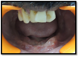
FIGURE 1 (a). Pre-Operative

FIGURE 1 (b). After Extraction

FIGURE 1 (c). Post-Operative image (After Denture placement)
FLEXIBLE DENTURES
Undercut is considered to be an aid for retention but only in unilateral cases. Severe unilateral or bilateral undercuts instead of enhancing retention interfere with the path of insertion and removal of complete denture. Conventional management techniques include modification in denture bearing area, denture base adaptation and relining.
Flexible denture base material is a bio-compatible nylon- based thermoplastic resin.
Some of the commercially available products are Valplast, Duraflex, Flexite, Lucitone, Impak, Proflex. These materials are strong that it can be made very thin, which make it comfortable to wear and aesthetically pleasing. Flexible dentures by Valplast is commercially available in USA since 1954, which is injection moulded, at above 300 degree C.It is denser than acrylic thus has lower water sorption. Its flexure modulus is 400Mpa at body temperature. It is virtually unbreakable material.
Valplast's Key Benefits Are:
- Retention - flexes into a retentive position, below the undercut.
- Comfort - thin, lightweight & flexible.
- Aesthetics - pink shades that allow your patient's natural tissue tone to appear through the material.
- Ease - no tooth or tissue preparation is required so you can offer patients a conservative & pain-free solution.
- Strength - clinically unbreakable, more durable than acrylic & won't absorb stains or odors.
Applications:
- Bilateral inoperable undercuts that cannot be managed by acrylic dentures without resorting to surgery.
- Unpigmented form used for TMJ Splints.
- For patients who are allergic to acrylic monomer.
- Can be used in combination with a metal frame to allow the aesthetic benefit of Valplast replacing the buccal arm.
- Used in cosmetic veneers to mask gingival recession in variety of situations, including dental implants.
- When history of frequent breakage is seen.
- In cleft palate and bulbous torus condition.
- Patients with limited mouth opening.
Advantages:
- The translucency of the material picks up underlying tissue tones, making it almost impossible to detect in the mouth.8
- Dentures can be made very thin and light weight, which makes it comfortable to wear that promotes better adaptation of the tongue and cheek to the denture base and aesthetically pleasing.
- Dentures are fabricated using advanced and improved technique, they exhibit better accuracy compared to conventional techniques.
- The amount of adjustment required at time of denture insertion is greatly reduced.
- Also, this reduces post-insertion complaints of denture-induced trauma [ulceration].
- Absolute biocompatibility because of its metal free and monomer free characteristics (less allergic reactions)
- Can be used as temporary dentures during the therapeutic episodes after surgical reconstruction of jaw.
- These dentures have advantage in overcoming midline denture fractures.
Due to their ability of excellent mouldability, light weight to density ratio and high thermal strength, flexible dentures have been proven as an excellent treatment option for complete and partial edentulism. However, careful case selection and clinical judgment is required to use flexible dentures in appropriate situations in order to obtain a successful treatment outcome.
Disadvantages:
- Flexibility is normally, not an advantage in complete dentures as retentive peripheral seal can be broken in function (though thinner & lighter acrylic denture can help its cause).
- Also a greater than normal shrinkage makes it difficult to fabricate
Contraindications:
- Bilateral distal extension maxillary & mandibular ridges
- Deep overbite
- For vertical stop e.g. occlusal rest

FIGURE 2 (a). Edentulous ridges

FIGURE 2 (b). Processed dentures with sprue

FIGURE 2 (c). Finished and polished denture

FIGURE 2 (d). Denture placement
HOLLOW DENTURE
For the patients with severely atrophic residual ridge or large maxillofacial defect, the conventional denture fabricated proves to be both bulkier and heavier and so compromises retention and stability of the prosthesis. Extreme reduction in the residual ridge height leads to increased space between maxillary ridge and mandibular ridge which ultimately increase the amount of denture base material which results in a less retentive denture. With the aim to reduce the weight, hollow dentures can offer solution for this situation.9-11 Care should be taken to exclude the denture base from the planned hollow cavity of the prosthesis.
Materials utilized for this purpose included a solid three dimensional spacer including dental stone (Ackermen, 1955), cellophane, wrapped asbestos (Worley & Kniejski, 1983), silicone putty (Holt, 1981) or modelling clay (DaBreo, 1990) during laboratory processing.

FIGURE. 3 (a). 2 openings were created into the denture base distal to the most posterior teeth.

FIGURE. 3 (b). Attach the clear resin covers by bonding them into position using auto polymerizing resin or light-polymerizing gel
SECTIONAL COMPLETE DENTURE
Oral submucous fibrosis is a precancerous condition, caused by prolonged use of tobacco, arecanut, spices, etc. It affects any part of the oral cavity and sometimes the pharynx. The fibrosis involves the lamina propria and the submucosa and may often extend into the underlying musculature resulting in the deposition of dense fibrous bands giving rise to the limited mouth opening which is a hallmark of this disease.12
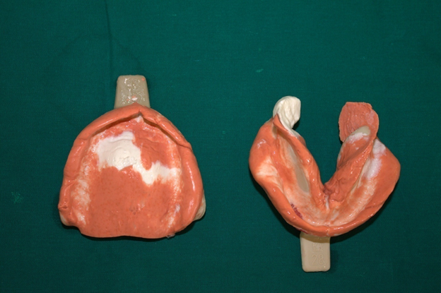
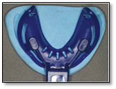
Microstomia is defined as an abnormally small oral opening, caused by number of reasons like scleroderma, oral submucous fibrosis, sequeale of burns, surgical resection of facial and oral neoplasms and temporomandibular joint disorders. Since they have a limited mouth opening, conventional method for taking impression and denture placement and removal is difficult and challenging. Hence it is necessary to modify the technique.12 It is advisable to follow sectional impression tray and technique for facilitating easy insertion and removal i.e. tray should be cut into two section.12 Final impressions were made with Non-Eugenol impression material as Eugenol would cause burning sensation to the patient.


FIGURE: 4. (a & b). Maxillary sectional denture base after wax occlusal rim fabrication

FIGURE: 4. (c).Intra oral view during Try In

FIGURE: 4. (d).Extra oral view after denture placement.
LIQUID SUPPORTED COMPLETE DENTURE
Conventional complete denture lacks retention and stability in patients with fibrous hyperplastic tissue. Several techniques have been employed for managing such cases. One of those techniques is to provide flexible tissue surface in complete denture (Liquid supported denture).13 This denture is based on the theory that when the force applied on the denture is absent, the base assumes its preshaped form that is the one during processing.14 The principle of this design was that a liquid-supported denture is flexible and continuously adapts itself to the mucosa. However, it is also rigid enough to support the teeth during actual use. Thus, the denture base is covered with a close-fitting flexible foil to keep a thin film of liquid in its place.13
|
Polythene sheet |
|
Liquid(glycerine) |
|
Denture base |
|
Denture teeth |
Table: Layers in liquid supported denture
In the absence of forces the foil remains in the resting position acting as a soft liner and when the dentures are in use, masticatory loads are distributed in all directions by the liquid resulting in even stress distribution. This helps in long-term preservation of bone and soft tissues. Apart from the combined benefits of tissue conditioners and soft liners, load from biting forces and even bruxism, will be distributed over a larger surface (Chase, 1961).
Advantages of liquid supported denture include:
- Good base adaptation due to hydrodynamics of the liquid improving support, retention and stability
- Optimal stress distribution of masticatory forces over a larger area which reduces tissue overloading, prevention of soreness.
- Increased comfort level and patient acceptance
- Optimal atmospheric pressure, adhesion, cohesion and mechanical interlocking in undercuts.
Precaution:
- Thickness of denture base should be at least 3mm.
- Seal should be perfect & should be checked for micro leakage.
- In case liquid leaks out, patient should inform the dentist & denture should be refilled.
- Denture care instructions should be given to dentist.
- Repair is possible if sheet gets ruptured & can be replaced over preserved stone replica.

FIGURE. 5 (a). Holes made in denture for injecting fluid

FIGURE. 5 (b). Glycerine incorporated as a liquid medium

FIGURE. 5 (c). Liquid supported denture with shock absorbing effect

FIGURE. 5 (d). Unidirectional loading of denture resulting in multidirectional distribution of hydrostatic pressure & clasping pressure at its borders
METAL REINFORCED DENTURE BASE
Routine Poly methyl methacrylate (PMMA) denture bases have good mechanical, biological and aesthetic properties. But, they may fail because of excessive para functional and/or functional forces (in cases of bruxism and/or complete dentures opposing natural mandibular teeth). Metal based denture can be used to strengthen the denture bases.14
These thin metallic bases have several advantages, besides rigidity and fracture resistance, like: excellent strength to volume ratio, good adaptation to the supporting tissues, enhanced control of denture plaque, high thermal conductivity, high biocompatibility, no dimensional changes in time through fluids absorption and no interferences with phonation. The fracture of acrylic resin denture is an unresolved problem in removable prosthodontics.14
Midline fracture of an acrylic denture base results from flexural fatigue failure, acrylic deformation of the base during function, sharp changes in contour, pin holes and residual processing stresses. Various approaches such as use of metal bases, wires, bars, & high impact acrylic resin reduces the incidence of midline fracture.4
The fracture of acrylic resin denture is an unresolved problem in removable prosthodontics.
Midline fracture of an acrylic denture base results—
- Flexural fatigue failure
- Acrylic deformation of the base during function
- Sharp changes in contour, pin holes
- Residual processing stresses
Various approaches such as use of metal bases, wires, bars, & high impact acrylic resin reduces the incidence of midline fracture
Indications:
- Deep palatal vault, prominent residual ridges, when additional strength is needed because stresses are concentrated over small parts of denture.
- Shallow flat palates and mentally compromised patients (poor neuro muscular coordination who may drop their denture.
Materials used for metal denture base4
- Cr-Co – most retentive
- Al
- Ni - Cr
- Titanium
- Gold
Cr – Co most often used because cost is reasonable & technique is commonly used in most dental laboratories.
Al - has advantage of being less dense, but construction technique less available & Al toxicity may be contributor to Alzheimer disease.
Gold - less retentive for maxillary denture but may be a value for mandibular denture due to its viscosity.

FIGURE. 6 (a). Refractory cast with wax pattern
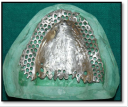
FIGURE. 6 (b). Finished and polished framework

FIGURE. 6 (c). Intra oral view
DENTURE WITH CHARACTERIZATION
Complete denture may not resemble the previous anatomic morphology of teeth or oral mucosa. Many patients demand more natural like appearance of complete denture such as spacing between incisor, fractured incisal edge, stained teeth, proclined profile, etc. So special considerations should be employed in modification of denture base and teeth. This modifications are called “characterization”. Complete denture can be characterized by two basic methods.
1. Characterization by selection, arrangement and modification of artificial teeth.
2. Characterization by tinting the denture bases.15
Indication for charcterization of denture base
- Patients with an active upper lip.
- Patients with a prominent pre-maxillary process.
- Actors, singers and others who may expose gum tissues areas during their performances.
- The psychological acceptance of the dentures by the patient

FIGURE. 7. Characterization (staining the denture base)
REFERENCES
- Glossary of Prosthodontic Terms. J Prosthet dent.
- Bruce R W. Immediate denture service designed to preserve oral structures. J Prosthet Dent 1966; 16:811-821.
- Jainiji,Immediate denture with unusual bone morphology: a clinical report. IAJD 2003; 2:23-25.
- Nitesh Rai, Shiva Shankar M, Jagadeesh, Shraddha Rani Unconventional Complete Dentures. JDOB 2012.
- Chierici G, Parker M L, Hemphill C D. influence of immediate dentures on oral motor skill and speech. J Prosthet Dent 1978; 39:21-28.
- Heartwell C M, Salisbury F W. immediate complete dentures: an evaluation. J Prosthet Dent 1965; 15:615-624.
- Passamonti. Immediate denture prosthesis. Dent Clin North Am. 1964; 8:781-800.
- Dr. Sunitha N Shamnur, Dr. Jagadeesh KN,Dr. Kalavathi SD, Dr. Kashinath KR. “Flexible dentures” – an alternate for rigid dentures?Journal of Dental Sciences & Research 1:1:74 –79.
- Naylor WP, Manor RC. Fabrication of flexible prosthesis for the edentulous scleroderma patient with microstomia. J Prosthet Dent. 1983 Oct; 50(4):536-8.
- Antonelli JR, Hottel TL. The “flexible augmented flange technique” for fabricating complete denture record bases. Quintessence Int. 2001 May; 32(5):361-4.
- Lowe LG. Flexible denture flanges for patients exhibiting undercut tuberosities and reduced width of the buccal vestibule: a clinical report. J Prosthet Dent. 2004 Aug; 92(2):128-31.
- Anupama Prasad D, Krishna Prasad D, ChetanHegde. Prosthodontic rehabilitation of a patient with oral submucous fibrosis and microstomia: a case report.NUJHS 2012; 2(3); 39-41.
- Nandita Nitin Keni, Meena Ajay Aras, VidyaChitre. Management of flabby ridges using liquid supported denture: a case report. J AdvProsthodont 2011; 3:43-6.
- Corina Marcauþeanu, LucianaGoguþa, AncaJivanescu, Bratu. Titanium complete denture base in a patient with heavy bruxism, year xv · nr.3/2008 · page. 96-99
- Rajeev Srivastava, VivekChoukse. Characterization of Complete Denture, International journal of dental clinics 2011:3(1):56-59.
Disclaimer from Guident
The views and opinions expressed in articles published in Guident are solely those of the authors and do not necessarily reflect the views of the editors, the editorial board or the publisher, Ivory India. Guident does not endorse any product, technique or treatment described. Clinical content is intended for qualified dental and medical professionals and is not a substitute for individual clinical judgement. Guident, its editors and the publisher accept no liability for any consequence arising from the use of information published in the journal.
How to cite this article
Dr. Shweta SainiSenior lecturer Guru Govind Singh Dental College, Dr. Pawanjeet Singh Chawla Reader, Dr. Mahinder Singh Chauhan. Complete Dentures- the Unconventional One a Review Part- I. Guident. 2016 Sep;9(10). Available from: https://ivoryindia.in/articles/prosthodontics/complete-dentures-the-unconventional-one-a-review-part-i.html