Post Orthodontic Esthetic Smile Designing
Abstract
Since the introduction of adhesive dentistry in the early 1980s1,porcelain veneers have been widely used in dentistry2. The ability of ceramics to match natural dentition and the good physical and optical properties of ceramics make them the material of choice for patients with high esthetic expectations3. However the definitive results of the restoration can only be accurately predicted after an estheticanalysis of the existing restorationsThisclinical report describes a conservative treatment of veneer replacement where spacing between teeth, malalignment, and midline shift were the main concerning factors for teeth.
INTRODUCTION
Dental ceramic veneers were introducedcommercially several yearsago and have been indicated with for treatmentof discoloration,stains,diastemasand abnormal tooth shape. Whenteeth are prepared for ceramic andmetal ceramic crowns,63% to 72% of the coronal toothstructure is removed4. Heat pressed lithium disilicate glass ceramic, consists of approximately 65% lithium disilicate in the form of crystalline structures5 and is a relatively strong ceramic with high flexural strength and excellent translucency6. Lithiumdisilicate restorations,compared with other materials, for example, leucite reinforced ceramics,have greater biaxial strength and fracturetoughness. Complete crystallization of lithiummetasilicate is achieved during firingand is associated with achievement ofthe final strength of the ceramic7.They can be etched and bonded to both enamel and dentin and can be used for anterior and posterior single crowns, anterior fixed dental prostheses, and laminate veneers, all of which provide excellent esthetics8.
A carefully developed diagnosis is necessary to formulate a conservative and predictable treatment plan. Providing minimally invasive treatment and understanding the best options for tooth preservation are important aspects of the prognosis of teeth. A systematic esthetic analysis that progresses from facial to dentofacial to dentogingival and finally to dental analysis is needed for a successful esthetic outcome19. This is particularly true if the previous treatment goal was not accomplished through a minimally invasive approach. The purpose of this clinical report was to present a comprehensive treatment planning for improving patient’s esthetics and the clinical parameters that are essential to conservative treatment planning for veneer replacement.
CLINICAL REPORT
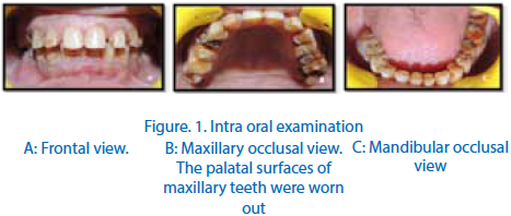
A 22years old male patient reported to the department of prosthodontics, ITS dental college,hospital and research centre, he was not satisfied with his appearance.The patient was determined to be at low caries risk with good oral hygiene. Intraoral examination revealed congenitally missing lateral incisors. There was spacing between the canines and the first premolars on both sides. The patient underwent orthodontic treatment for the same and the canines and were shifted to take the space of lateral incisors. The patient was not satisfied with the esthetic results and wanted smile correction.
Diagnostic impressions were made and wax up was done.The treatment plan was to provide all ceramic crowns by changing the morphology of canines into lateral incisors and that of the first premolars to canine. Veneers were planned for the central incisors in order to achieve a proportion between all teeth. By this the smile line could be madeesthetically pleasing for the patient. Intentional root canal treatment was done with respect to 13 14 23 and 24. Minimally invasive preparation for veneers was done with the central incisors. Gingival retraction was done with retraction cord dipped in a haemostat (Viscostat,ultradent) and final impression was made with dual step putty wash impression technique in a stock tray with a spacer. Provisional restorations were fabricated with the help of putty index of the wax up done on the diagnostic cast. Bi phasic CAD/CAM zirconia crowns were fabricated for the canines and the first premolars that were changed into the morphology of lateral incisors and canines respectively. Veneers were fabricated for the central incisors form lithium disilicate (IPS e.max,IvoclarVivadent). Before cementation, the all ceramic crowns were surface treated first by sandblasting with 50 micron units. The veneers were etched with hydrofluoric acid(Pulpdent) for 60 seconds followed by application of silane coupling agent(Pulpdent). Final cementation of both the veneers and crowns was done with self etchself adhesiveresin cement (U200,Rely X).Tack curing was done for three seconds followed by excess removal of cement. Dental floss was also used between the veneers to ensure proper removal of excess cement and then the prosthesis were light cured, 20 seconds on each surface.

CONCLUSION
All-ceramic crowns are durable and provide optimum estheticand predictable alternative for anterior and posterior locations—provided that consistent attention is paid to proper tooth preparation, cementation, and occlusal adjustment. In addition to optimum esthetics, these crowns have success rates that equal or exceed those of ceramometal crowns.Inthe presence of diastemas and/or malalignment of the anterior teeth, orthodontic correction can be the most conservative treatment option. Sometimes restorative procedures are necessary after orthodontics to provide the optimal outcome. Treatment plans should not be selected empirically but should be based on thorough documentation, measurements,casts and photographs as part of adequate treatmentplanning. As illustrated for this patient, his lateral incisors were missing along with spacing between the canines and premolars,planning veneers for the central incisors and all ceramic crowns by changing the morphology of the canines into laterals and that of premolars into canines was the best possible treatment option keeping in mind the esthetics and functional requirements.
REFERENCES
- Karine T. L et alCeramic materials for porcelain veneers: Part II. Effect of material, shade, and thickness on translucencyJ Prosthet Dent 2014;112:864-870.
- Christensen GJ. Thick or thin veneers. J Am Dent Assoc 2008;139:1541-3.
- Kelly JR, Nishimura I, Campbell SD. Ceramics in dentistry: historical roots and current perspectives. J Prosthet Dent 1996;75:18-32.
- Edelhoff D, Sorensen JA. Tooth structure removal associated with various preparation designs for anterior teeth. J Prosthet Dent2002; 87:503-9.
- Albakry M, Guazzato M, Swain MV. Biaxial flexural strength, elastic moduli, and x-ray diffraction characterization of three pressable all-ceramic materials. J Prosthet Dent 2003;89:374-80.
- Höland W, Schweiger M, Frank M, Rheinberger V. A comparison of the microstructure and properties of the IPS Empress 2 and the IPS Empress glass-ceramics. J Biomed Mater Res 2000;53:297-303.
- Schmitter M and Seydler B.B Minimally invasive lithium disilicate ceramic veneers fabricated using chairside CAD/CAM: A clinical report.J Prosthet Dent 2012;107:71-74
- ZandinejadA et al.Virtually designed and CAD/CAM-fabricated lithium disilicate prostheses for an esthetic maxillary rehabilitation: A senior dental student clinical report.J Prosthet Dent 2015;113:282-288
- ArmstrongS.G and Maia.R.Conservative treatment planning in veneer replacement.J Prosthet Dent 2015;1-4
Disclaimer from Guident
The views and opinions expressed in articles published in Guident are solely those of the authors and do not necessarily reflect the views of the editors, the editorial board or the publisher, Ivory India. Guident does not endorse any product, technique or treatment described. Clinical content is intended for qualified dental and medical professionals and is not a substitute for individual clinical judgement. Guident, its editors and the publisher accept no liability for any consequence arising from the use of information published in the journal.
How to cite this article
Dr.Roopashi Berry, Dr.AmitJayna, Dr.Aditya Chaudhary. Post Orthodontic Esthetic Smile Designing. Guident. 2016 May;9(6). Available from: https://ivoryindia.in/articles/prosthodontics/post-orthodontic-esthetic-smile-designing.html