Rehabilitation of Complete Maxillary Arch
Corps Dental Unit, P-8, Khulna Marg, SI Lines, Bhopal, MP, India
ABSTRACT
The gradual wear of the occlusal surfaces of teeth keep on happening throughout life. However, excessive occlusal wear may lead to pulpal trauma, occlusal disharmony, impaired function, and esthetic disfigurement. Depending on the cause, tooth wear can be classified as attrition, abrasion, and erosion. Therefore, it is important to identify the factors which contribute to excessive wear and reduce the vertical dimension of occlusion. This case report presents the rehabilitation of complete maxillary arch in a 55 yrs old patient with severe attrition. The patient was treated using gnathological approach with Porcelain fused to metal crowns on all maxillary teeth.
Key words:- Full Mouth, Vertical Dimension, PFM
INTRODUCTION
The gradual wear of the occlusal surfaces of teeth keep on happening throughout life of patient. However, excessive occlusal wear may result in pulpal trauma, occlusal disharmony, impaired function, and esthetic disfigurement1. Tooth wear can be in form of attrition, abrasion, and erosion. Therefore, it is important to identify the factors which contribute to excessive wear and reduce the vertical dimension of occlusion2. Mostly, the vertical dimension of occlusion (VDO) is maintained by continuous tooth eruption and alveolar bone growth. The alveolar bone undergoes an adaptive process and compensates for the loss of tooth structure to maintain the VDO as the teeth are worn3,4.
However, the rehabilitation of the severely worn dentition becomes more challenging when insufficient space for restoration is present. Proper diagnosis and careful comprehensive treatment plan is required in managing such patients. Full mouth rehabilitation enhances patient’s self esteem, confidence and quality of life. Full mouth rehabilitation is the procedure when restoration of the missing teeth involves the complete rehabilitation of the oral cavity, in terms of function and aesthetics.
CASE REPORT
A 55-year-old male patient reported with the chief complaints of difficulty in eating and unaesthetic appearance of his face because of excessive wear of upper teeth. Intraoral examination revealed a generalized loss of dental substance that was greater in the maxillary anteriors
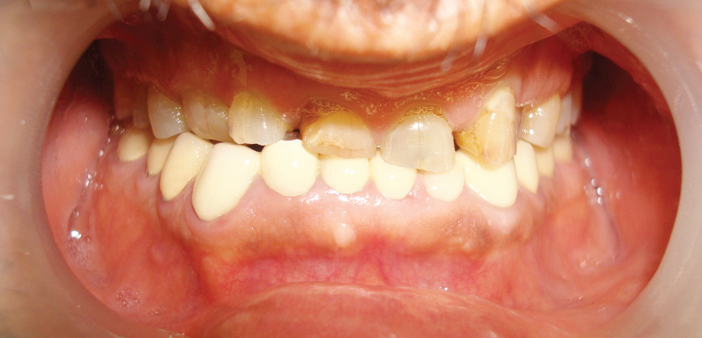
Fig 1
up to the gingival level [Fig 1]. All mandibular teeth were restored with crowns 5 years back. The anterior teeth had sharp enamel edges, dentinal craters, and attritional wear due to the loss of posterior support. The patient did not have temporomandibular joint disorder history and soreness of the masticatory muscles. Loss of 7mm of vertical dimension was evident and his phonetics was also altered.
Based on the findings, the treatment plan of full maxillary arch rehabilitation with porcelain fused to metal crowns at increased vertical dimension of 4mm with group function occlusion.
Case was taken for following objectives of treatment to
- Attain functional harmony.
- Reestablish form, function and esthetics.
- Establish a group function occlusal scheme.
Endodontic treatment of all maxillary teeth except all molars was carried out. Severely attrited maxillary incisors had got post and core to obtain a sufficient clinical crown length and ferrule effect. Splint made up of hard sheet was given to the patient for 03 weeks. After that another splint of 4mm biostar sheet was given to the patient for another 02 weeks. The adaptation of patient to the increased VDO was evaluated during 02 months trial period. Muscle tenderness and temporomandibular discomfort were not found.
A lucia jig was fabricated at an established increased VDO at 4mm. Interocclusal and protrusive records were made using polyvinylsiloxane occlusal registration material (Jetbite). The diagnostic impressions were made using irreversible hydrocolloid. Patient’s casts were mounted on a semi-adjustable articulator (Hanau H2) using a face-bow record. With the help of protrusive record, the condylar guidance of patient was noted to be 250 and lateral condylar guidance was calculated to be 150. Diagnostic wax-up was done. Provisional crowns were fabricated with autopolymerizing resin using a vacuum formed matrix produced from the diagnostic wax-up. Adjusted occlusion was transferred to customized anterior guide table, which was made with acrylic resin (PATTERN RESIN; GC Corp, Tokyo, Japan). The post cementation and core build up was done in maxillary anteriors [Fig 2]. All maxillary teeth were prepared and definitive two stage double mix putty light body impression was made with polyvinylsiloxane impression material (Aquasil, Dentsply) [Fig 3]. The interocclusal record was made with interocclusal recording material at established VD and face bow was used fro recording of orientation jaw relation. Provisional crowns were cemented with soft zinc polycarboxlate temporary cement (Hybond) [Fig 4]. After die cutting, the casts were
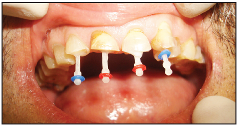
Fig 2

Fig 3

Fig 4

Fig 5
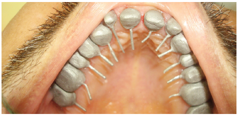
Fig 6

Fig 7

Fig 8
mounted on semiadjustable articulator (Hanau H2). The wax patterns were fabricated, invested and cast [Fig 5]. The metal copings were retrieved; finished and metal try-in was done [Fig 6]. After the metal try-in, the ceramic was applied and bisque try-in was completed. Porcelain fused to metal restorations were made using customized anterior guide table [Fig 7] as well as premeasured horizontal and lateral condylar guidance of 250and 150 respectively. The amount of occlusal adjustment on the lingual surface of maxillary anterior teeth was minimal because the patient’s anterior guidance table was used in the production of definitive restoration. The group function occlusion was checked in mouth and after verification, crowns were cemented with temporary polycarboxylate cement. After two weeks, once patient was comfortable all crowns were cemented with resin modified glass ionomer cement (FujiCEM; GC America, Alsip, USA) [Fig 8]. Oral hygiene instructions and regular check-up were administered.
DISCUSSION
Such cases requires the development of sufficient restorative space, which may lead to fulfilling the esthetics, functional and occlusal parameters essential to long term success. Success in maintaining severe wear cases depends on the development of proper incisal guidance to allow for proper disocclusion within patient’s envelop of motion5.
The etiology of tooth wear is multifactorial, and clinical controlled trials of restorative and prosthodontic approaches are limited in quantity and quality. In this case, the remaining tooth structures were too small to have sufficient retention of composite resin. Therefore, the conventional treatment modality including a trial overlay splint, provisional restoration, careful monitoring, and definitive prosthesis, was chosen.
The trial period of overlay prostheses which are reversible and conservative is between 3 weeks and 5 months, and that of intensive fixed provisional prostheses is 2 – 6 months6.
In this case, the patient was carefully monitored for 2 months to evaluate the adaptation to the removable occlusal overlay splints. No discomfort, wear, and muscle fatigue were observed during that period. The increase of VDO was determined by patient’s physiologic factors like interocclusal rest space and speech. Arbitrary increase of VDO would lead to multiple complications.
The anatomy of the cusps is created by establishing the appropriate form of the posterior cusps aligned to the condylar path; thus it also contributes to posterior disclusion. Posterior disclusion is crucial in controlling harmful lateral forces. This case has demonstrated that the required space may be obtained by seating the condyles in centric relation position. The maintenance of severe wear cases can be ensured by the development of proper anterior guidance that allows for posterior disclusion within the patient’s envelope of function. Taking this guidance into account during provisionalization ensures minimal adjustments in the definitive restorations and a greater long-term predictability of the case.
CONCLUSION
Dental problems are often multi-factorial, and restorative treatment may not only be satisfactorily. Multidisciplinary approach, and meticulous treatment planning is required for creating the perfect esthetic with health. Full Mouth Rehabilitation corrects imperfections in bite position and enhances the appearance. Full Mouth Rehabilitation creates a smile that is functional, comfortable and esthetic but proper knowledge of neuromuscular dentistry with the artistry of cosmetic dentistry is required.
REFERENCES
- Turner KA, Missirlian DM. Restoration of the extremely worn dentition. J Prosthet Dent 1984;52:467-74.
- Prasad S, Kuracina J, Monaco EA Jr. Altering occlusal vertical dimension provisionally with base metal onlays: a clinical report. J Prosthet Dent 2008;100:338-42.
- Dawson PE. Functional Occlusion - From TMJ to smile design.1st ed. New York; Elsevier Inc.; 2008. p. 430-52.
- Jahangiri L, Jang S. Onlay partial denture technique for assessment of adequate occlusal vertical dimension: a clinical report. J Prosthet Dent 2002;87:1-4.
- Hoyle DE. Fabrication of a customized anterior guide table. J Prosthet Dent 1982;48:490-1.
- Yunus N, Abdullah H, Hanapiah F. The use of implants in the occlusal rehabilitation of a partially edentulous patient: a clinical report. J Prosthet Dent 2001;85:540-3.
Disclaimer from Guident
The views and opinions expressed in articles published in Guident are solely those of the authors and do not necessarily reflect the views of the editors, the editorial board or the publisher, Ivory India. Guident does not endorse any product, technique or treatment described. Clinical content is intended for qualified dental and medical professionals and is not a substitute for individual clinical judgement. Guident, its editors and the publisher accept no liability for any consequence arising from the use of information published in the journal.
How to cite this article
Dr. Ashish Kalra, Dr. E Mahesh Gowda, Dr. Shikha Punjani. Rehabilitation of Complete Maxillary Arch. Guident. 2016 Jul;9(8). Available from: https://ivoryindia.in/articles/prosthodontics/rehabilitation-of-complete-maxillary-arch.html