Cheiloscopy and Dermatoglyphics as Genetic Markers and Role of Family Pedigree in the Inheritance of Non Syndromic Cleft Lip and Palate
Department of Public Health Dentistry, Coorg Institute of Dental Sciences, Virajpet Karnataka, India
ABSTRACT
Introduction: Clefting is a condition formed by the failure or improper fusion of tissues during the development process. In the oral region, clefts can occur in the lips or palate or both simultaneously Cleft Lip with or without Cleft Palate (CL/P) belongs to a heterogeneous group of disorders that affect the oral cavity and the lips with a substantial rate of dysmorphogenesis. They are one of the most common birth defects worldwide.
Objective: To analyze the familial pedigree and various pattern types of lip prints and dermatoglyphics in parents of CL(P) children to detect if any specific type can be contemplated as a genetic marker in the transmission of CL(P) and to compare these patterns with that of parents of unaffected children.
Methods:Parents of 50 children with CL /P or both as cases. Parents of 50 children (age and gender matched) without CL /P or both as controls. Family history was taken including first and second degree relatives. Pedigree was constructed through interview of the parents of study group and control group. Cheloscopic analysis was done according to the Suzuki & Tsuchihashi lip print classification and Demetoglyphic analysis were classified as arches (A), loops (L) or whorls (W) based on Galton (1982) classification.
Results:An increase frequency of type II lip patterns among parents of CL(P) affected children as compared to that of parents of unaffected children. Increased frequency of loops pattern in parents CL(P) affected children as compared to that of parents of unaffected children.
Based on the present study results, specific type cannot be contemplated as a genetic marker in the transmission of CL(P). But, dermatoglyphics and chelioscopy can be used as important screening markers to predict the occurrence and inheritance of cleft lip and cleft palate. 10% 0f the cases presented family history with more than one occurrence of CL/P.
Conclusion: For a complex trait like CL(P), these noninvasive tools are an effort to take a step forward in shedding light on the prediction of transmission of CL(P) deformity to the offsprings.
INTRODUCTION
Clefting is a condition formed by the failure or improper fusion of tissues during the development process. In the oral region, clefts can occur in the lips or palate or both simultaneously. Cleft Lip with or without Cleft Palate (CL/P) belongs to a heterogeneous group of disorders that affect the oral cavity and the lips with a substantial rate of dysmorphogenesis. They are one of the most common birth defects worldwide. CL/P can present in isolation or as a part of other Mendelian syndromes, with gene - environment interactions or from teratogenic agent.1
Epidemiologically, CL/P has a wide variability across different geographical, racial and ethnic groups with a high prevalence rate of about 1 in 500 Asian and Native American population and low prevalence of 1 in 2500 among the African ancestry populations. The etiology of non-syndromic forms of clefts is controlled by both genetic and environmental components, making it a complex trait of study.2,3
In humans, the development of the primary palate and the lip is completed by the 7th week of intra uterine life and that of secondary palate by 12th week. The dermal ridges develop in relation to the volar pads, which are formed by the 6th week of gestation and reach maximum size between 12th and 13th weeks. This means that the genetic message contained in the genome – normal or abnormal is deciphered during this period and is also reflected by dermatoglyphics.4
Dermatoglyphics has been a useful tool in understanding basic questions in biology, medicine, genetics and evolution in addition to being the most widely accepted method for personal identification. Cummins and Midlo (1926) were the first to coin the term Dermatoglyphics. It is the science and art of the study of surface markings / pattern of ridges on the skin of the fingers, palm, toes and soles.5
Cheiloscopy (Quiloscopy) can be defined as a method of identification of a person based on the characteristic arrangement of lines appearing on the red part of lips, or as a science dealing with the lines appearing on the red part of lips. It was first described by R. Fischer in 1902 6 (a). Lip prints are unique and do not change during life of a person. Lip print pattern is an anatomical character of the human lips, which may be useful in identification and diagnosis of congenital diseases and anomalies.6 Several studies have associated altered dermatoglyphic patterns with congenital defects, syndromes and other types of developmental disorders. The excessive asymmetry between dermatoglyphic patterns of the left and right hands may signify relatively unstable genetic control during embryogenesis and in turn may contribute to the development of congenital malformations.7
Nonsyndromic CL/P is a complex multifactorial disease and the genetic linkage and association analyses have provided evidence to support the involvement of several genes and chromosomal regions.8 Recent advances in gene mapping have resulted in the identification of genes responsible for Mendelianinherited clefting syndromes.9,10,11 It is likely that mutations having a mild effect on the function of genes causing the syndromes could cause phenotypes not distinct from nonsyndromic CL/P, which, in turn, means that those genes could be involved in the etiology of nonsyndromic CL/P.
The risk of developing clefts is influenced by generalized embryological instabilities. Our hypothesis is that, due to a shared embryological chronology between the formation of the lip/palate and fingerprints in the first trimester, individuals with oral clefts may also show abnormal cheiloscopy and dermatoglyphics. Hence, this study was aimed to analyze the familial pedigree and various pattern types of lip prints and dermatoglyphics in parents of CL(P) children to detect if any specific type can be contemplated as a genetic marker in the transmission of CL(P) and to compare these patterns with that of parents of unaffected children.
Aim
- To analyze the familial pedigree and various pattern types of lip prints and dermatoglyphics in parents of CL(P) children to detect if any specific type can be contemplated as a genetic marker in the transmission of CL(P) and to compare these patterns with that of parents of unaffected children.
Objectives of the study
- To analyze the various pattern types of lip prints and dermatoglyphics in parents of CL(P) children and of unaffected children.
- To compare types of lip prints and dermatoglyphics among parents of CL(P) children and parents of unaffected children.
- To detect if any specific type can be contemplated as a genetic marker in the transmission of CL(P).
- To study family pedigree of families with more than one occurrence of CL/P.
MATERIALS AND METHODS
Study design
- Case-Control study
Study source
Cleft of the lip with or without cleft palate is typically classified in syndromic and nonsyndromic forms. Syndromic forms of CL/P (5%-7% of cases) consists of more than 200 different conditions and is characterized by the presence of a specific malformation pattern involving the presence of other associated anomalies in addition to CL/P.1 These syndromes are mostly due to monogenic diseases or chromosomal disorders, although in some cases, the origin of the disease is of environmental nature. On the other hand, nonsyndromic CL/P (93%-95% of cases) consists of isolated, nonspecific malformations and shows a multifactorial etiology due to the interaction of a genetic background of susceptibility with environmental factors. Recurrence risk of nonsyndromic CL/P has been evaluated in 4% to 10%12
Study population
Cases: Parents of Non syndromeic CL/P cases who reported in the department of Oral and Maxillofacial surgery from different institutions/centers.
Controls: Parents of unaffected CL/P who were general population.
Method of collection of data (including sampling procedure if any)
- Permission from the Cleft centres such as Coorg Institute of Dental Sciences, A. B. Shetty Memorial institute of Dental Sciences, Yenepoya Institute of dental science, St. Joseph’s Hospital Mysore, was taken prior to the start of the study.
- The nature and purpose of the study was explained to all the participants.
- Informed consent was obtained from the all the study participants.
SAMPLE SIZE
Parents of 50 children with CL /P or both as cases.
Parents of 50 children (age and gender matched) without CL /P or both as controls.
SAMPLING METHOD
Inclusion criteria
- Parents of children presenting Nonsyndromic Cleft lip, cleft palate and Cleft lip/Palate on Clinical examination(cases group)
- Parents of children without any medical history or congenital anomalies.
Exclusion criteria
Cleft lip/palate associated with any:-
- History of developmental disabilities, including learning disabilities, attention deficits and hearing impairment, as they may be the first indication of an underlying syndromic genetic disorder.
- History of maternal illnesses.
- Medication (e.g., anticonvulsants and retinoic acid derivatives), vitamins taken before and after conception and during pregnancy.
- Tobacco chewing, smoking and alcohol intake during Pregnancy.
- Any lesion or inflammatory state of lips or previous history of surgery to lips and individuals with known hypersensitivity to lipstick.
Methodology:
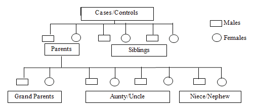
- After obtaining the written informed consent from the parents, the case history with the complete family history upto 3 generations of the cleft lip and/or cleft palate patient was recorded as per the proforma. Family history was taken including first (Parents, Siblings) and second degree relatives (Grandparent, Aunt, Uncle, Niece and nephew). Pedigree was constructed through interview of the parents of study group and control group.
Cheloscopy and Demetoglyphic analysis
Lip prints were taken using following methodology: First, the upper and lower lips of the subject were cleaned thoroughly with sterile cotton with Povidone-Iodine cleansing solution with the cotton swab. Petroleum jelly was applied on the lips if the lips are dry as this would hamper the uniform spread of lipstick. After about 2-3 minutes, excess petroleum jelly was wiped off the lip surface using cotton wool. Lipstick application was done stepwise. Firstly the boundaries of the lips were marked with a red-colored (Lakme) lip liner to confine the lipstick application within the marked area. The tip of the lip liner was cleansed with a cotton ball dipped in hand cleanser, prior to using it on the next person. Lipstick is then uniformly applied with the help of a lipstick application brush in a systematic manner to avoid the smudging of the lipstick in the grooves. Once the application of the lipstick is over, the subjects were made to wait for 30 sec for drying of the lipstick. He or she was asked to keep the teeth in occlusion and gently retract the opposing lip when the print of the concerned lip was being taken. Lip print was then obtained using 14-mm wide and 50-mm long strip of cellophane tape (on the glued portion). Lip impression is made with gentle pressure on either side. Since the human lips are so mobile that the strength or the direction of the pressure applied and even the minimal movement of the lip can affect the accuracy of the lip print taken, the subject is strictly advised not to change the position and avoid any movement of the lips during the procedure of recording the lip print. The cellophane tape is then stuck onto a proforma where the patient details are recorded. The lips of the subjects were thoroughly wiped off the lipstick colour after completion of the procedure using cotton wool. This procedure was repeated for each subject. The lip prints are repeated if the ones obtained were not satisfactory. The lipstick applicator brush was disinfected using a disinfectant spray in the interval of usage between consecutive subjects for hygienic purpose. The lip prints was then visualized using a magnifying glass and analysed according to the Suzuki & Tsuchihashi lip print classification (1970).
Type I Complete Straight Grooves (SG)
Type II Branched Grooves (BG)
Type III Intersected Grooves (IG)
Type VI Reticular Grooves (RG)
Type V Undifferentiated Grooves (UG)
Finger prints of each subject were recorded. The subjects were asked to wash their hands with detergent and after thorough drying; their finger prints were recorded using standard ink method. Blue duplicating ink was used on which the distal phalanx of the each finger was rolled gently. Rolled prints of each finger digits impressions were transferred to a proforma. Then, the strip was labelled by sides (right or left) and by each digit using Roman numerals (customarily, the thumb I and the little finger V). After the completion of printing, finger print patterns were classified as arches (A), loops (L) or whorls (W) based on Galton (1982) classification. Arch has no triradius and is the simplest pattern, the loop has one triradius and one core, and the whorl has usually two triradii.
RESULTS


Table 5: Distribution of study subjects based on Father’s Finger print


DISCUSSION
While the etiology of cleft lip and palate is still poorly understood, many researchers believe that this anomaly is a consequence of genetic and environmental factors that affect facial development.12,13 During early embryonic development, pathological genetic factors, which disrupt the development of craniofacial structures, could also influence the early formation of dermatoglyphics in cleft lip and palate patients.14 The epidermal ridges of the fingers and palms as well as facial structures such as lips, alveolus and palate form from the same embryonic tissues (ectoderm) during the same embryonic period; thus, these features may serve as proxy markers altering early development in cleft lip and palate.15 Despite the intense interest in cleft lip and palate research, a paucity of attention has been given to the role of dermatoglyphic traits of parents (especially in sporadic cases) as a risk marker.16
In the present study among cases, 4 (8%) children had cleft lip only, 13 (26%) children had cleft palate only and 33 (66%) children had cleft lip and palate (Unilateral/Bilateral). The distribution is similar to the study conducted by Manasa Ravath CJ et al17, where 9 (18%) children had cleft lip only, 7 (14%) children had cleft palate only and 34 (68%) children had cleft lip and palate (Unilateral/Bilateral). In the study conducted by Neda Eslami et al, 55 study subjects were with unilateral/bilateral cleft lip and palate18.
Considering the lip print patterns, the wrinkles on lips have unique individual characteristics that are not only markers of identification but also of genetic correlation to an individual’s identity.19 In the present study, it was found the father’s upper lip print pattern among cases showed high prevalence of Type II (branched grooves) 46% and among controls high prevalence of Type III (intersected grooves ) 36%. These results are in accordance with the study conducted by Priyankar Singh et al20, wherein father’s lip prints were Type II (branched grooves) 40% among cases and Type I (straight grooves) 43% among controls. The results are in contrast with the study conducted by Manasa Ravath CJ et al17, wherein among fathers upper lip pattern Type I (straight) 28% was highly frequent.
In the present study, mothers lip print distribution showed the prevalence of Type II (branched grooves) 48% among cases and 52% among controls. These results are in accordance with the study conducted by Priyankar Singh et al20, in which mother’s lip prints were Type II (branched grooves) 32% among cases and Type I (straight grooves) 42% among controls. The results are in contrast with the study conducted by Manasa Ravath CJ et al17 in which Type I (straight grooves) are frequently found (38%) among cases.
The present study showed type II patterns (branched grooves) among 75.3% of all cases. This is similar to the results of the study by Saad et al5, where 62% type II patterns (branched grooves) were seen in the study group among both the parents. In the study conducted by Saujanya K et al21 in Andhra Pradesh, India, Type O (unique type) was observed among 3.3% cases at the centre of the upper lip, which was not found in any of the cases in present study and the authors have attributed the results may be due to geographical variation.21 All the above mentioned studies have used Suzuki & Tsuchihashi lip print classification (1970). Another unique lip print pattern was observed by the study conducted by Katherine Neiswanger et al22, Whorl lip print patterns which are circular grooves on the central upper lip and/or the left and right lower lip. Whorls were more frequent in CL/P families from Argentina than in CL/P families from the U.S. or Hungary. If these results are confirmed, whorl lip print patterns could be part of an expanded phenotypic spectrum of nonsyndromic CL/P. As such, they may eventually be useful in a clinical setting, allowing recurrence risk calculations to incorporate individual phenotypic information in addition to family history data.22
Both dermatoglyphics and chelioscopy have proved their worth as key tools for the prediction of congenital disorders along with their genetic basis, and indeed they have been used as markers for the inheritance of cleft lip and cleft palate.7,17,22,23 Dermatoglyphic analysis in the present study revealed that among fathers finger print, Loop pattern was found to be significantly higher (p<0.01) among cases in digit one (84%), two (80%) and five (82%) of the right hand. Among mothers finger print pattern, Loop pattern was found to be to be significantly higher (p<0.01) among cases in digit one (66%), three (50%), four (50%) and five (66%) and Whorl pattern was found to be higher in digit two (62%) of the right hand. These results are in accordance with the study conducted by Naveen RA et.al7, where overall finger print of fathers showed the high occurrence of Loops (48%) among cases, but whorls (59%) among controls.
Finger print pattern of mothers also showed high occurrence of loops (51%) among cases. The results are in accordance with the study conducted by Saujanya K et al21 in which it was found that loop pattern was mostly observed among fathers (58%) and mothers (52%) of the cases. Among control mothers, arch pattern was highly observed (55%).
In the present study, there was greater frequency of loops than whorls and arches (L-63%>W-28%>A-9%) among parents of cases. Study by Balgir et.al24, showed a greater frequency of loops than arches and whorls in CL(P) parents (L-57%>A-24%>W-19%). Admala et al25 concluded in their study that increased dermatoglyphic asymmetry with higher loop patterns were seen in the parents with clefted children and increased whorl patterns in parents with normal children traits are genetically controlled and are passed from one generation to the other which is governed by laws of inheritance propounded of Mendel and the features of dermatoglyphics and cleft deformities are not an exception.26 Threshold theory which was put forth by Carter and Wilkinson stated that abnormalities are the result of both genetic and environmental factors, but expressed only after exceeding threshold limit.27 According to this statement, before they could be physically expressed, there is an inevitable chance for few variations to occur in dermatoglyphics due to their correlated formation at intrauterine life. So as an attempt to derive the proportion and intensity of these genetic variations with reference to NSCL/P in the previous generations, the present study was planned with unaffected parents of cleft progeny. The pattern types can easily be detected on the fingertips and any variations can be easily determined.28 Hence, pattern types were included in the study. In the present study, there was statistically significant difference seen between individual pattern types, where loops being more frequently seen, next being whorls and finally the arches in all the four groups. These results are in accordance with the study conducted by Harika et al29 where loop pattern was found among 61% of participants. This indicates the overall genetic predilection for loop pattern. In contrary to the present study, studies conducted by Woolf et al30 found that fathers of cleft patients had an increased frequency of arches.
Since the risk of recurrence of CL/P within a family is 28-40-fold greater than that for the general population, genetic factors are thought to contribute to the development of this disorder. It has been suggested that nonsyndromic clefting in humans is most likely due to a combination of genetic and environmental factors as described by the multifactorial threshold
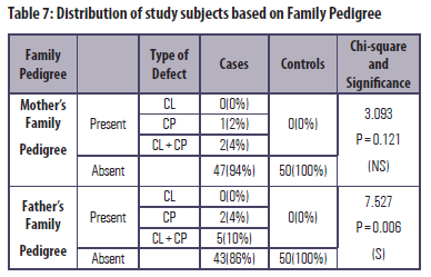
model.31 In the present study results, mother’s family pedigree showed 3(6%) affected family members and father’s family pedigree showed 7(14%) affected family members. In the study conducted by Charles M W32 showed 3% of cases with family history. Scott et al33 suggested that accurate phenotyping is crucial to understanding both the epidemiology and aetiology of any congenital malformation because the power to detect effects is weakened when heterogeneous groups are treated as a single entity. Although clefts of the lip and palate show a range of phenotypic expression, they are generally defined as qualitative traits. A range of study designs will be needed to achieve this level of documentation, including family studies, case–control studies and eventually prospective cohort data. Many family-based studies of
CONCLUSION
Based on the observations from our study the following conclusions have been drawn:
- An increase frequency of type II lip patterns among parents of CL(P) affected children as compared to that of parents of unaffected children.
- Increased frequency of loops pattern in parents CL(P) affected children as compared to that of parents of unaffected children.
- Based on the present study results, specific type cannot be contemplated as a genetic marker in the transmission of CL(P). But, dermatoglyphics and chelioscopy can be used as important screening markers to predict the occurrence and inheritance of cleft lip and cleft palate.
- 10% 0f the cases presented family history with more than one occurrence of CL/P.
CL/P have demonstrated significant genetic associations in some populations, whereas other studies have found no association.34 For example, the transforming growth factor alpha locus (TGFA) has been found to be associated with CL/P in U.S. whites, in Australian whites, in West Bengal, India, and in Japanese (and others); yet no associations were found in Filipinos, U.S. Hispanics, Danish whites, Shanghai Chinese, and others33. It has been suggested that the differences in association represent the action of different genes in different environments and populations. Because CL/P is known to be a genetically heterogeneous entity, related phenotypic traits, such as dermatoglyphics, may be similarly heterogeneous.
REFERENCES
- Stanier P, Moore GE. Genetics of cleft lip and palate: syndromic genes contribute to the incidence of non-syndromic clefts. Human molecular genetics. 2004 Jan 13;13(suppl_1):R73-81.
- Scott NM, Weinberg SM, Neiswanger K, Daack-Hirsch S, O'brien S, Murray JC, Marazita ML. Dermatoglyphic pattern types in subjects with nonsyndromic cleft lip with or without cleft palate (CL/P) and their unaffected relatives in the Philippines. The Cleft palate-craniofacial journal. 2005 Jul;42(4):362-6.
- Dixon MJ, Marazita ML, Beaty TH, Murray JC. Cleft lip and palate: understanding genetic and environmental influences. Nature Reviews Genetics. 2011 Mar;12(3):167.
- Balgir RS. Congenital oral clefts and dermatoglyphics. Israel journal of medical sciences. 1984 Jul;20(7):622-4.
- Saad WM, Kamel AH, Hassan FZ, Elotiefy MA. Genetic studies on the inheritance of lip prints in cleft lip and palate. Egypt J Plast Reconstr Surg. 2005;29(1):9-12.
- Jahanbin A, Mahdavishahri N, Naseri MM, Sardari Y, Rezaian S. Dermatoglyphic analysis in parents with nonfamilial bilateral cleft lip and palate children. The Cleft Palate-Craniofacial Journal. 2010 Jan;47(1):9-14.
- Admala NR, Arjunan S, Adusumilli G, Thirumala JR, Devanna R, Pichai S. Dermatoglyphics and cheiloscopy in the inheritance of cleft lip and palate: Unraveling the mystery. Journal of Indian Orthodontic Society. 2014 Jan 1;48(3):175-9.
- Carinci F, Pezzetti F, Scapoli L, Martinelli M, Avantaggiato A, Carinci P, Padula E, Baciliero U, Gombos F, Laino G, Rullo R. Recent developments in orofacial cleft genetics. Journal of Craniofacial Surgery. 2003 Mar 1;14(2):130-43.
- Celli J, Duijf P, Hamel BC, Bamshad M, Kramer B, Smits AP, Newbury-Ecob R, Hennekam RC, Van Buggenhout G, Van Haeringen A, Woods CG. Heterozygous germline mutations in the p53 homolog p63 are the cause of EEC syndrome. Cell. 1999 Oct 15;99(2):143-53.
- Kondo S, Schutte BC, Richardson RJ, Bjork BC, Knight AS, Watanabe Y, Howard E, de Lima RL, Daack-Hirsch S, Sander A, McDonald-McGinn DM. Mutations in IRF6 cause Van der Woude and popliteal pterygium syndromes. Nature genetics. 2002 Oct;32(2):285.
- Suzuki K, Hu D, Bustos T, Zlotogora J, Richieri-Costa A, Helms JA, Spritz RA. Mutations of PVRL1, encoding a cell-cell adhesion molecule/herpesvirus receptor, in cleft lip/palate-ectodermal dysplasia. Nature genetics. 2000 Aug;25(4):427.
- Blomberg S. Influence of maternal distress during pregnancy on fetal malformations. Acta Psychiatrica Scandinavica. 1980 Oct;62(4):315-30.
- Millicovsky G, Johnston MC. Hyperoxia and hypoxia in pregnancy: simple experimental manipulation alters the incidence of cleft lip and palate in CL/Fr mice. Proceedings of the National Academy of Sciences. 1981 Sep 1;78(9):5722-3.
- Peterka M, Tvrdek M, Likovský Z, Peterkova R, Fara M. Maternal hyperthermia and infection as one of possible causes of orofacial clefts. Acta chirurgiae plasticae. 1994;36(4):114-8.
- Mathew L, Hegde AM, Rai K. Dermatoglyphic peculiarities in children with oral clefts. Journal of Indian Society of Pedodontics and Preventive Dentistry. 2005 Oct 1;23(4):179.
- Jahanbin A, Mahdavishahri N, Naseri MM, Sardari Y, Rezaian S. Dermatoglyphic analysis in parents with nonfamilial bilateral cleft lip and palate children. The Cleft Palate-Craniofacial Journal. 2010 Jan;47(1):9-14.
- Manasa Ravath CJ, Girish HC, Sanjay Murgod RB, Savita JK. Lip prints and inheritance of cleft lip and cleft palate. Journal of clinical and diagnostic research: JCDR. 2014 Jul;8(7):ZC28.
- Eslami N, Jahanbin A, Ezzati A. Palm and finger print characteristics in nonfamilial cleft lip and palate patients and their parents. Journal of Craniofacial Surgery. 2013 May 1;24(3):769-72.
- Dineshshankar J, Ganapathi N, Yoithapprabhunath TR, Maheswaran T, Kumar MS, Aravindhan R. Lip prints: Role in forensic odontology. Journal of pharmacy & bioallied sciences. 2013 Jun;5(Suppl 1):S95.
- Singh P, Nathani DB. Dermatoglyphics and Cheiloscopy as Key Tools in Resolving the Genetic Correlation of Inheritance Patterns in Cleft Lip and Palate Patients: An Assessment of 160 Patients. The Cleft Palate-Craniofacial Journal. 2017 Sep;54(5):588-94.
- Niranjani K, Prasad MG, Vasa AA, Divya G, Thakur MS, Saujanya K. Clinical Evaluation of Success of Primary Teeth Pulpotomy Using Mineral Trioxide Aggregate®, Laser and BiodentineTM-an In Vivo Study. Journal of clinical and diagnostic research: JCDR. 2015 Apr;9(4):ZC35.
- Neiswanger K, Chirigos KW, Klotz CM, Cooper ME, Bardi KM, Brandon CA, Weinberg SM, Vieira AR, Martin RA, Czeizel AE, Castilla EE. Whorl patterns on the lower lip are associated with nonsyndromic cleft lip with or without cleft palate. American Journal of Medical Genetics Part A. 2009 Dec;149(12):2673-9.
- Siraz MA, Akshay HS. Comparative study of some of the qualitative and quantitative dermatoglyphic patterns among the patients of cleft lip and cleft palate against normal population. MedPulse–Int Med J. 2015;2:381–385.
- Balgir RS. Dermatoglyphics in cleft lip and cleft palate anomalies. Indian Pediatr 1993;30:341-6.
- Admala NR, Arjunan S, Adusumilli G, Thirumala JR, Devanna R, Pichai S. Dermatoglyphics and cheiloscopy in the inheritance of cleft lip and palate: Unraveling the mystery. Journal of Indian Orthodontic Society. 2014 Jan 1;48(3):175-9.
- Abhilash PR, Divyashree R, Patil SG, Gupta M, Chandrasekar T, Karthikeyan R. Dermatoglyphics in patients with dental caries: A study on 1250 individuals. J Contemp Dent Pract. 2012 May 1;13(3):266-74.
- Carter CO, WILKINSON JA. 10 Genetic and Environmental F actors in the Etiology of Congenital Dislocation of the Hip. Clinical Orthopaedics and Related Research®. 1964 Jan 1;33:119-28.
- Mulvihill JJ, Smith DW. The genesis of dermatoglyphics. JPediatr 1969; 75: 579-89.
- Harika DJ, Sridevi E, Sankar AS, Pranitha K, Gosla SR, Kiran GB. Dermatoglyphic analysis in parents with cleft children: A comparative study. Contemporary clinical dentistry. 2018 Sep;9(Suppl 2):S291.
- Woolf CM, Gianas AD. Congenital cleft lip and fluctuating dermatoglyphic asymmetry. American Journal of Human Genetics. 1976 Jul;28(4):400.
- Ardinger HH, Buetow KH, Bell GI, Bardach J, VanDemark DR, Murray JC. Association of genetic variation of the transforming growth factor-alpha gene with cleft lip and palate. American journal of human genetics. 1989 Sep;45(3):348.
- Woolf CM, Gianas AD. Congenital cleft lip and fluctuating dermatoglyphic asymmetry. American Journal of Human Genetics. 1976 Jul;28(4):400.
- Scott NM, Weinberg SM, Neiswanger K, Brandon CA, Daack-Hirsch S, Murray JC, Liu YE, Marazita ML. Dermatoglyphic fingerprint heterogeneity among individuals with nonsyndromic cleft lip with or without cleft palate and their unaffected relatives in China and the Philippines. Human biology. 2005 Apr 1:257-66.
- Marazita ML, Hu DN, Spence MA, Liu YE, Melnick M. Cleft lip with or without cleft palate in Shanghai, China: evidence for an autosomal major locus. American journal of human genetics. 1992 Sep;51(3):648.
Disclaimer from Guident
The views and opinions expressed in articles published in Guident are solely those of the authors and do not necessarily reflect the views of the editors, the editorial board or the publisher, Ivory India. Guident does not endorse any product, technique or treatment described. Clinical content is intended for qualified dental and medical professionals and is not a substitute for individual clinical judgement. Guident, its editors and the publisher accept no liability for any consequence arising from the use of information published in the journal.
How to cite this article
Dr. Pooja M. R., Dr. Jithesh Jain, Dr. Ananada S. R.. Cheiloscopy and Dermatoglyphics as Genetic Markers and Role of Family Pedigree in the Inheritance of Non Syndromic Cleft Lip and Palate. Guident. 2020 Mar;13(4). Available from: https://ivoryindia.in/articles/research/cheiloscopy-and-dermatoglyphics-as-genetic-markers-and-role-of-family-pedigree-in-the-inheritance-of-non.html