Microbiology of Endodontic Faliure Cases
Gian Sagar Dental College, Banur, India
ABSTRACT
The aim of this study was to detect the various microorganisms present in the root canal of endodontic failure cases. 30 patients with teeth showing symptoms of root canal failure were selected. Sample was collected using paper points after removal of the root canal filling. The collected sample was evaluated for the presence of aerobic and anaerobic microorganisms using culturing technique. Enterococcus feacalis and Peptostreptococcus were the most frequently found aerobe and anaerobe respectively.
Keywords: retreatment, E. Faecalis, microflora, root canal.
INTRODUCTION
The influence of bacterial persistence in the root canals on treatment outcome is an important issue in endodontics because bacteria have been shown to play a major role in persistence or emergence of apical periodontitis lesions after root canal treatment. Root canal infections have a different nature than that of caries or periodontitis because they become established in originally sterile compartments of the oral cavity. In many cases, this led to the concept that the etiology of root canal infections involves only a single pathogen. For example, the predominance of certain proteolytic black-pigmented anaerobic organisms in cultures from infected root canals associated with acute symptoms suggested that these organisms are foremost etiological agents in such cases.1
The persistence of disease in periapical tissues is most commonly associated with difficulties or mishaps which occur during or after initial endodontic treatment. Inadequate aseptic control, poor access cavity design, missed canals, inadequate instrumentation and leaking temporary or permanent restorations are the main factors that may be critically important in post-treatment disease.2
Root canal infections have a different nature than that of caries or periodontitis because they become established in originally sterile compartments of the oral cavity. In many cases, this led to the concept that the etiology of root canal infections involves only a single pathogen. For example, the predominance of certain proteolytic black-pigmented anaerobic organisms in cultures from infected root canals associated with acute symptoms suggested that these organisms are foremost etiological agents in such cases.3 Complete elimination of microbes in the teeth with an infected dental pulp is the main objective of the root canal treatment.4
Despite thorough mechanical instrumentation and disinfection of the root canal system, microbes have been recovered at subsequent treatment sessions and at the end of the treatment. Such residual organisms and the leakage of bacterial products are likely to play a role in treatment failures.5,6
The purpose of this study was to detect the various aerobic and anaerobic microorganisms present in the root canals of endodontic failure cases.
MATERIALS AND METHOD
Thirty patients were selected for the study. The inclusion criteria of patients for the study was:-
1. Patients with root canal filled teeth and having a history of pain.
2. Patients with root canal filled teeth and having tenderness to percussion on clinical examination.
3. Patients with root canal filled teeth having pain and a periapical radiolucency on radiographic examination. (PHOTOGRAPH 1)
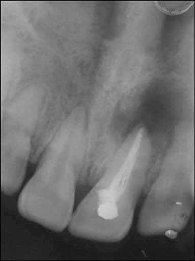
Fig 1: - Radiograph of tooth included in the study
The exclusion criteria of the patients for the study was:-
1. Any patient who was on antibiotics.
2. Any patient having a severe systemic disease.
3. Any tooth without a coronal seal
4. Any tooth with large intraradicular post.
SAMPLING PROCEDURE
Firstly, the tooth to be re-treated was isolated from oral cavity with a rubber dam. The coronal restoration was removed, access cavity was prepared and the entire area was disinfected with 5.25% sodium hypochlorite solution. The solution was inactivated with sodium thiosulphate in order to avoid interference with bacteriological sampling. The gutta-percha from the root canal was removed using sterile Gates-Glidden drill and sterile endodontic files without the use of chemical solvents. Irrigation with sterile saline solution was performed in order to remove any remaining material and to moisten the canal prior to sample collection.
For microbial sampling from root canal, a sterile paper point was introduced to the full length of the root canal and kept in place for 60 seconds. As many paper points as possible were introduced in the canal to absorb all the fluid inside the canal.
For the culture of aerobic organisms, one sample from each tooth was transferred to Peptone water and for the culture of anaerobic organisms, sample from each tooth was transferred to Thioglycollate medium. Both the samples were transported to microbiology laboratory immediately.(PHOTOGRAPH 2)

Fig 2: – Sample kept in thioglycollate medium and Peptone water
In the laboratory, a gram stained smear was prepared from each specimen. The samples were directly inoculated onto blood agar and MacConkey agar plates and incubated aerobically at 37o C for 24 hours. For anaerobes, specimens were directly inoculated on blood agar and incubated anaerobically in Gaspak jar at 37o C for 48 hours. The plates were observed upto 7 days for any growth. The colonies were further identified as aerobic and anaerobic bacteria by standard laboratory testing techniques (Photograph 3 & 4).

Fig 3: – Anaerobic Jar used for anaerobic culturing

Fig 4: - Anaerogas Pack and indicator tablet used in anaerobic jar
The tests carried out to study the biochemical reactions of aerobes were Carbohydrate fermentation reaction, Catalase test, Coagulase test, Indole test, Methyl red test, Voges – Proskauer test, Nitrate reduction test, Test for citrate utilisation, Test for urease production, H2S production and Oxidase test.
The tests done to study the biochemical reactions of anaerobes were Carbohydrate fermentation reactions, Catalase test, Indole test, Nitrate reduction test, Aesculin hydrolysis test, Growth stimulation test, Gelatin liquefaction test, Stromy clot reaction (Litmus milk test) and Growth inhibition test.
The antibiotic sensitivity of aerobic organisms was determined against different antibiotics by the modified Kirby-Bauer’s method and for anaerobic organisms by disc diffusion method. For the purpose of test, nutrient agar plates were used for aerobes and Brain heart infusion agar plates were used for anaerobes. A standard control strain of Escherichia coli used was NCTC 10418, of Staphylococcus aureus NCTC 6571 and of Pseudomonas aeruginosa NCTC 10662.
RESULTS
In the present study, Enterococcus faecalis was the most commonly found aerobe which was seen in 18 samples out of 30 samples. (Photograph 5) Other aerobic microorganisms found in group I were Diptheroids, Pseudomonas, Nisseria, Streptococcus mutans, Streptococcus sanguis, Enterobacter, Coagulase –ve staphylococci, Staphylococcus epidermidis, E. coli, Candida albicans(Photograph 6) and Citrobacter freundii (Table 1) . Anaerobes were seen in 10 of the 30 samples. The anaerobic microorganisms present were Peptostreptococcus, Fusobacterium nucleatum and Fusobacterium nerophorum Bacteroides fragilis, Bacteroides melanogenicus, Veillonella, Peptococcus and Lactobacillus. (Table 2)

Fig 5: - Culture plate showing growth of E. faecalis

Fig 6: – Budding yeast form of C. albicans under microscope
Table1

Table II

DISCUSSION
Success in endodontics depends on a series of well planned and executed steps that start with a proper diagnosis and end with a hermetic seal of the root canal. The term failure consequently may be defined as a treatment that did not reach the objective or fell short of the acceptance level. From a biological perspective, an endodontic failure is associated with an inflammatory process in the periradicular supporting structures of a tooth.1
The influence of bacterial persistence in the root canals on treatment outcome is an important issue in endodontics because bacteria have been shown to play a major role in persistence or emergence of apical periodontitis lesions after root canal treatment. This indicates that persisting bacteria can survive in treated canals and are able to induce or sustain periradicular tissue inflammation, underpinning the concept that the eradication of bacteria from the root canal system should be the ultimate goal of the endodontic treatment of teeth with apical periodontitis.7
In the present study, E. faecalis was the most commonly found aerobic microorganism. Enterococci possess a number of virulence factors that permit adherence to host cells and extracellular matrix, facilitate tissue invasion, effect immunomodulation and cause toxin-mediated damage. These factors include:
(1) aggregation substance,
(2) enterococcal surface proteins such as esp,
(3) gelatinase,
(4) a cytolysin toxin,
(5) extracellular superoxide production,
(6) capsular polysaccharides and
(7) antibiotic resistance determinant.8
Once established in the root canal, E. faecalis faces several challenges for survival, including an ability to withstand antimicrobial agents used during treatment and endure potential starvation in the cleaned and obturated canal. E. faecalis appears to be highly resistant to the medicaments used during treatment and is known to resist the antibacterial effect of dressing with calcium hydroxide.9
Peptostreptococcus was the most common anaerobe isolated from the endodontic failure cases. Genus Peptostreptococcus includes the species formerly known as Peptococcus, with the exception of Peptococcus niger. Extracellular proteinase appears to be a major virulence determinant and is designated the name gingipain. Gingipains seem to be key players in subverting host defense systems with the complement and neutrophils as the main targets. The direct extracellular matrix proteolytic activity exhibited by the Peptostreptococcus spp. is limited. However, Peptostreptococcal production of proteolytic enzymes may have an important adjunctive effect on the pathogenesis of certain soft tissue infections.10
Citrobacter freundii was found in 3 samples in this study. It is a common opportunistic pathogen which causes nosocomial infections. Its occurrence may be because of contamination of the sample during processing. Other microbes frequently observed were streptococcus, staphylococcus and fusobacterium. Candida albicans was also found in 5 samples suggesting the role of fungi in endodontic failures.
Chavez de Paz LE, et al.11 (2003) investigated the bacteria recovered from 200 teeth with apical periodontitis after antimicrobial endodontic treatment. A total of 248 strains were isolated from 107 teeth giving bacterial growth. Lactobacillus spp. (22%), nonmutans streptococci (18%), and Enterococcus spp. (12%) were the most common isolates. Gram-negative anaerobes were relatively sporadic. Large radiographic bone lesions, persistent pain and use of intracanal calcium hydroxide dressing correlated with bacterial presence.
Mindere A, et al.12 (2010) investigated the microbial flora of 33 root filled teeth with apical periodontitis in Latvian patients and determined the prevalence of β-lactamase producing bacteria associated with such lesions. Bacteria were found in 32 (97%) of initial specimens from the teeth. 79% of the isolated microbial species were Gram-positive bacteria. The most common isolates were Streptococcus (27%), Actinomyces (27%), Staphylococcus (18%), Enterococcus (18%) and Lactobacillus (18%) spp. Yeasts were found as four isolates in 3 cases (9%). β-lactamase-producing bacterial strains were detected in 12 specimens (36% of the patients). The most common enzyme-producing bacteria belonged to Actinomyces and Staphylococcus spp.
Various other species of microorganisms have been isolated from endodontic failure cases. Those isolates were not recovered from the samples of the present study because of the limitations of the culturing technique and the various recent methods of microbial isolation used in other studies.
Knowledge of the microbial flora of endodontic failure cases may lead to many changes in the treatment strategies during root canal treatment and may help to eradicate the resistant microflora more effectively and efficiently from endodontic failure cases.
CONCLUSION
Within the limits of this study, Enterococcus feacalis and Peptostreptococcus were the most commonly found aerobe and anaerobe respectively. Isolation of Candida albicans, supported the role of fungi in endodontic failures. . Based on the knowledge of the bacterial diversity and persistence in post-treatment endodontic infections, future studies should focus on the bacterial profiles of a significant number of endodontic samples using molecular methods such as the reverse-capture checkerboard DNA–DNA hybridization, microarray technology etc.
BIBLIOGRAPHY
- Lin LM, Skribner JE, Gaengler P. Factors associated with endodontic treatment failures. J Endod 1992;18:625–7.
- Peciuliene V, Maneliene R, Balcikonyte E, Drukteinis S, Rutkunas V. Microorganisms in root canal infections: a review. Stomatologija, Baltic Dental and Maxillofacial Journal. 2008; 10: 4-9.
- Haapasalo M, Ranta H, Ranta K, Shah H. Black-pigmented Bacteroides spp. In human apical periodontitis. Infect Immun 1986;53:149 –53.
- Trope M, Bergenholtz G.Microbiological basis for endodontic treatment: can a maximal outcome be achieved in one visit? Endodontic Topics. 2002; 1: 40-53.
- Molander A, Reit C, Dahlen G, Kvist T. Microbiological status of root-filled teeth with apical periodontitis. Int Endod J. 1998; 31: 1-7.
- Sundqvist G, Figdor D, Persson S, Sjogren U. Microbiologic analysis of teeth with failed endodontic treatment and the outcome of conservative re-treatment. Oral Surg Oral Med Oral Pathol Oral Radiol Endod. 1998; 85: 86-93.
- Pinheiro ET, Gomes BPFA, Ferraz CCR, Sousa ELR, Teixeira FB, Souza- Filho FJ. Microorganisms from canals of root-filled teeth with periapical lesions. Int Endod J. 2003; 36: 1-11.
- Rocas IN, Siqueira JF Jr, Aboim MCR, Rosado AS. Denaturing gradient gel electrophoresis analysis of bacterial communities associated with failed endodontic treatment. Oral Surg Oral Med Oral Pathol Oral Radiol Endod. 2004; 98: 741-9.
- Siquiera JF Jr, Rocas IN. Clinical implications and microbiology of bacterial persistence after treatment procedures. J Endod. 2008; 34: 1291–1301.
- Sundqvist GK, Eckerbom MI, Larsson AP, Sjogren UT. Capacity of anaerobic bacteria from necrotic dental pulps to induce purulent infections. Infect Immun. 1979; 25(2): 685-93
- Evans M, Davies JK, Sundqvist G, Figdor D. Mechanisms involved in the resistance of Enterococcus faecalis to calcium hydroxide. Int Endodontic J. 2002; 35: 221-8.
- Olsen I, Dahlen G. Salient virulence factors in anaerobic bacteria, with emphasis on their importance in endodontic infections. Endodontic Topics. 2004; 9: 15–26.
- Chavez de Paz LE, Dahlen G, Molander A, Moller A, Bergenholtz G. Bacteria recovered from teeth with apical periodontitis after antimicrobial endodontic treatment. Int Endod J. 2003; 36: 500-8.
- Mindere A, Kundzina R, Nikolajeva V, Eze D, Petrina Z. Microflora of root filled teeth with apical periodontitis in Latvian patients. Stomatologija, Baltic Dental and Maxillofacial Journal. 2010; 12: 116-
Disclaimer from Guident
The views and opinions expressed in articles published in Guident are solely those of the authors and do not necessarily reflect the views of the editors, the editorial board or the publisher, Ivory India. Guident does not endorse any product, technique or treatment described. Clinical content is intended for qualified dental and medical professionals and is not a substitute for individual clinical judgement. Guident, its editors and the publisher accept no liability for any consequence arising from the use of information published in the journal.
How to cite this article
Dr. Munish Batrav, Dr. Rajveer Kaur Department of Conservative Dentistry & Endodontics. Microbiology of Endodontic Faliure Cases. Guident. 2015 Jan;8(2). Available from: https://ivoryindia.in/articles/research/microbiology-of-endodontic-faliure-cases.html