Apical Surgery with Retrograde MTA as a Filling Material
Department of Conservative Dentistry and Endodontics Seema Dental College And Hospital, Rishikesh A Case Report Apical surgery
Abstract
Apical surgery is considered often a last resort to treat a tooth with a periapical lesion that cannot be managed with conventional endodontic treatment or retreatment. The goal of apical surgery is to prevent bacterial leakage from the root-canal system into the peri radicular tissues by placing a tight root-end filling following root-end resection. In addition, its objective is to promote tissue regeneration by removal of the periapical pathologic tissue and by exclusion of any irritants within the physical confines of the affected root. The application of microsurgical techniques in apical surgery, i.e., incision and flap elevation, production of a small osteotomy, and the use of sonic- or ultrasonic driven microtips, results in less trauma to the patient and faster postsurgical healing. A major step in apical surgery is to identify possible leakage areas at the cut root face and subsequently to ensure adequate root-end filling. Only a tight and persistent apical obturation will allow periapical healing with good long-term prognosis. Mineral Trioxide Aggregate (MTA) has since long been successfully used by dentists in the management of teeth.
Keywords- Mineral Trioxide Aggregate, Root canal Treatment, Root End Resection , Apical surgery.
INTRODUCTION
Periapical pathology could be the fate of untreated or unsatisfactorily treated traumatically injured teeth. Surgical endodontics is a reliable method for the treatment of teeth with periapical lesions which do not respond to conventional root canal treatment.1 Apical surgery belongs to the field of endodontic surgery, which also includes incision and drainage, closure of perforations, and root or tooth resections. The goal of a periapical surgery is to gain access to the affected area, evaluate the root circumference and root canal anatomy, and place a biocompatible seal in the form of root end filling that stimulates the regeneration of periapical hard and soft tissues. Its success rate varies from 86 –92% while failure rate is 4.7% 2,3. The prognosis depends on several factors such as: different surgical procedures and materials, clinical and radiographic evaluation, systemic conditions, local factors such as involved teeth and their anatomy, previous treatment and its quality.4,5,6 It is therefore of clinical relevance to perform a thorough clinical and radiographic examination of the tooth before apical surgery (including adjacent and opposing teeth), in order to decide whether surgical or nonsurgical endodontics should be considered. According to the updated guidelines by the European Society of Endodontology, indications for apical surgery comprise (1) radiological findings of apical periodontitis and/or symptoms associated with an obstructed canal (the obstruction proved not to be removable, displacement did not seem feasible or the risk of damage was too great), (2) extruded material with clinical or radiological findings of apical periodontitis and/or symptoms continuing over a prolonged period, (3) persisting or emerging disease following root-canal treatment when root canal retreatment is inappropriate, and (4) perforation of the root or the floor of the pulp chamber and where it is impossible to treat from within the pulp cavity.7 Various studies have shown that MTA is an excellent material for use in apexification, as a root end filling material for immature permanent teeth, as a pulp capping and pulpotomy agent and for repair of perforations. Besides these uses, Mineral trioxide aggregate (MTA) has also emerged as a reliable bioactive material with extended applications in endodontics that include the obturation of the root canal space.8,9,10 The present paper describes the successful outcome of apical surgery performed in maxillary right and left central incisor followed by retrograde filling by MTA.
CASE REPORT
A thirty -year-old female patient reported to the Department of Conservative Dentistry &Endodontics, Seema Dental College And Hospital, Rishikesh with the chief complaint of tender on percussion in front region of upper jaw for 15 days. The patient gave history of trauma to the upper front teeth 6-7 years back. The medical history was non-contributory.
Clinical examination revealed endodontically treated 11,21. The tooth No.11,21 was tender on percussion, showed negative response to vitality test and was non-carious with no visible fracture line in the crown. The periodontal health of the patient was satisfactory and free from traumatic occlusion.
The treatment plan included gross debridement and disinfection and obturation of root canal (figure-1) followed by Root end resection (figure-3) and subsequent sealing of the root end with MTA (figure-4).
In the first appointment, access opening was done in relation to 11,21 and the canal was cleaned and shaped. Copious irrigation was performed for complete cleaning of the canal. After thorough cleaning and drying, the canal was obturated with gutta percha.Gutta percha points were extended beyond the apex so that it can be located easily during surgery.
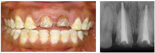
Fig-1 preoperative

Fig-2 Flap retarction
Local anesthesia was administered and a full thickness trapezoidal mucoperiosteal flap was raised extending from distal aspect of 12 to mesial aspect of 22 using Bard Parker (B.P) blade size #15 (BD, Sao Paulo, Brazil). Apical curettage was performed for the removal of granulation tissue. Surgical site was irrigated using normal saline. Following the raising of a flap, surgical osteotomy was done to get access to the root end (figure-2). Horizontal surgical excision of 3.0 mm of the apex was done using a Lindeman Surgical Bur (Brasseler USA) and the root end was exposed. Inverted cone bur were used to make a 3.0-mm deep retro preparation. Hemostasis was obtained in the osseous crypt, and then the retro preparation was disinfected and dried. The retrograde filling was done with MTA (Angelus, Londrina, PR Brazil). The flap was then repositioned and approximated with moderated digital pressure and moist gauze and then finally sutured (figure-5). Post-operatively antibiotics and analgesics were prescribed to the patient along with 0.12% chlorhexidine mouth rinse for maintenance of oral hygiene.

Fig-3- Retro Preparation i.r.t 11 and 21

Fig- 4 – Retrograde-filling with MTA

Fig-5 – Suture Placed
DISCUSSION
Root End Resection is a surgical procedure which involves the excision of pathological periapical tissue from root surface (including apical accessory canals) and sealing the canals against pathological agents thereby aiding in tissue regeneration and creation of new structural support to the tooth. It is the principal modality available to manage failure of conventional orthograde endodontic treatment of a large non- healing periapical lesion. The reported success rate for periapical surgery varies between 44% and 90%. Harty et al. (1970) found that the success rate of 1016 cases of root end resectionm was 90%.
Oginni and Olusile concluded that the success rate of Root End Resection of anterior teeth was 71.9%. Peterson and Gutmann reported a success rate of 64%. 11-15 The reason for this great variation may be the lack of agreement on a definition of success and failure of surgery, the difference in the expertise of those who perform the operations, and the decision to operate or not to operate on teeth with unfavorable prognosis. better case selection can be achieved through obtaining quality information presurgically using digital imaging, which helps establish a more accurate prognosis. Recent advancements in high- resolution, 3-D digital imaging using limited Cone Beam radiography techniques have helped improve surgical case selection through better visualization of the operating field and the determination of the exact location of the associated pathology prior to the actual surgical procedure. This information improves case selection prior to the surgery, which in turn, helps improve case outcome. The advent of ultrasonic devices with surgical preparation tips that allow conservative bony access cavities while still allowing for deep retro preparation depths (following root resection) have further advanced the Root End Resection procedure.16-19
Various cements have been used as root end filling materials. The choice of a root end filling material could be governed by handling properties, biocompatibility, apical seal, and long term clinical success. MTA has been investigated and used as a root end filling material since its introduction. Despite its good physical and biological properties and it being hydrophilic in nature; its use has always remained a challenge because of its technique sensitivity, prolonged setting time, and high cost. The search for an alternative material was with the aim to reduce cost and increase the feasibility for both professionals and patients.
In this case, we performed Root End Resection in maxillary left central incisor followed by MTA filling. In accordance with the results of the aforementioned studies, we obtained good results along with patient satisfaction. Post operatively, the tooth remained asymptomatic.
CONCLUSION
Based on the contemporary understanding of endodontic concepts for success and failure, assessment and subsequent treatment of Root End Resection procedures have greatly improved. Advances in Root End Resection armamentaria and materials (including bioceramic retrofilling materials and clinical techniques for their efficient use) have enabled endodontists to treat challenging cases with much greater efficiency. While successful outcomes are still predominantly a function of proper case section and triage, using the novel retrofilling technique, as described in this article, can make this previously challenging aspect of surgical endodontics much easier for clinicians
REFERENCES
- Ahmed HM, Al Rayes MI, Saini D. Management and prognosis of teeth with traumatic induced crown fractures and large periapical cyst like lesions following apical surgery with and without retrograde filling. J Conserv Dent 2012; 15:77-9.
- Girish CS, Ponappa K, Girish T, Ponappa M. Sealing ability of mineral trioxide aggregate, calcium phosphate and polymethylacrylate bone cements on root ends prepared during Erbium: Yttrium aluminium garnet laser and ultrasonics evaluated by confocal laser scanning microscopy. J Conserv Dent 2013; 16:304-8.
- Tsesis I, Faivishevsky V, Kfir A, Rosen E. Outcome of surgical endodontic treatment performed by a modern technique: a meta-analysis of literature. J Endod 2009; 35:1505-11.
- Tsesis I, Faivishevsky V, Kfir A, Rosen E. Outcome of surgical endodontic treatment performed by a modern technique: a meta-analysis of literature. JEndod 2009; 35:1505-11.
- Friedman S. Considerations and concepts of case selection in the management of post treatment endodontic disease (treatment failure). Endod Topics 2002; 1:54-78.
- Gutmann JL, Harrison JW. Surgical endodontics. Blackwell Scientific Publications.1991:362-8.
- Thomas von Arx. Apical surgery: A review of current techniques and outcome. The Saudi Dental Journal. 2011; 23(1): 9-15.
- Boqen G, Kuttler S. Mineral trioxide aggregate obturation: a review and case series. J Endod 2009 Jun;35(6):777-90.
- Hayashi M, Shimizu M, Ebisu S. MTA for obturation of mandibular central incisors with open apices: case report. Journal of Endodontics 2004;30(2):120-122.
- O’Sullivan S.M. and G.R. Hartwell. Obturation of a retained primary mandibular second molar using mineral trioxide aggregate: a case report. J Endod 2001; 27:703–705.
- Strindberg LZ. The dependence of the results of pulp therapy on certain factors. An analytic study on radiographic and clinical follow–up examinations. Acta Odontol Scand. 1956; 14 (suppl 2): 1-175.
- Sjögren U, Haggland B, Sundgvist G, Wing
- K. Factors affecting the long–term results of endodontic treatment. J Endod. 1990; 16: 498-504.
- Ørstavic D. Time–course and risk analyses of the development and healing of chronic apical periodontitis in man. Int Endod J. 1996; 29: 150-155. [14]. Ray HA, Trope M. Periapical status of endodontically treated teeth in relation to the technical quality of the root filling and the coronal restoration. Int Endod J. 1995; 28: 12-18.
- Sudqvist G, Figdor D, Sjögren U. Microbiologic analysis of teeth with failed endodontic treatment and the outcome of conservative retreatment. Oral Surg Oral Med Oral Pathol Oral Radiol Endod. 1998; 85: 86-93.
- Kim S, Kratchman S. Modern endodontic surgery concepts and practice: a review. J Endod. 2006; 32:601-623.
- Gutmann JL, Harrison JW. Surgical Endodontics. St. Louis, MO: Ishiyaku EuroAmerica; 1991
- Stübinger S, Kuttenberger J, Filippi A, et al. Intraoral piezosurgery: preliminary results of a new technique. J Oral Maxillofac Surg. 2005; 63:1283- 1287.
- Glickman GN, Koch KA. 21st-century endodontics. J Am Dent Assoc. 2000;131(suppl):39S- 46S.
Disclaimer from Guident
The views and opinions expressed in articles published in Guident are solely those of the authors and do not necessarily reflect the views of the editors, the editorial board or the publisher, Ivory India. Guident does not endorse any product, technique or treatment described. Clinical content is intended for qualified dental and medical professionals and is not a substitute for individual clinical judgement. Guident, its editors and the publisher accept no liability for any consequence arising from the use of information published in the journal.
How to cite this article
Dr. Annil Dhingra, Dr. Anshdeep Singh, Dr. Praveen Singhal. Apical Surgery with Retrograde MTA as a Filling Material. Guident. 2019 Apr;12(5). Available from: https://ivoryindia.in/articles/endodontics/apical-surgery-with-retrograde-mta-as-a-filling-material.html