Nonsurgical Healing of a Large Periapical Lesion
Department of Conservative Dentistry and Endodontics, Seema Dental College And Hospital, Rishikesh
INTRODUCTION
Periapical lesions develop as sequelae to pulp disease. They often occur without any episode of acute pain and are discovered on routine radiographic examination. It is accepted that all inflammatory periapical lesions should be initially treated with conservative nonsurgical procedures. Studies have reported a success rate of up to 85% after endodontic treatment of teeth with periapical lesions.
It is an accepted fact that a periapical lesion cannot be differentially diagnosed as a cyst, granuloma, or abscess based on a radiograph.3,4 Based on conservative endodontic treatment there are various intracanal medicaments like calcium hydroxide, antibiotics, steroids, etc for the management of periapical abscess whereas calcium hydroxide based intracanal medicaments are considered as the gold standard. Calcium hydroxide is widely used as an intracanal endodontic material, due to its high alkalinity tissue dissolving effect, causes induction of repair by hard tissue formation and bactericidal effect. Several works have been done studied the mixture of other substances to calcium hydroxide with the purpose of improving some of its properties. Among these additional substances are vehicles that can speed up or slow down ionic dissociation, substances that aid the filling of pulpal cavity by means of their consistency, substances used as antimicrobial medium and media that enhance radiopacity. Metapex, a silicone oil-based calcium hydroxide paste containing 38% iodoform is very popular. With the increased knowledge, skill and materials more of these periapical lesions are now treated nonsurgically.
This case report shows how a correct diagnosis of root canal anatomy, combined with thorough debridement, and disinfection of a root canal healed a large periapical lesion.
CASE REPORT
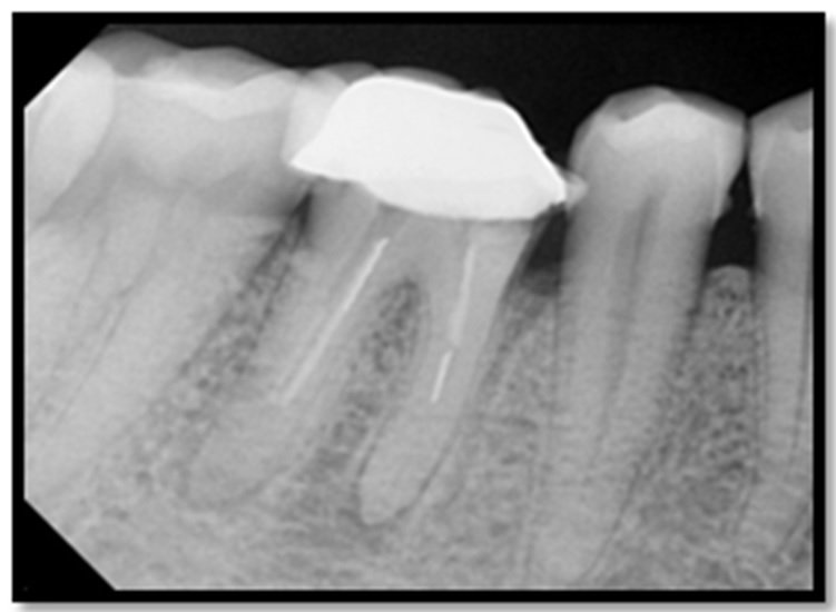
A 20 year old male patient with non contributory medical history reported to the Department of Conservative Dentistry and Endodontics with a chief complaint of pain in his lower right back tooth since 10 days. Extra oral examination revealed no sign or symptom. Intraoral examination revealed a minor firm swelling of the vestibule above the molar. Pain was spontaneous and throbbing in nature. The tooth right mandibular molar was tender on percussion. The radiograph revealed a periapical radiolucency in relation to 46. The margins of radiolucency were diffuse.
Root canal therapy was initiated in 46. Access cavity was cut with endo access bur at high speed. The access cavity was modified and the canals were explored with the number 15K file [Figure 2]. Combined hand and rotary Protaper files were used in root canal preparation. The biomechanical preparation with the first few files was intentionally done extending a few millimeters beyond the apical foramen. Copious irrigation with 3% sodium hypochlorite was used during biomechanical preparation. The canals were prepared up to F3 Protaper files.

Figure-1: Pre operative radiograph

Figure-2: Working length
After bio-mechanical preparation of canals, a non-setting calcium hydroxide paste (Metapex, Meta Biomed Co. Ltd.) was dispensed into the canals and temporary restoration was placed.

Figure 3: 2 months follow-up

Figure 4: 4 months follow-up

Figure 5: 6 months follow-up

Figure 6: 8 months follow-up

Figure 7: 12 month follow up
Follow up was done after one week and when the patient was asymptomatic, tooth was restored with Composite. Further follow-up radiographs were taken at 2 months (Fig-3), 4 months (Fig-4), at 6 months follow-up(Fig-5) and at 8 months follow-up the tooth was obturated,12 month follow up visit it was seen that there was resolution of the radiolucency w.r.t 46 (Fig- 7).
Radiographic changes showed in series of follow ups that there was significant reduction in lesion and appearance of new trabeculae pattern and increased bone density in the periapical area. Finally, tooth was obturated with lateral condensation with gutta percha points, followed by permanent restoration. Recall appointments consisted of reinforcement of oral hygiene instructions, scaling if required and periapical radiographs of the involved tooth.
DISCUSSION
In the present case on clinical examination shows caries which involves the pulp leading to periapical infection. The tooth is non vital and on radiographically shows radiolucency on mesial root of the tooth 46. The perfect mechanism involved in formation of periapical lesions is not fully understood. It is considered that if the pulp becomes necrotic the environment around the periapical area become suitable to allow microorganisms to multiply and various toxins enter into the periapical tissue initiating inflammatory reaction which leads to formation of periapical lesions.
Recent studies have shown, however, that intracanal application of certain medicaments prior to the completion of endodontic therapy may produce highly favorable results when followed by conventional therapy, even when the periapical area is very large. Metapex was used which consist of Ca (OH)2 and iodoform, as intra canal medicament in the above cases.
It has been advocated that direct contact between the calcium hydroxide and the periapical tissue was necessarily beneficial for Osseo inductive reasons. It is suggested that if the calcium hydroxide is confined to the root canal, it is possible that the inflammation created by the diffusion of the calcium hydroxide through the apical foramen may be sufficient to cause break-up of the abscess epithelial lining, thereby allowing a connective tissue invagination into the lesion with ultimate healing suggested that the action of calcium hydroxide beyond the apex may be fourfold: (i) anti inflammatory activity; (ii) neutralization of acid products; (iii) activation of the alkaline phosphatase, (iv) antibacterial action. Even copious amounts of sodium hypochlorite are necessary to utterly dissolve all remnants of pulp tissue as well as completely destroy all microorganisms.
CONCLUSION
Non-surgical healing with metapex is a nontraumatic and cost effective option that can be employed in cases of periapical lesions. It is a good alternative treatment option in cases where peri-apical surgery is indicated but patients want to avoid intraoral surgery.
The result confirms demonstrating that even large peri-radicular lesions can respond successfully to non-surgical endodontic treatment.
REFERENCES
- Fernandes M, Ataide D. I. Nonsurgical management of periapical lesions. J Conserv Dent.2010;13(4):240-5.
- Kritigya Gupta et al, a clinical study on management of mandibular molar with large periapical abscess and furcation involvement: a conservative approach. Annals and Essence of dentistry, Vol. IV Issue3 Jul-Sep2012,26- 29
- Dua KK, Kaur Atwal PK. Nonsurgical healing of a large periapical lesion associated with a two-rooted maxillary lateral incisor. CHRISMED J Health Res 2018;5:48-50.
- Endodontic treatment of a large periradicular lesion: A case report
- Masoud Saatchi.Healing of large periapical lesion: A nonsurgical treatment approach.m Aust Endod J 2007;33:136-140
- Hoen MM, LaBounty GL, Strittmatter EJ. Conservative treatment of persistent periradicular lesions using aspiration and irrigation. J Endod 1990;16:182?6.
Disclaimer from Guident
The views and opinions expressed in articles published in Guident are solely those of the authors and do not necessarily reflect the views of the editors, the editorial board or the publisher, Ivory India. Guident does not endorse any product, technique or treatment described. Clinical content is intended for qualified dental and medical professionals and is not a substitute for individual clinical judgement. Guident, its editors and the publisher accept no liability for any consequence arising from the use of information published in the journal.
How to cite this article
Dr. Akanksha Bhatt, Dr. Anil Dhingra. Nonsurgical Healing of a Large Periapical Lesion. Guident. 2022 Dec;16(1). Available from: https://ivoryindia.in/articles/endodontics/nonsurgical-healing-of-a-large-periapical-lesion.html