
Surgical Management of a Large Inflammatory Periapical Lesion with MTA
Seema Dental College and Hospital, Rishikesh, India
INTRODUCTION
Endodontic treatment of a pulpless tooth with periapical radiolucency of a considerable size either cyst or granuloma always has a question of success. For such cases, earlier the endodontic treatment followed by surgery, i.e., root end resection was the treatment procedure to be done to remove the pathology. However, in modern days, this surgical process is avoided, and this may be successfully managed by orthograde root-end filling material placement in the apical third of the root followed by obturation.
An ideal root-end filling material is necessary to seal the pathway between the root canal and its’ periapical tissues. These materials should be biocompatible and should not easily disintegrate. Moreover, it should be strong enough for obturating the rest of the root canal later on. Otherwise, this root-end filling material may be displaced to the periapical region during condensing forces. Placement of this material in the apical third area is also crucial; that is why conventionally we are placing the material in retrograde way and the root end resection procedure is mandatory. However, in case of maltreated teeth and posterior teeth, this procedure is troublesome or cumbersome. In this respect, the choice of root-end filling material should be as such that it is an ideal endodontic repair material and it will seal the pathway of communication between the root canal and surrounding tissues.
Currently, mineral trioxide aggregate (MTA) is one of the best root-end filling materials amongst the available materials that is used for this purpose.1,2 Amalgam, super ethoxy benzoic acid (EBA), intermediate restorative material (IRM), glass ionomer cement (GIC), and composite resins are also used as retrograde filling materials.3 Disadvantages of these materials are moisture sensitivity, percolation, and to some extent the cytotoxicity.4
It is also in practice to place the Ca(OH)2 in the apical third area of the root canal by orthograde method in these types of cases. Considering different studies and review of literatures, it has been established that MTA is the best root-end filling material. This material is almost non-resorbable and non-toxic, having bacteriostatic properties to heal, and offers a good apical seal.5
Histological examination showed the stimulation of cementum and hard tissue formation with minimal inflammatory response after using this hydrophilic material MTA.6 A recent investigation comparing MTA with Ca(OH)2 showed that the calcified barrier in the former group is more tubular than in the later one.
An intra-group comparative evaluation revealed that MTA healed the periapical radiolucency in a significantly shorter time in comparison to Ca(OH)2. On the basis of available information, it appears that MTA can be used as root-end filling material with high success rate.
MTA is a hydrophilic material and it needs moisture for its setting reaction. The manufacturer recommends sealing a moistened cotton pellet into the root canal for at least 4 h. Many studies supported this recommendation.
Therefore, here in our study we used the MTA in the apical third of root having considerable size of periapical pathology, which was diagnosed on IOPA radiographs. To evaluate the periapical pathology of posterior teeth using MTA in orthograde way and avoiding traumatic surgical exploration, i.e., apicectomy was the prime object of our study.7
CASE REPORT
Case
A 18-year male patient came to the Department of Conservative Dentistry and Endodontics, Seema dental college and hospital with the complaint of pain in relation to the maxillary right anterior region. On diagnosis patient gave history pain i.r.t 11 and 12 last 3month.On clinical examination, there was grade I mobility. On radiographic examination revealed a well-defined periapical radiolucency of about 1.1 × 1.0 cm around the apices of maxillary right central and lateral incisors. Both the teeth tested responsive to thermal and electric pulp testing. There was only mild tenderness to percussion and palpation. The diagnosis was irreversible puilpitis with chronic apical abscess with respect to maxillary right central and lateral incisors.
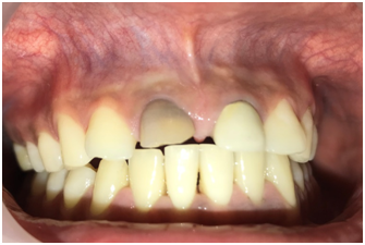
PRE OPERATIVE
MANAGEMENT
The treatment plan was decided as root canal therapy in the maxillary right central and lateral incisors followed by root end resection.
There is internal resorption i.r.t 11 so,the MTA plug was made i.r.t 11 and obturated with lateral compaction.whereas in 12 canal was obturated with lateral compaction,using gutta-percha points of 20 0.6%taper(Dentsply.Maillefer,Ballaigues, Switzerland) and Apexit plus (ivoclarvivadent) under rubber dam isolation, 1 day prior to the surgery. Guttapercha was extended beyond the apex to locate the extent of lesion.
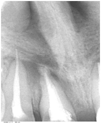
RADIOGRAPHIC VIEW OF OBTURATED CANALS
After effective local anesthesia with 2% lignocaine,bleeding points were marked after that a full-thickness LuebkeOchsenbein flap was reflected. A small defect was visible in the cortical plate with the central incisor, which was enlarged to aid in complete curettage of the granulation tissue (which was sent for biopsy). This was followed by irrigation with betadine (Win Medicare; Win Medicare Dental Products, New Delhi, India) and sterile saline solution. Then the extendedguttapercha was cut.Root end resection done. After that MTA was placed into the defect and completely used as root end filling material in teeth. The flap was repositioned and closed with 4-0 silk suture.
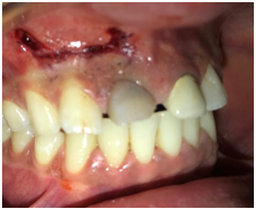
BLEEDING POINTS MARKED
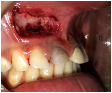
FLAP RAISED

MTA PLACED
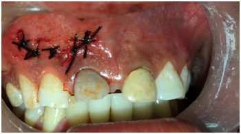
SUTURE PLACED
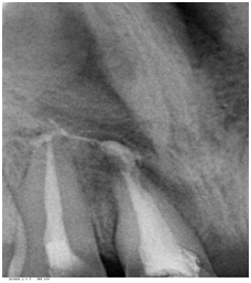
AFTER ROOT END RESECTION

AFTER 2 MONTH FOLLOW UP
DISCUSSION
Orthograde root canal therapy should be the first option for treatment of all inflammatory periapical lesions which have 85% of success rate. Periapical surgery remains the last resort when orthograde treatment fails or is not possible. After a surgical procedure, healing usually occurs by repair or regeneration.5 Regeneration has been defined as the reproduction or reconstitution of a lost or injured part to restore the architecture and function of the periodontium. Regenerative surgery with the use of MTA can support the formation of new tissue and allows for regenerative rehabilitation with functional reconstruction.4 The four critical factors that influence bone regeneration after the periapical surgery are primary wound closure, angiogenesis as a blood supply and source of undifferentiated mesenchymal cells, space maintenance, and stability of the wound (PASS principle).6
The present case report evaluated the clinical efficacy of MTA in the treatment of intrabony defect. MTA is a root end filling material,which has 96% of success rate. In vitro sealing ability and biocompatibility studies comparing root-end filling materials have shown MTA to be superior to other commonly used materials.
The main constituents of this material are calcium silicate (CaSiO4), bismuth oxide (Bi2O3), calcium carbonate (CaCO3), calciumsulfate (CaSO4), and calcium aluminate (CaAl2O4). MTA has a pH of 10.2, which rises to 12.5 at 3 hours after mixing.503 The pH has been reported to be approximately 9.5 at 168 hours after mixing.145 MTA is less cytotoxic than amalgam, SuperEBA, or IRM root-end fillings. This is seen most frequently in sections where MTA was used as the filling material. MTA appears to be able to induce cementoblastic cells to produce hard tissue. Recently, cementogenesis in the presence of MTA has been evaluated by assessment of the expression of osteocalcin (OCN), cell growth, and the morphology of cementoblast-like cells.496 Scanning electron microscope (SEM) analysis indicated that cementoblasts could attach to and grow on MTA. In addition, strong expression of the OCN gene was seen after application of MTA. MTA can also increase the production of both proinflammatory and anti-inflammatory cytokines from osteoblasts.2
MTA need wet environment to set,whenMTA used in root end filling it comes in contact with blood and set.In this case report size of lesion decreses after 2 month of follow up.
REFRENCES
- 1.Rafter M. Apexification: a review. Dent Traumatol. 2005;21(1):1-8.
- 2. El-Meligy OA, Avery DR. Comparison of apexification with mineral trioxide aggregate and calcium hydroxide. Pediatr Dent. 2006;28(3):248-53.
- 3. Witherspoon DE, Ham K. One-visit apexification: technique for inducing root-end barrier formation in apical closures. PractProcedAesthet Dent. 2001;13(6):455-60; quiz 62.
- 4. Evidenced-based review of clinical studies on the root apex. J Endod. 2009;35(8):1158-9.
- 5.Endodontic Surgery of a Symptomatic Overfilled MTA Apical Plug: A Histological and Clinical Case Report.SaeedAsgary a, Sara Fayazi.Iranian Endodontic Journal 2017;12 (3): 376-380
- 6.Surgical Management of a Large Inflammatory Periapical Lesion with Platelet‑rich Fibrin Nalam N. V. D.2017 Journal of Interdisciplinary Dentistry
- 7.Advantages of ProRoot MTA in treating periapical lesions. Clinical cases.Marta Girdeae,et al.OHDMBSC - Vol. V - No. 4 - December, 2006
Disclaimer from Guident
The views and opinions expressed in articles published in Guident are solely those of the authors and do not necessarily reflect the views of the editors, the editorial board or the publisher, Ivory India. Guident does not endorse any product, technique or treatment described. Clinical content is intended for qualified dental and medical professionals and is not a substitute for individual clinical judgement. Guident, its editors and the publisher accept no liability for any consequence arising from the use of information published in the journal.
How to cite this article
Dr.Annil Dhingra Dr. Mayuri Dr.Neha Arya Department of Conservative and Endodontics. Surgical Management of a Large Inflammatory Periapical Lesion with MTA. Guident. 2018 Jun;11(7). Available from: https://ivoryindia.in/articles/endodontics/surgical-management-of-a-large-inflammatory-periapical-lesion-with-mta.html


