Wonders That Maxillary Molars Can Do-Case Series on Multiple Extra Canals in Maxillary First Molars
Department of Conservative Dentistry and Endodontics, Seema Dental College And Hospital, Rishikesh
ABSTRACT
Introduction: There are cases in the endodontic treatment which are a real challenge. There are teeth with diferent anatomies of the root canals of diferent teeth and their treatment. There are teeth with multiple canals not visible easily and which are carious, Patients usually lose their teeth because these problems.
Purpose: The aim of this study is to investigate several cases with different root canal anatomies like in multiple accessory canals in maxillary first molars and manage them.
Material and methods: one case with severly decayed upper maxillary molar i.e 26, one case of maxillary first molar with severly decayed crown structure i.e 16 ,a case of severly decayed maxillary first molar i.e 16 .
Results: Results are observed after several years. In performing a root canal treatment on such teeth, one should appreciate the complexity of the root canal system and use proper magnification to manage these kind of cases.
Conclusions: Knowledge of the phenomenon of the complexity of the root canal system, multiple Accessory canals in different teeth, knowledge and practice to use proper magnification in Endodontics , this will improve the medical practice of the general dental practitioner.
CASE-1
A 37-year-old male with a noncontributory medical history was referred with a complaint of severe discomfort with his right maxillary teeth. The clinical and radiographic examinations revealed a maxillary right first molar with deep occlusal caries with tenderness on percussion. The clinical findings, radiographic findings, and pulp sensibility test led to a diagnosis of irreversible pulpitis with acute apical periodontitis with maxillary right first molar. Radiographic evaluation of the involved tooth did not reveal any unusual anatomy at the first but while negotiating the canals MB on mesial side was found, with DB on the distal side of the chamber and two palatal canals were found. Total 4 orifices were found which were confirmed radiographically and prepared with protaper gold rotary files after preparation of the canal the which were then finally obturated.
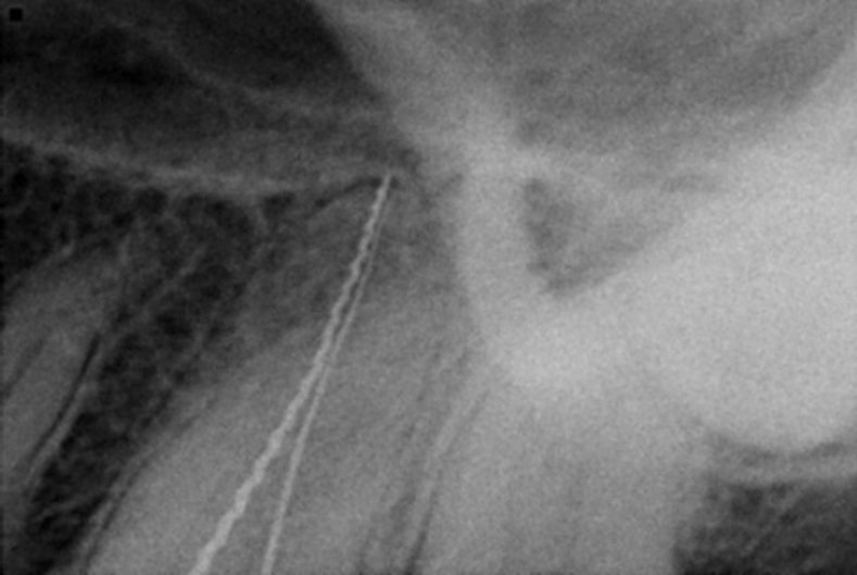
Working length for two palatal canals

Working length for all 4 canals recorded

Master cone selected

Obturation done
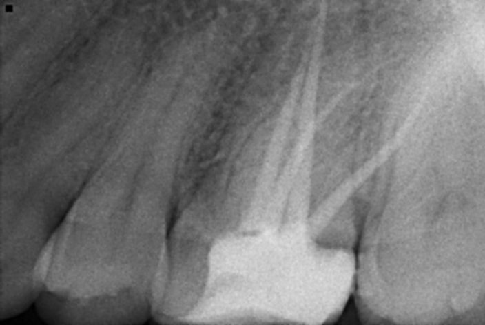
Post operative radiograph

Orifices for 4 canals
CASE-2
A 20-year-old female with a noncontributory medical history was referred with a complaint of severe discomfort with his right maxillary teeth. The clinical and radiographic examinations revealed a maxillary right first molar with deep occlusal caries with tenderness on percussion. The clinical findings, radiographic findings, and pulp sensibility test led to a diagnosis of irreversible pulpitis with acute apical periodontitis with maxillary right first molar. Radiographic evaluation of the involved tooth did not reveal any unusual anatomy at the first but while negotiating the canals MB1,MB2 on mesial side was found, with DB on the distal side of the chamber and two palatal canals were found. Total 5 orifices were found which were confirmed radiographically and prepared with protaper gold rotary files after preparation of the canal the MB1 & MB2 were merged together which were then finally obturated.

Pre op radiograph

Working length

Working length for two diff. Palatal canals
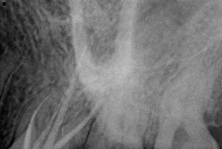
Master cone mesial view

Master cone distal view

obturation

Post op radiograph

Image of two palatal canal orifices.
CASE-3
A 37-year-old male with a noncontributory medical history was referred with a complaint of severe discomfort with his right maxillary teeth. The clinical and radiographic examinations revealed a maxillary right first molar with deep occlusal caries with tenderness on percussion. The clinical findings, radiographic findings, and pulp sensibility test led to a diagnosis of irreversible pulpitis with acute apical periodontitis with maxillary right first molar. Radiographic evaluation of the involved tooth did not reveal any unusual anatomy at the first but while negotiating the canals MB1,MB2 along with MB3 on mesial side was found, with DB1 and DB2 on the distal side of the chamber and one palatal canal. Total 6 orifices were found which were confirmed radiographically and prepared with protaper gold rotary files after preparation of the canal the MB1 & MB2 were merged together which were then finally obturated.


Pre op radiograph

Working length MB1, MB2, MB3

Working length of DB1, DB2

Master cone selected

Final obturation

Post op radiograph
CONCLUSIONS
Knowledge of the phenomenon of the complexity of the root canal system, multiple Accessory canals in different teeth, knowledge and practice to use proper magnification in Endodontics , this will improve the medical practice of the general dental practitioner.
REFERENCES
- DIFFICULT CASES IN ENDODONTICS – PROGNOSIS AND PROPHYLAXIS OF COMPLICATIONS Janet Kirilova, Snezhanka Topalova-Pirinska. Department of Conservative Dentistry, Faculty of Dental Medicine, Sofia, Bulgaria
- Mayhall JT. Taurodontism: a review of the condition and endodontic treatment challenges. Inter Endod J. 2008 May; 41(5):375-88. [PubMed] [CrossRef]
- R. M. Al Shalabi, O. E. Omer, J. Glennon, M. Jennings, and N. M. Claffey, “Root canal anatomy of maxillary first and second permanent molars,” International Endodontic Journal, vol. 33, no. 5, pp. 405–414, 2000.View at: Google Scholar
Disclaimer from Guident
The views and opinions expressed in articles published in Guident are solely those of the authors and do not necessarily reflect the views of the editors, the editorial board or the publisher, Ivory India. Guident does not endorse any product, technique or treatment described. Clinical content is intended for qualified dental and medical professionals and is not a substitute for individual clinical judgement. Guident, its editors and the publisher accept no liability for any consequence arising from the use of information published in the journal.
How to cite this article
Dr. Rishika Luhach, Dr. Annil Dhingra. Wonders That Maxillary Molars Can Do-Case Series on Multiple Extra Canals in Maxillary First Molars. Guident. 2022 Feb;15(3-4). Available from: https://ivoryindia.in/articles/endodontics/wonders-that-maxillary-molars-can-do-case-series-on-multiple-extra-canals-in-maxillary-first-molars.html