Cleidocranial Dysplasia
Guru Nanak Institute of Dental Science & Reasearch, Kolkata-114
Abstract
Cleidocranial dysplasia, also known as cleidocranial dysostosis is a rare disorder resulting in skeletal, facial, dental deformities. It is usually caused due to mutations in Cbfa1 gene, also known as Runx2, producing difficulties in bone ossification resulting in hypoplasia or aplasia of clavicles, short stature, delayed closure of fontanelles, disorders of jaws and dentition such as multiple supernumerary teeth, and delayed eruption of permanent teeth. A male patient aged 19 years has reported to our department with complaints of mobility of posterior teeth. On examination, the typical features of cleidocranial dysplasia were found such as hypoplasia of clavicles, delayed eruption of permanent teeth, multiple impacted supernumerary teeth etc. This paper will enlighten the views regarding the presence of peculiar oro-dental features in this type of patient and suggested that a multidisciplinary approach is needed to provide a healthy oral environment to the patient.
Keywords: Hypoplasia of clavicles, Supernumerary teeth, Permanent teeth.
INTRODUCTION
Cleidocranial dysplasia, also known as cleidocranial dysostosis or Marie and Sainton disease is a rare developmental disorder resulting in skeletal, facial, dental deformities.1-4 It is autosomal dominant pattern in heritance but may also occur due to unknown etiology.5 It mainly occurs due to mutations in the Cbfa1 gene, also known as Runx2, located on the short arm of choromosome6, which encodes the transcription factors essential for osteoblast differentiation.2,3,6,7 The patient having cleidocranial dysplasia usually presents with the typical features of hypoplasia or aplasia of clavicles (absence of collar bones),short statures, delayed closure of fontanelles, larger head with prominent bulging of forehead, multiple wormanian bones.8,9 Presence of hypoplastic maxilla with underdeveloped maxillary sinuses, prognathic mandible, with delayed eruption of permanent teeth and multiple impacted supernumerary teeth are noted in this type of patient.2-4 Delayed eruption mainly attribute due to the absence of cellular cementum and an increase in the amount of acellular cementum of the roots of the affected teeth.1,3,10
CASE REPORT
A male patient aged 19 years has reported to our department with chief complaints of mobility of posterior teeth for last 3-4 months. Past medical history revealed that there was delayed closure of fontanelles as told by his mother. On examination, the following observations are summarized as stated below:
General examination
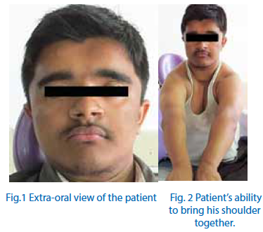
It was found that the patient has got unique ability to bring his shoulders together (Fig.1 and Fig.2).
Extra-oral examination
Presence of frontal bossing, hypoplastic maxilla, depressed nasal bridge, and relatively prognathic mandible were noted (Fig.1).
Intra-oral examination
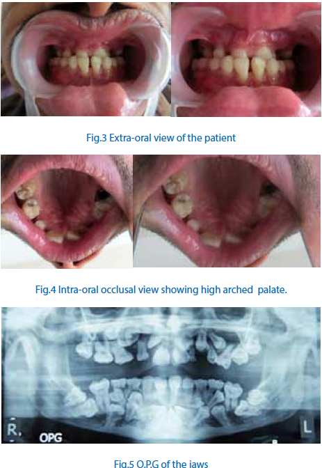
Presence of anterior and posterior crossbite, malocclusion, altered eruption pattern, multiple retained deciduous teeth, absence of several permanent teeth, and high arch palate were also observed (Fig.3, Fig.4). The oral hygiene of the patient was found to be good with very minimal amount of plaque and calculus within the oral cavity. Gingival sulcus depth was found to be around 3 mm throughout the dentition. Grade II mobility was noted in the retained deciduous teeth namely 75, 84, 85.
Radiographic examination
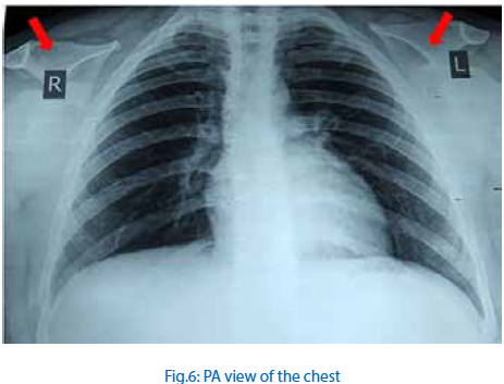
Presence of multiple impacted supernumerary teeth especially in the lower mandibular region was observed in the O.P.G radiograph (Fig.5).The impacted teeth in the lower mandibular arch were likely to be sixteen in number as revealed through radiograph. Similarly, the impacted teeth in the upper maxillary arch were likely to be eight in number. The numbers of impacted teeth with completely formed root were likely to be ten in the lower mandibular arch and six in the upper maxillary arch. Hypoplasia of clavicles and bell shaped rib cage was noted in the postero-anterior [PA] view of the chest (Fig.6).
Thereby, based on clinical and radiographic finding a diagnosis of cleidocranial dysplasia was made. Then, after completion of initial checkups patient was referred to the other departments for execution of a proper treatment plan and periodic checkups was advised.
DISCUSSION
Cleidocranial dysplasia is a rare inherited skeletal disorder. Clinically, it predominately affects bones that undergo intra-membranous ossification, generally the skull and clavicles11. However, it is evidenced from several clinical studies that it usually affects the entire skeleton therefore; it is considered to be a dysplasia rather than a dysostosissup>12.
The narrow, highly arched palate, the retention of deciduous teeth, delayed eruption of permanent teeth, and the presence of large numbers of impacted supernumerary teeth are all classical oral findings in cleidocranial dysplasia3.Lack of cellular cementum and increase in the amount of acellular cementum may be responsible for delayed eruption of permanent teeth.1,3,10 But, some literatures evidenced that osteoclastic resorption may be defective.3 It may be hypothesized that the crowding of the impacted supernumerary teeth may prevent the eruption of permanent teeth. Thereby, simple extraction of primary teeth may sometimes insufficient to promote eruption of permanent teeth.2,3
Therefore, a multidisciplinary approach is needed to treat such patients such as extraction of retained deciduous teeth and multiple impacted supernumerary teeth after which orthodontic extrusion of the permanent teeth will be carried out depending upon the clinical situation Sometimes implant placement4 will be advocated to provide a stable occlusion and comfortable oral health to the patient.
CONCLUSION
Although cleidocranial dysplasia is a rare developmental disorder, it has got immense impact on dental and oral health as it usually produces various skeletal and dental malformations. Thereby, a detailed understanding of the general clinical features and sequence of treatments will be needed to treat such patients.
REFERENCES
- C et al.: Cleidocranial dysplasia presenting with retained deciduous teeth in a 15-year-old girl: a case report. Journal of Medical Case Reports .2012; 6:25.
- Callea et al.: A Case of Cleidocranial Dysplasia with Peculiar Dental Features: Pathogenetic Role of the RUNX2 Mutation and Long Term Follow-Up. OHDM.2014 June; 13(2):548-551
- Grover et al.: Cleidocranial Dysplasia Revisited: A Closer View from a Periodontal Perspective
- J Life Sci, 2011;3(2): 141-145.
- Roberts et al.: Cleidocranial dysplasia: a review of the dental, historical, and practical implications with an overview of the South African experience. Oral Surg Oral Med Oral Pathol Oral Radiol.2013 Jan;115(1):46-55.
- Tanaka JL, Ono E, Filho EM, Castilho JC, Moraes LC, Moraes ME : Cleidocranial dysplasia: importance of radiographic images in diagnosis of the condition". J Oral Sci 2006;48 (3): 161–6
- Chen S, Santos L, Wu Y, Vuous R, Gay I, Schulze J, Chuang HH, MacDougall M: Altered gene expression in human cleidocranial dysplasia dental pulp cells. Arch Oral Biol 2005, 50:227-236.
- Otto F, Thornell AP, Crompton T, Denzel A, Gilmour KC, Rosewell IR, et al. Cbfa1, a candidate gene for cleidocranial dysplasia syndrome, is essential for osteoblast differentiation and bone development. Cell. 1997; 89, 765–771.
- Brueton LA, Reeve A, Ellis R, Husband P, Thomson EM, Kingston HM: Apparent cleidocranial dysplasia associated with abnormalities of 8q22 in three individuals. Am J Med Genet 1992, 43(3):612-618
- De Nguyen T, Turcotte JY: Cleidocranial dysplasia: review of literature and presentation of a case. J Can Dent Assoc 1994, 60(12):1073-1078.
- Counts AL, Rohrer MD, Prasad H, Bolen P: An assessment of root cementum in cleidocranial dysplasia. Angle Orthodont 2001, 71:293-298.
- Romeo U, Galluccio G, Palaia G, Tenore G, Carpenteri F, Barbato E, Polimeni A: Cleidocranial Dysplasia: Maxillary Alterations on the Transverse Plane. Presence of Crown-radicular Anomalies and Multidisciplinary Approach of a Clinical Case. OHDM.2014 June;13(2):529-535.
- Lo Muzio L, Tetè S, Mastrangelo F, Cazzolla AP, Lacaita MG, Margaglione M, Campisi G. A novel mutation of gene CBFA1/RUNX2 in cleidocranial dysplasia. Annals of Clinical and Laboratory Science. 2007; 37: 115-120.
Disclaimer from Guident
The views and opinions expressed in articles published in Guident are solely those of the authors and do not necessarily reflect the views of the editors, the editorial board or the publisher, Ivory India. Guident does not endorse any product, technique or treatment described. Clinical content is intended for qualified dental and medical professionals and is not a substitute for individual clinical judgement. Guident, its editors and the publisher accept no liability for any consequence arising from the use of information published in the journal.
How to cite this article
Dr.Sohini Banerjee Prof, Dr.T.K.Pal Dr.Ravi Prakash B.S Department of Periodontics. Cleidocranial Dysplasia. Guident. 2015 Apr;8(5). Available from: https://ivoryindia.in/articles/general/cleidocranial-dysplasia.html