3 Years Follow of Implantswiss Short Implant in Posterior Maxilla: A Case Report
Wisdom Dental Clinics ®, Jaipur, India, The Tooth Clinic, Bareilly, Uttar Pradesh, India
ABSTRACT
Placement of the dental implants in posterior maxilla requires maxillary sinus floor elevation. However, this is avoided by placing short implant, these implants are increasingly being seen as an alternative to more extensive bone augmentation procedures. Long term follows up revealed survival of the implants. This case report describes placement of short implantswiss implant in posterior maxilla followed by digital impression .
keywords:posterior maxilla,bone augmentation,digital impression
INTRODUCTION
Dental implants have been an ideal treatment solution for patients who require permanent replacement of their missin teeth. Dental implants can be placed successfully in most cases, however, certain clinical situations, such as reduced bone dimension, may pose a significant challenge. This situation is frequently encountered in posterior maxillary bone, where, following tooth extraction, sinus pneumatization often results in inadequate vertical bone height for implant placement. Sinus floor elevation using lateral window and bone grafting before implant placement has been the gold standard with highly predictable results.1
However, sinus floor elevation should be avoided as much as possible due to systemic problems and associated delayed healing. In such situations, a less invasive surgery such as the use of short implant is more suitable.
An alternative method for treatment of atrophic bone in posterior maxilla Involves the use of short dental implants. The use of short dental implants may limit the needs for bone augmentation. The predictability of short implants has been assessed in a recent survey of randomized clinical trials of implants placed in augmented sinus.2 Short (length ≤ 8 mm) implants have presented a predictable survival rate and have resulted in three times lower intraoperative complications compared to long implants.3
Short implants placed in a posterior partial edentulous region have presented a high initial survival rate, which is similar to long implants. As mentioned above, shorter dental implants may represent the preferred treatment alternative in atrophic alveolar bone since they have been associated with lower biological complications, decreased morbidity, costs and surgical time.4
The CAD-CAM technology is a computerized technique that allows to obtain a three-dimensional object from a vector drawing performed on the computer.Both CAD and CAM are acronyms, respectively, for Computer Aided Design and Computer Aided Manufacturing. Born in the 60s of last century, this technology is now used in industry for the production of an infinite number of objects also in the dental field. In the 80s the first systems in the dental industry, CEREC have been commercialized.5
The evolution of techniques and of implant components is making the implant-prosthetic rehabilitation increasingly complete and reliable. The increased demands of rehabilitation can be processed more serenely by operators in the industry. In this regard a valuable aid for the prosthodontist comes from a rapidly expanding sector which is that of the CAD-CAM applications in dentistry.6
This case report aims to provide information on the successful treatment involving the use of short dental implants in posterior maxilla. In the present case we have loaded implantswiss short implant at the time in posterior maxilla .
CASE REPORT
A 59-year-old male reported to the clinicwith the com plaint of missing tooth 26 (left maxillary first molar).The tooth had been extracted 6 months earlier due to gross decaying since then he did not wear any prosthesis and was having difficulty in chewing, so he wanted to have a fixed solution.
After intraoral examination, treatment options available were:fixed partial denture and an implant-retained restoration/crown; patient agreed with the implant-retained restoration/crown as this was more predictable and conservative to teeth and long term solution.[Figure 1a,b]

Figure 1a,1b : implant retained restoration /crown, fixed partial denture
To check for the bone level for implant selection CBCT (cone Beam computed tomography) was done, which showed deficient 3D bone i.r.t 26.Looking at the compromised bone level,he was offered treatment options with dental implants and bone augmentation. The patient refused bone augmentation and chose the option of short implant placement in the posterior maxilla.After measuring the bone level clinically and radiographically, it was decided to place a bone level short implant (4.8×5 mm, implantswiss Implant system) by flapless surgical procedure.[figure 2]

Figure 2: Short implantswiss system
Implant placement was performed under local anaesthesia; a tissue punch was used to perforate the gingival tissue so as to gain access to bone without elevating the flap. The osteotomy procedure was initiated with a pilot drill at the punch site under copious irrigation;the final osteotomy was prepared by sequential drilling. The proper angulations and depth of osteotomy were evaluated with a depth gauge and radiographs. After the final osteotomy preparation, implantswiss implant was placed with an insertion torque of 30Ncm by a torque measuring wrench posing good primary stability,parallel to the roots of the adjacent teeth. ISQ measurements were in the range of 75-78, a healing abutment was then placed on the implant to facilitate the development of a gingival emergence profile for the anticipated restoration. Post-operative OPG was taken to check for the placement of the implant. Postsurgical instructions were explained to control postoperative pain.Antibiotics and analgesics were prescribed to the patient for 5 days. The patient was recalled after 1 week for a routine check-up; there was no extraoral swelling or discomfort. [Figure 3a,b]

Figure 3a,b: Postoperative OPG and IOPA after implant
The patient was recalled after 3 months for carrying out the prosthetic procedures.ISQ test was done again, which showed excellent biological stability with reading measuring 84, showing excellent secondary stability.
For taking impressions to avoid discomfort and save time and improve accuracy, we took direct impressions using the CAD/CAM technique using CEREC, Dentsply-Sirona workflow. Procedure for chairside screw retained prosthesis began with the use of a scanbody over ti-base.Digital model were prepared and prosthesis designing was done chairside only by CEREC workflow with the help of Artificial Intelligence (A.I) .
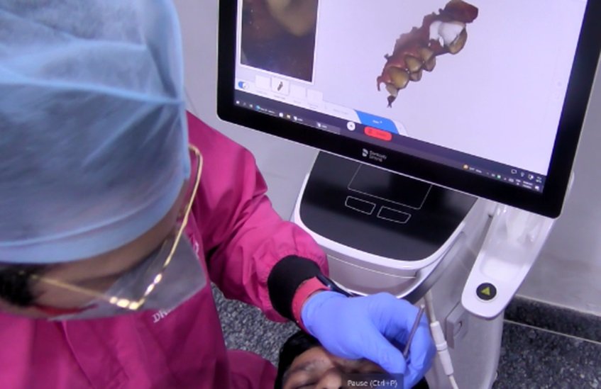
Figure 4: Digital impression in process
Manufacturing was done with Sirona CEREC Zirconia A3 meso block which included milling, sintering and glazing done chairside only on Primemill & Speedfire .[Figure 4a,b,]
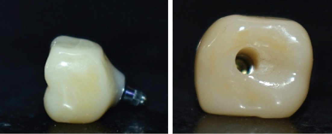
Figure 4a,b: Prepared crown
Crown was cemented extraorally over ti-base with dual-cure resin cement.Finally, within 1hour chair side screw retained prosthesis was prepared which was ready to be delivered. After final placement of prosthesison its desired site, intraorally photographs [Figure 5a,b,c,d,e] post-operative radiograph[Figure 6a,b] was taken.

Figure 5a,b,c,d: Intraoral photographs

Figure 6: After crown placement OPG
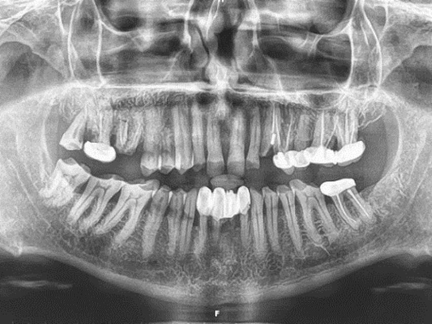
Patient was asked to come for timely follow-ups for 3years, and radiographs were taken respectively, which showed long term success with short implants and two new implants were placed.[Figure 7]

Figure 7: Follow up OPG after 3 years
DISCUSSION
Short dental implants have been proposed as an alternative treatment to simplify implant placement procedures in severely defective alveolar ridge, to avoid vital structures, minimize surgical trauma and the risk of complications associated with invasive surgical procedures. With this in mind, short dental implants would be more suitable in present case.7
Many factors such as implant surface, implant primary stability, bone density and quantity, surgical preparation techniques used, management of the soft tissue and prosthodontic design have been related to implant survival rates. Biologically, a good primary implant stability is important, which can be achieved by under-prepared osteotomy site with the drilling stopped at diameter below the implant diameter. When using short dental implants, one should be aware of some risk factors that may increase stress such as a higher crown-implant length ratio, high bone density in the region, and higher bite force. Some methods are, therefore, required to decrease stress by minimizing the size of the crown in the bucco-palatal dimension to decrease the lateral force on the restoration, avoiding cantilever as much as possible, and splinting multiple implants together with splinted crowns.8
An additional aspect to this treatment, which is relatively current, is the digital impression technique, rather than traditional, rubber-based impressions. The accuracy of digital scanners when measuring soft-tissue parameters was recently demonstrated by Schneider etal.9 Ramsey and Ritter8 demonstrated the digital fabrication of a 3-dimensional model, facilitating fabrication of a virtual abutment with the production of a CAD/CAM-designed custom abutment and all-ceramic crown, delivered to the clinician, seated on a stereolithographic model.
The obvious advantages to the digital impression technique include patient and operator comfort. The unpleasantness of rubber-based impressions is eliminated with this technology. The impression procedure is also of shorter duration, resulting in a more pleasant patient experience. The distortion of conventional impression materials is not a factor with digital impressions, contributing to their accuracy. Also, distortion related to contraction of gypsum-based stone models is obviated. The virtual reproduction of a working cast, which facilitates virtual abutment design, negates the need to pour traditional models, shortening laboratory working time, and avoids the possible dimensional changes associated with these materials.10
As with any procedure, case selection is critical to achieving optimal results. The patient featured in this case report was one with a thick periodontal biotype, lowering the risks of significant postoperative recession.11The extraction socket was intact after tooth removal, and his occlusal scheme permitted the placement of a provisional restoration out of contact with the mandibular antagonist teeth. Had any of these criteria not been met, a deviation from the current procedure may have been necessary. This may have included flap reflection and concurrent guided bone regeneration, delayed placement with socket augmentation, or the need for a transitional removable partial denture.12
CONCLUSION
Dental implant treatment with short implant is suitable for patients as it is considered minimally invasive surgery which can reduce the risks of surgical complications and compromised post-operative healing. The sinus augmentation procedure could be avoided by placement of small dental implants in posterior maxilla. To achieve long term short implant survival, many factors such as good surgical technique, primary implant stability and prosthetic modifications to allow for stress reduction during mastication, should be considered.
REFERENCES
- P Papaspyridakos A De'Souza K Vazouras H Gholami S Pagni H P Weber Survival rates of short dental implants (≤6mm) compared with implants longer than 6mm in posterior jaw areas: A meta-analysisClin Oral Implants Res201829682010.1111/clr.13289
- DS Thoma M Zeltner J Husler CH Hammerle RE Jung EAO Supplement Working Group 4 - EAO CC 2015 Short implants versus sinus lifting with longer implants to restore the posterior maxilla: a systematic reviewClin Oral Implants Res201526111546910.1111/clr.12615
- N P Lang L Pun K Y Lau K Y Li M C Wong A systematic review on survival and success rates of implants placed immediately into fresh extraction sockets after at least 1 year Clin Oral Implants Res2012235396610.1111/j.1600-0501.2011.02372.
- MR Norton The influence of insertion torque on the survival of immediately placed and restored single-tooth implantsInt J Oral Maxillofac Implants2011266133343
- Murata H, Kawamura M, Hamada T, Saleh S, Kresnoadi U, Toki K. Dimensional stability and weight changes of tissue conditioners. J Oral Rehabil. 2001;28(10):918–23. [PubMed] [Google Scholar]
- Guzzo F, DE Luca G, Barnaba P, Severino D. Cad-cam procedure and implant-prosthetic rehaBilitation. Case report. Oral Implantol (Rome). 2016 Nov 13;9(1):27-32. doi: 10.11138/orl/2016.9.1.027. PMID: 28042428; PMCID: PMC5159934
- A Mokcheh H Jegham S Turki Short implants as an alternative to sinus lift for the rehabilitation of posterior maxillary atrophies : Systematic review and meta-analysisJ Stomatol Oral Maxillofac Surg201912012837
- M Srinivasan L Vazquez P Rieder O Moraguez JP Bernard UC Belser Survival rates of short (6 mm) micro-rough surface implants: a review of literature and meta-analysisClin Oral Implants Res201425553945
- Brunski, J. B., In vivo bone response to biomechanical loading at the bone/dental-implant interface. Adv Dent Res, 1999. 13: p. 99-119.
- Kan JY, Roe P, Rungcharassaeng K, et al. Classification of sagittal root position in relation to the anterior maxillary osseous housing for immediate implant placement: a cone beam computed tomography study. Int J Oral Maxillofac Implants. 2011;26(4):873-876.
- Kan JY, Rungcharassaeng K, Lozada JL, Zimmerman G. Facial gingival tissue stability following immediate placement and provisionalization of maxillary anterior single implants: a 2- to 8-year follow-up. Int J Oral Maxillofac Implants. 2011;26(1):179-187.
- Gimenez B, Ozcan M, Martinez-Rus F, Pradies G. Accuracy of a digital impression system based on parallel confocal laser technology for implants with consideration of operator experience and implant angulation and depth. Int J Oral Maxillofac Implants. 2014;29(4):853-862
Disclaimer from Guident
The views and opinions expressed in articles published in Guident are solely those of the authors and do not necessarily reflect the views of the editors, the editorial board or the publisher, Ivory India. Guident does not endorse any product, technique or treatment described. Clinical content is intended for qualified dental and medical professionals and is not a substitute for individual clinical judgement. Guident, its editors and the publisher accept no liability for any consequence arising from the use of information published in the journal.
How to cite this article
Dr. Gaurav Gupta, Dr. D.K. Gupta, Dr. Richa Gupta. 3 Years Follow of Implantswiss Short Implant in Posterior Maxilla: A Case Report. Guident. 2024 Dec;18(1). Available from: https://ivoryindia.in/articles/implantology/3-years-follow-of-implantswiss-short-implant-in-posterior-maxilla-a-case-report.html