Implant Surfaces-Revisited
Thai Moogambigai Dental College and Hospital Chennai, India.
ABSTRACT
The aim of this article is to review and to discuss the surface modifications of implant biomaterials . Osseointegration occurs around the implants through the tissue growth or through a direct apposition between the tissue and the implant surface. The implant bone attachments is based on the tissue in growth into roughened implant surface . The characteristic feature of various implant surface modifications are illustrated in this article.
INTRODUCTION
The titanium and titanium alloys are the best standard materials in dental implants because of their mechanical strength, chemical stability, and biocompatibility Integration of titanium implants with the surrounding bone structure is critical .
It has been shown that after loading there are more implant failures in implant with relatively smooth surface ,in comparison with rough –surface implant .Implant surface modification are required because of immediate loading implants are subjected to biomechanically challenging situation .It has also been suggested that texturing the implant surface may not directly contribute to initial stability but it may reduce the risk of stability loss and also helps in wound healing (secondary osseointegration)1.
OSSEOINTEGRATION
The term osseointegration was first used by Prof P-I Branemark ,since then it has the been used to describe the procedure of bone attachment with titanium .He and his colleagues in 1950 &1960,while examining microcirculation of bone & wound healing in rabbits through means of vital microscopy accidentally discovered the process of osseointegration and a new dimension in field of implantology . In this process the jawbone attaches directly to dental implant and grows up to the implant surface without intervening soft tissue ,which could not be removed from bone without fracture.17
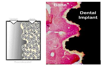
OSSEOINTEGRATION IS DEFINED AS
- By Branemark as a direct structural and functional connection between ordered living bone and the surface of a load-carrying implant .
- By Albrektsson in 1981 as a direct on light microscopical level ,contact between living bone implant
- By Steinemann in 1986 as a bony attachment with resistance to shear and tensile force16
SURFACE ROUGHNESS
When the implant is placed in the body, there will be a biological response that will be mediated by the interaction of the implant through its surface. Micro-level features are included to impart osseointegration (or) direct bone to implant contact.
As osseointergration plays a key role in the successful clinical outcome of an implant. The implant surface roughness has been identified as an important parameter for implant and its capacity for being anchored into bone tissue(2;3). The implant surfaces are evolved by generations The most important factors that affect the quality and speed of osseointegration are the physical and chemical nature of the implant surface . These properties also have an effect on the maintenance of soft tissue and surrounding bone around the implant as the contact between cells and biomaterials occurs11 . The first step in this response involves the adsorption of specific proteins, lipids, sugar, and ions that can activate cells mechanisms to induce either acceptance or rejection of the implant by determining which and how many cells populate the surface. Therefore, a high percentage of bone-implant contact(BIC) is necessary to create sufficient anchorage of the implant, which is a determinant factor for osseointegration.17
In order to increase the success rate of dental implants, research has focused on the control of surface properties of implant it has shown that osteoblastic cells adhere more quickly to rough surfaces than to smooth surfaces (3,4) This surface roughness has a positive influence on cell migration and proliferation ,which in turn leads to better BIC results. suggesting that the microstructure of the implant influences biomaterial –tissue interaction .
Goals of implant surface modification
1)To achieve initial stability
2)To stable the blood clot favoring healing process
3)To stimulate the bone formation5
IMPLANT SURFACE MODIFICATION TECHNIQUE
Implant surface irregularities can be produced by 2 methods
- Ablative (or) subtractive procedure
a) sand blasting
b) acid etching
c) anodic oxidation
d) laser treated
- Additive procedures
a) Hydroxyappatite coated
b) Plasma protein coated
c) Ceramic coated
d) Bisphosphate coated
e) Fluoride coated
- ABLATIVE PROCEDURES :-
In these procedure the implant is subjected with various physical and chemical agents ,which makes the surface rough by removing the implant biomaterial from its surface . The surface roughness has a positive influence on cell migration and proliferation ,which in turn leads to better BIC result suggesting that the microstructure of implant influences biomaterial –tissue interaction, Which enhances cellular activity and bone apposition. Osteogenesis at the implant surface occurs by several mechanism including cell differentiation ,transformation of osteoblast and bone tissue formation.4;7

The histological picture shows bone is in close contact with the implant surface with no intervening gap and a rim of osteoblast is depositing osteoid matrix ,bone grows inside the irregularities of the implant surfaces
- .a) SURFACE BLASTING :-
In these procedure the implant surface bombarded with small hard ,biologically inert ceramic particle like alumina ,silicon carbides, titanium oxide and calcium phosphate.Compressed air is used to project the particles through a nozzle at high velocity; the surface roughness depends upon the size of the particles ,time of blasting, pressure and distance of particle to the implant surface.
Advantages
It improves adhesion, proliferation and differentiation of osteoblasts.
Disadvantages
Particles are left on the surface after blasting exhibit a greater bone response as determined by removal torque8,1

1 . b) ACID ECTHING :-
This technique consists on increasing the thickness of the oxide layer and the roughness by immersing the metallic implant into an acidic solution like hydrochloric and sulphuric acid. it induce higher adhesion of cells and expression of genes involved in the promotion of osseointegration. . Acid etched implants have been seen to have a higher implant stability .As etched surfaces has roughness of 0.6 μm to 0.9 μm that have higher removal torque values and higher bone implant contact area9,2 .
Advantage
They provide homogeneous roughness, increased active surface area and improve cells adhesion, and therefore rapid osseoinegration in early stage of peri- implant healing
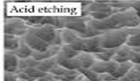
Research has shown that , The average roughness of sandblasted and acidetched surfaces was about 2.15 μm. Cytotoxicity tests showed that sandblasted and acid-etched implants had non-cytotoxic cellular effects and appeared to be biocompatible. Scanning electron microscopic examination showed that the surface roughness produced by sandblasting and acid etching could affect cell adhesion mechanisms. Osteoblast-like cells adhering to the machined implants presented a very flat configuration, while the same cells adhering to the sandblasted and acid-etched surfaces showed an irregular morphology and many pseudopodi. These morphologic irregularities could improve initial cell anchorage, providing better osseointegration for sandblasted and acid-etched implants18
1. c) ANODIC OXIDATION :-
Anodic oxidation on the Ti implant surface increases not only the roughness of the surface but also the thickness of the Ti surface oxide, which is important for the biologic bone response .It is an electrochemical process where the implant is immersed in an electrolyte and current is applied, the implant becomes anode resulting in micropores of variable diameter and an increase of the oxide layers .Here micro or nanoporous surfaces is produced by potentiostatic or galvanostatic anodization of titanium in strong acids at high current density .
In a study evaluating the human jawbone response to the oxidized Ti surface, histomorphometry resulted in significantly more favorable bone response (higher bone-to-implant contact) to the oxidized Ti surface at both the maxilla and the mandible, compared with the turned Ti surface
Advantages
Anodization technique include improved biocompatibility, increased cell attachment and proliferation1,7
Disadvantages
Anodic oxidation itself is considered to have a limitation in bone response from the results that an electropolished (very smooth) Ti surface tends to decrease bone formation rate despite that the surface is anodized .

1 . d) LASER TREATED :-
The implant is subjected to laser beam ;Laser is an emerging field for use as a micromachining tool to produce a 3-D structure at micrometer metrices.
This technique generates short pulses of light of single wavelength, providing energy focused on one spot. It is rapid, extremely clean, and suitable for the selective modification of surfaces and allows the generation of complex microstructures and has high resolution.6
THERE ARE VARIOUS LASER SOURCE
- Co2
- Argon
- Diode
- Erbium
- Neodymium-yittrium- aluminium(NdYag)
The common and effective laser source used in this procedure are Nd;YAG(Neodymium-yittrium-aluminium) 50kw of power ;frequency of 7.5 khz

Nd:YAG lasers for the ablation is used in dentistry from 1989,
Diode –pump consist of water-free glass fibers (KaVo, DELight), articulated arms with mirrors (Smart 2940D, Fidelis Plus II) or flexible hollow fibers They have a limitation of maximal power transmission at about 6 W. Therefore, more powerful Nd:YAG lasers need a hollow transmission system for their light.6,12
Laser surface modification allows the surface to retain the base material properties The changes that does occur comes from the morphing of the micro structure laser- implant surface with 200-microm pores resulted in a profound improvement in osseointegration .
In this process the implant surface in bombarded with small spherical media called Shot Each piece of shot striking the implant acts as a tiny hammer ,imparting to the surface small indentation(or)dimple This produces a honeycomb like uniform surfaces with small pores. 11

Bone-to-implant contact was better on the surfaces subjected to laser treatment than on the machined titanium implants. The laser treated surface, promote the alterations in the surface energy as well as in the macro- and microstructures of titanium,and lead to improved bone-to-implant contact
Study had showed that the decontamination of the laser implant surface is the important feature ,which is caused by the physical properties of the laser energy and its interaction with tissues, due to reflection, scattering, transmission, absorption and slight temperature elevation ;which is used in peri - implantitis treatments Only the valley and parts of the flank of the implant threads can be laser treated while the remaining part was left as-machined. As the flank portion of the implant thread might have the higher risk to expose to the microorganism and plaque12

ADVANTAGES OF LASER SURFACE
- The laser technique has add no chemical
- corrosion resistant
- Non complicate procedure
- Have high fatigue strength
- Laser implant surface have bactericidal effect and it is used in peri- implantitis therapy.11
2.ADDITIVE PROCEDURE
In these procedure the implant surface is coated with various biomaterials which improves the biological and bio-mechanical properties of implant4,7
a) Hydroxyappatite coated
b) Plasma protein coated
c) Ceramic coated
d) Bisphosphonate coated
e) Fluoride coated
The dental implants are coated with high hydrophilic bio material ,This hydrophilic surfaces have better blood coagulation than hydrophobic surface, The hydrophilic surface improves the absorption of protein such as fibronectin and vitronectin on the surface of dental implant could promote cell adhesion and osseointegration13

The histological picture shows close adaptation of bone to implant surface and no gap (or) connective tissue are presented at the bone – implant interfaces
2.a) HYDROXYAPPATITE COATED
It was first introduced by DeGroot, HA coated implant surfaces improves the bone implant attachment compared with machined surfaces,They have excellent reliability in penetration of bone into implant surface ;which enchances fixation,especially in areas of limited initial bone contact .The hydroxyappatite coated implant surfaces are oseoinductive material with good bio –intergration 6,10

Advantages of HA coated implant surfaces
1) Faster healing of bone interface
2)stronger interface
3)less corrosion
4)increases surface area
5)increase initial stability
Studies had shown Histologically significant differences in defect osseointegration were seen between treatment groups. HAcoated implants had significantly greater osseointegration within the defect than Ti implants . Average trabeculation of newly formed bone in the defect after healing was significantly greater for HA-coated implants than for titanium . Finally, there were significantly less residual allograft particles in defect areas adjacent to HA-coated implants than Ti implants. The use of HA-coated implants in large size defects with demineralized freeze-dried bone allograft(DFDBA) and expanded polytetrafluoroethylene membrane (ePTFEM) produced significantly more osseointegration.19
2. b) PLASMA PROTEIN COATED
The plasma protein coated surface is made by plasma spraying a powder form of molten droplets at high temperature of 15,000â°c and plasma is sprayed with nozzle to provide high velocity 600 m / sec ,of 0.005 -0.1mm diameter particle size,which results in irregular pores ,pits and depression ,forming a rough implant surface.
Advantage
These plasma coated surface have high wettability,they improve oseointergation by controlling both cell growth and differentiation of osteoblast which attaches cell to implant surfaces ,have more bone implant contact 14.

An animal study had shown the histomorphometric evaluation showed significantly more bone-to-implant contact after topical PRP application in the early healing phase (6 weeks). Topical(Platelet – rich plasma) PRP application significantly increased the activity of bone regeneration at implant host sites during early healing.20)
2.c) CERAMIC COATED
The ceramic coated implant surfaces was introduced by Dressman in 1980
The bioceramics are categorized into 3 groups based on the interation with host tissue
A) Bioactive
B) Bioresorbable
C) Bioinert
Bioactive
They are bioglass/glass ceramic,posses osteoconduction bone growth along the bone implant interface and osteoproduction , Bioglass is a cleverly designed glass that contain silicon, calcium and sodium oxides . Presnt in proportions of 45:25:25 % of these components. In the body - the sodium surface dissolves. The remaining material is not stable and reorganizes into silica ( some of this dissolves) and tiny crystals of hydroxy - carbonate apatite (HCA) .This porous surface layer is a very favorable substrate for the re -growth of bone tissue.
Bioresorbable
The 1st bioresorbable implant material used are plaster of paris but due to poor mechanical properies its application are not wide. Calcium sulfate ,calcium alumina & calcium phosphate are apparently resorbed over a period of time and replaced by living bone, They are dissolved quick by the body and they supplies the needed minerals to the body. They do not have any particular interaction with the tissues. Many products today combine these ceramics with proteins that encourage bone growth.
Bioinert
They are alumina ,zirconia and carbon , Which are highly biocompatible and do not degrade - but they are very expensive , excellent corrosion resistant ,high strength and so hard they do not wear.
Methods of ceramic coating
i)compression method and
ii)sintering method
Compression method
Powder compression is made in a die by means of lower and upper punch ;Stearic acid in alcohol is used as lubricant ,after powder compact is pushed out ,it is placed in a rubber tube which is brought under vaccum and is isostatically compressed for 100MN/M in a oil containing pressure vessel ,heated at 100â°c in a wet oxygen atmosphere for 6 hours and cooled over slowly
Sintering method
Hot setting pressing is done in this method which is introduced by Oademan (1969), The highest dentisty and strength which are possible in crystalline ceramic material are formed by both heating and pressure used to enchance the ceramic density the sentering is occurred at 900â°c and under pressure of 50 MN /M5,14
Advantages
They are highy biocompatible and esthetics
High resistance to corrosion ,flexion and fracture
High bone implant contact

2. d) BISPHOSPHONATE COATED
They have high resonance, bisphosphonate reduces osteoclast activity by impair the osteoclast to form ruffled border and inhabits the bone resorbtion, thereby changing the balance of bone turnover in favor of bone formation, leading to a net gain in local bone density . The improved early fixation might reduce the rate of failures and could also facilitate a shorter time to full loading.
They also reduce capacity to resorb bone impedes the removal or separation of bacterially contaminated bone from adjacent sterile bone. Bisphosphonate used to coat dental implants are bandronate and zoledronate. (15) .Studies have shown that osteoconductive calcium phosphate coatings promote bone healing and apposition, leading to the rapid biological fixation of implants.21

2. (e) FLUORIDE COATED
The fluoride-modified titanium implants demonstrated a firmer bone anchorage than the unmodified titanium implants. These implants achieved greater bone integration than unmodified titanium implants after a shorter healing time.The fluoride has specific attraction to calcium and phosphate,they increses the trabecular bone density and induces the calcification of bone .The presence of fluoride induces the bone cell growth factor which is required for osteogenesis FHA (fluorapatite)coatings showed good integration in the bone tissue , 15,22

DISADVANTAGES OF COATED IMPLANT SURFACES
- Flaking ,cracking of coated surfaces
- Increase the plaque retention
- Increase the bacterial and nidus for infection
- Complication of treatment of failing impant
- Low mechanical tensile & shear strength under fatigue loading
SANDBLASTED ,LARGE – GRIT ,ACID –ETCHED(SLA) - IMPLANT SURFACE;
Recently introduced sandblasted,large –grit, acid-etched (SLA) surface. Cell culture demonstrate that the SLA surface stimulates bone cell differentiation and protein production, has large amounts of bone-to-implant contact, and results in large removal torque values in functional testing of the bone contact , The microroughened surface of sandblasted ,large – grit ,acid –etched(SLA) implant have been shown to have even better early osseointegration ,Animal studies have shown mean bone – implant contact of 30%-40% for titanium plasma sprayed implant as compared to 50%-60% for SLA.and the healing was significantly greater for SLA Implants than for (TPS) implant; It was also shown that SLA implant has more favourable clinical outcome during early loading 6 weeks after implant placement23

Studies have shows that various implant surface modifications have enhanced the bone –implant contact and reported that highest extent of bone – implant interface was observed in sand-blasted and acid etched surface with mean valueof 50% - 60% and hydroxyapatite (HA) coated implant with 60%-70% . It was minimum for electropolished ,as well as the sand – blasted acid pickled implant surface with mean values ranging between 20-25%,for sand –blasted implants, with a large grit and titanium plasma –sprayed implant the mean bone contact was 30-40%18,24.
IMPLANT NANOTITES
In recent years, the enhancement of bone formation at the bone-implant interface has been achieved through the modulation of osteoblasts adhesion and spreading, induced by structural modifications of the implant surface, particularly at the nanoscale level ,Which was introduced by Richard Feynman and K. Eric Drexler. Nanotechnology has been defined as ‘‘the creation of functional materials, devices and systems through control of matter on the nanometer length scale (1–100 nm), and exploitation of novel phenomena and properties (phycical,biological and chemical) at the length scale.
“Coating implants with nanotextured titanium, hydroxyapatite, and pharmacological agents like bisphosphonates” results in Osteoblast adhesion By Increasing Vascularity & fibroblast activity which Mimics the cellular environments .
NANOSURFACE IMPLANT – The stream of the hydroxyapatite (or) calcium phosphate powder is blown through a very high temperature flame that partially melts and ionizes the powder, which emerges from the flame, hitting the metallic surface which has to be coated.

calcium phosphate material on a Nanotite Implant is so small that it weighs less than 20 micrograms.This is in contrast to the 20,000 micrograms of CaP on the typical plasma sprayed surface “A one thousand fold difference”. Platelet activation upregulates the healing response and the amount of RBC agglomeration on nanotite surface was 54% greater than as seen in smooth machined surface, Platelet adhesion enhanced by 110% in comparison to a smooth machined surface
Advantages
Advantages of nano-structured implants include selective cellular behaviors and controlled protein adsorption leading to a “tailored” biological regulation.Once osseointegration is established, bone-implant mechanical interlocking may be better maintained on the nano-structured implant, potentially contributing to the long-term stability25.
CONCLUSION
The osseointegration rate of titanium dental implants is related to their composition and surface roughness. Rough-surfaced implants favor both bone anchoring and biomechanical stability of an implant.
The increasing the surface roughness increases the bone-to-implant contact area and the osseointergration ;The ideal surface morphology for dental implant is not identified . Dental implant Surfaces has been constantly improving in the recent years, providing better osseintergaration.
REFERENCE
1) Rama Krishna Alla1, Kishore Ginjupalli2et al . Surface roughness implant trends : Trends Biomater. Artif. Organs, 25(3), 112-118 (2011)
2) Gaviria L, Salcido JP, Guda T, Ong JL. Current trends in dental implant : journal of Korean asso oral maxilla – facial surgery 2014 (40)40- 65
3) John B David P. biomaterials &biomechanics of oral & maxilla facial implant current status and future development: IJOMI 2000;15(1) 15-38
4) Mish CE; Bidez M.WA. scientific rationale for dental implant design chapter 30;dental implant prosthetis 2005 ;MOSby pubilshing 322-348
5) Eldo koshy ,sunitha Raj Philip .Dental implant surface over view :journal of clinical implant dent 2015(1)14
6) Be klinfe A Gustafsson T Berglanda. Impant surfaces : journal clinical periodontal , 2002(29)213-235
7) Gustavo mendoa;Chniea ,fransciso ET AL . Advancing dental implant surface technology : Journal of biomaterial 2008(29)
8) Wennaberg A Albreksson . Torque & histomphometric evaluation of titanium screw blasted : journal of bio material research(2002)
9) Gintara jaodzbaly ; maria supragoniene Ann Wennerberg .New acid eched titanium dental implant surface : J Blatic dent &maxillofacial 2003 101-105
10) Radhika B paresh ,omkar shetty,Rubina tabassum . Surface modification for endosseous dental implant: international J oral implant clinical res 2012 (3) 116-121
11) Investigation on laser dental implant decontamination :journal of laser micr/nano enginerring vol 3 no 2, 2008
12) Karin E. Sisti, DDS, Fellow PhD, Rafael de Rossi, PhD et al . Evaluation of titanium implant with surface modification by laser beam bio mechanical study in rabbit :journal of braz oral res 2009 (23)2 137-143
13) Implant surface modification over view : Journal of cinical & diagnosis research 2012 vol 6(2)319-326
14) Modification of titanium implant surface: journal dent res 92 (3)279-283
15) Fluoride mg titanium implants international J of oral & maxilla facia implant 19 (5)655-664
16) Branemark R,Branemark P-I,Bjorn Rydevik ,RoberR,Myers . osseointegration in skeletal reconstruction and rehabilitation :A rewiew J of rehab research &development vol 38 no 2,2001;175-181
17) Steinemann SG ,EuenvergerJ,Maeusil PA,SchroederA :Adhesion of bone to titanium, J Adv biomater 1986
18) OrsiniG,AssenzaB,Scarano A,PiatteliM, ,Piatteli . A,Surface anasslysis of machined versus sand blasted and acid etched Ti : int j oral maxillofac impant implant 2000 ;15 (6)
19) William C. Stentz,* Brian L. Mealey,† John C et al .Effects of Guided Bone Regeneration Around Commercially Pure Titanium and Hydroxyapatite-Coated Dental Implants. II. Histologic Analysis : Journal of Periodontology October 1997, Vol. 68, No. 10, Pages 933-949
20) Zechner, Werner; Tangl, Stefan; Tepper, et al . Influence of Platelet-rich Plasma on Osseous Healing of Dental Implants: A Histologic and Histomorphometric Study in Minipigs: : International Journal of Oral & Maxillofacial Implants . Jan/Feb2003, Vol. 18 Issue 1, p15-22. 8p
Disclaimer from Guident
The views and opinions expressed in articles published in Guident are solely those of the authors and do not necessarily reflect the views of the editors, the editorial board or the publisher, Ivory India. Guident does not endorse any product, technique or treatment described. Clinical content is intended for qualified dental and medical professionals and is not a substitute for individual clinical judgement. Guident, its editors and the publisher accept no liability for any consequence arising from the use of information published in the journal.
How to cite this article
Dr. Shivagami, Dr .Rathika Rai, Dr.K.T.Dhivya Department of Prosthodontics. Implant Surfaces-Revisited. Guident. 2016 Jun;9(7). Available from: https://ivoryindia.in/articles/implantology/implant-surfaces-revisited.html