Implantswiss Implant with Chairside Screw Retained Crown by Cerec Workflow- a Case Report
Wisdom Dental Clinics, Jaipur, India
ABSTRACT
Implant therapy is reliable treatment option to replace missing teeth, both for function and esthetics. Dental implants may be used to replace single teeth, replace multiple teeth, or provide abutments for complete dentures or partials. The correct surgical placement of a implant is mandatory to obtain the ideal functional result. Through proper treatment planning the correct position and number of implants can be determined. Nowadays CAD/CAM is the recent dental restoration technology. Other dental restoration technologies are casting, pressing and 3D printing. The CAD/CAM technology can be used by dentist ("labside CAD/CAM systems") to manufacture restorative materials (e.g. crowns, bridges, and inlays) based on the specifications for individual patients and it provide chair side screw retained crown by digital impression. Hence it is one of the most convenient and reliable method than conventional method.
Key Words –implantswiss implant, digital impression, chair side screw retained crown
INTRODUCTION
The implant treatment is a therapeutic option to replace lost teeth due to various causes with high success and survival rates.1 The most frequent single molar to be replaced is the first mandibular molar because it’s the first tooth to be lost.
To treat posterior partial edentulism implant restoration a reliable solution despite of low rate of complications in addition to the high long-term success rate.2 Full and unlimited function of the masticatory system is the primary treatment goal of implants.
However, according to the Seventh Workshop on Periodontology3, the risk of complications of peri-implant infection exists and increases in subjects with periodontitis, with poor oral hygiene and smokers.4
The occlusion of dental implants characteristics inherently similar to natural and restored dentitions, but the bio physiological differences between a tooth and an implant make it impossible to apply the natural teeth occlusion concept to dental implants directly.
Compared with natural dentition supported by the periodontal ligament (PDL), the displacement of osseointegrated dental implants reported to be approximately 3–5 μm vertically and 10–50 μm laterally, while the values of natural teeth are 25–100 μm and 56–108 μm, respectively.5-7
Therefore, the control of these risk factors and periodontal therapy are key elements to achieve stable results over time.8
After implant placement, digital impressions are considered more convenient and reliable method than conventional method. Digital impression technique is time-efficient technique than conventional impressions.
Patient preferred the digital impression technique over conventional techniques. Digital impressions offer speed, efficiency, ability of storing captured information indefinitely and transferring digital images between the dental office and the laboratory.9
The advantages of the digital impressions and scanning systems are that they improve patient acceptance, reducing the distortion of impression materials, 3D pre-visualization of tooth preparations, and potential cost and time effectiveness.10
Several studies on the accuracy of intraoral scanners and digital impressions have been published, testing single-unit restorations11-14, several teeth in a row15-17, quadrants18, and full arch scans.19,20
Present case report illustrates implant placement in the mandibular posterior region to achieve functional stability with chair side screw retained crown by digital impression.
CASE REPORT
A 50-Year-old nonsmoking male patient reported to clinic with chief complain of difficulty in chewing food. Clinical examination showed missing lower right 1stmolar. OPG was not showing significant changes. It was planned to restore with implant retained prosthesis. CBCT was done to evaluate 3-D bone (Figure 1).
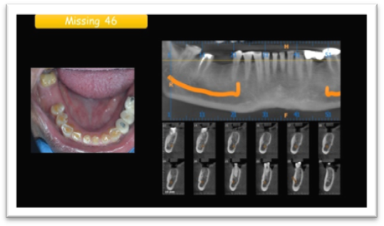
Figure 1: CBCT showing missing 46
ImplantSwiss Bone level implant of dimension measuring 4.3 x 12 was selected based on CBCT evaluation. Following an injection of 2% lidocaine with 1: 80,000 anaesthetic agent in the area of the missing 1st molar, free hand implant placement was done. Very good Primary stability was achieved with Insertion torque of more than 40 NCM. ISQ measurements was in range of 80-82, therafter healing abutment was placed (Figure 2)

Figure 2: X -ray showing placement of implant
Post-operative after 2 months ISQ test done again, which showed excellent biological stability with reading measuring in between 84-87.(Figure 2) Thereby showing excellent secondary stability or osseointegration .
Post-operative OPG was suggested after 2 months to check hard tissue architecture around implantswiss implant. (Figure 3)

Figure 3: 2 months post-operative OPG
Excellent soft tissue healing around Implantswiss implant. (Figure 4)

Figure 4: Soft tissue healing around Implantswiss Implant.
Digital impression were made by CEREC Workflow (Primescan). Implantswiss implant scan bodies were used & it was planned to give screw retained crown by using Implantswiss implant Ti-Base (Figure 5 a,b)

Figure 5 (a)

Figure 5 (b)
Figure 5: Digital impression made using primescan
Sirona meso block of A2 shade was used in Primemill machine. (Figure 6 a,b )

Figure 6 (a)

Figure 6 (b)
Figure 6: Milled Sirona Meso block of A2 shade
Sintering of Meso Sirona done using SpeedFire. (Figure 7)
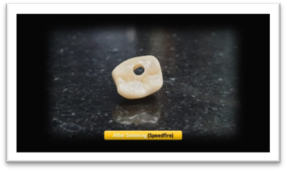
Figure 7: Sintering of Meso Sirona done using SpeedFire
Now Implantswiss implant Ti -Base Swiss was cemented extraorally with resin-based cement to prepared crown. (Figure 8 a,b )

Figure 8 (a)

Figure 8 (b)
Figure 8: Ti base implantswiss implant
IOPA X-ray showed Implant & Prosthesis in perfect 3-D position (Figure 9) ensuring very good prognosis.
Occlusion & proximal contacts were verified.

Figure 9: X- ray showing successful placement of screw retained crown
DISCUSSION
When teeth have to be replaced by oral implants, there are various factors that result in timing of implant placement after dental extraction, among these, the three-dimensional position of the tooth in the oral cavity, the hard and soft tissue contour of the site and the adaptive changes of the alveolar ridge after tooth extraction that may affect the outcome of the therapy.21
The outcome shown here was made possible by the technology of the "ImplantSwiss" implant.
Even though no technique has yet been identified as the gold standard, intraoral digital impressions can be considered a reliable alternative for fixed implant prosthetic restorations.22-24
The elaboration of a digital approach has been proven to be even more accurate25 and efficient than conventional materials. Since the digital impressions are instantly sent and saved/stocked electronically for the fabrication of definitive prosthetic restorations26, thus enhancing efficiency of the workflow, the IOS has proved the decrease of margin of error caused by traditional impression taking27 and cast production28 methods.
Furthermore, from a systematic review29 evaluating the advantages of digital technologies for the manufacturing of implant-supported rehabilitations, it emerged the increased efficiency of CAD/CAM systems compared to conventional fabrication procedures.
Technologies that include 3D radiographic imaging, intraoral optical scanners (IOSs), virtual implant planning software programs, computer-guided implant placement, and computer-aided design and computer-aided manufacturing (CAD-CAM) have streamlined dental implant therapy that enhanced the predictability and reproducibility of treatment outcomes.30-32
Digital scanning through the use of IOSs offers multiple benefits, including improved patient experience, reduced distortion, digital recordkeeping, pre visualization of the 3D preparation, improved communication with the dental laboratory technician, and a more effective teaching experience.33-36
CONCLUSIONS
Masticatory ability can be improved with the delivery of posterior implant restoration. The bite force that are distributed on the implant prosthesis inevitably increases after placement of implant prostheses, a routine follow-up and occlusal evaluation are strongly needed.
The technology of the Implantswiss® implant is one of best technology as all it requires is a stable second cortical for implant anchorage to provide a long-term successful function.
The primary aim of any dental implant is to achieve treatment and achieve occlusion on both sides as well as bilateral and unlimited masticatory function.
The digital scanning technique is more efficient than the conventional impression technique for single implant-supported restorations. Digital scans and CAD-CAM implant casts have far best accuracy comparable with that of conventional impressions and gypsum casts. Most participants preferred digital scans over conventional impressions.
The present case report showed that careful surgical and restorative interdisciplinary planning, along with the inclusion of the laboratory technician is key in decision-making and excellent motivation of the patient, resulted in a highly functional case as well as a satisfied patient.
REFERENCES
- T. Albrektsson and N. Donos, “Implant survival and complications. The Third EAO consensus conference 2012,” Clinical Oral Implants Research, vol. 23, supplement 6, pp. 63–65, 2012.
- Shah Z, Shah A, Raiyani P. Two narrow implants replacing a mandibular right first molar: A case study. J Dent Implant [serial online] 2016 [cited 2021 Nov 16];6:75-8. Available from: https://www.jdionline.org/text.asp?2016/6/2/75/202153
- N. P. Lang and T. Berglundh, “Periimplant diseases: where arewe now? Consensus of the seventh European workshop on periodontology,” Journal of Clinical Periodontology, vol. 38, supplement 11, pp. 178–181, 2011.
- L. J. A. Heitz-Mayfield, “Peri-implant diseases: diagnosis andrisk indicators,” Journal of Clinical Periodontology, vol. 35,supplement 8, pp. 292–304, 2008.
- Sheridan RA, Decker AM, Plonka AB, Wang HL. The Role of Occlusion in Implant Therapy: A Comprehensive Updated Review. Implant Dent. 2016;25(6):829–838. doi: 10.1097/ID.0000000000000488 . [PubMed] [CrossRef] [Google Scholar]
- Kim Y, Oh TJ, Misch CE, Wang HL. Occlusal considerations in implant therapy: clinical guidelines with biomechanical rationale. Clin Oral Implants Res. 2005;16(1):26–35. doi: 10.1111/j.1600-0501.2004.01067.x . [PubMed] [CrossRef] [Google Scholar]
- Gross MD. Occlusion in implant dentistry. A review of the literature of prosthetic determinants and current concepts. Aust Dent J. 2008;53 (Suppl 1): S60–8. doi: 10.1111/j.1834-7819.2008.00043.x . [PubMed] [CrossRef] [Google Scholar]
- J. Lindhe and J. Meyle, “Peri-implant diseases: consensus reportof the sixth European workshop on periodontology,” Journal ofClinical Periodontology, vol. 35, no. 8, pp. 282–285, 2008.
- Kim SY, Kim MJ, Han JS, Yeo IS, Lim YJ, Kwon HB: Accuracy of dies captured by an intraoral digital impression system using parallel confocal imaging. Int J Prosthodont 2013, 26(2):161–163.
- Christensen GJ: Impressions are changing: deciding on conventional, digital or digital plus in-office milling. JADA 2009, 140:1301–1304.
- Syrek A, Reich G, Ranftl D, Klein C, Cerny B, Brodesser J: Clinical evaluation of all-ceramic crowns fabricated from intraoral digital impressions based on the principle of active wavefront sampling. J Dent 2010, 38:553–559.
- Henkel GL: A comparison of fixed prostheses generated from conventional vs digitally scanned dental impressions. Compend Contin Educ Dent 2007, 28:422–424.
- Brawek PK, Wolfart S, Endres L, Kirsten A, Reich S: The clinical accuracy of single crowns exclusively fabricated by digital workflow the comparison of two systems. Clin Oral Investig 2013, 17(9):2119–2125.
- Seelbach P, Brueckel C, Wöstmann B: Accuracy of digital and conventional impression technique and workflow. Clin Oral Investig 2013, 17(7):1759–1764.
- Seelbach P, Brueckel C, Wöstmann B: Accuracy of digital and conventional impression technique and workflow. Clin Oral Investig 2013, 17(7):1759–1764.
- Luthardt RG, Loos R, Quaas S: Accuracy of intraoral data acquisition in comparison to the conventional impression. Int J Comput Dent 2005, 8:283–294.
- Güth JF, Keul C, Stimmelmayr M, Beuer F, Edelhoff D: Accuracy of digital models obtained by direct and indirect data capturing. Clin Oral Investig 2013, 17:1201–1208.
- Karl M, Shubinski P, Taylor T: Effect of intraoral scanning on the passivity of fit of implant-supported fixed partial prostheses. Quintessence Int 2012, 43:555–563.
- Mehl A, Ender A, Mörmann W, Attin T: Accuracy testing of a new intraoral 3D camera. Int J Comput Dent 2009, 12:11–28.
- Ender A, Melh A: Full arch scans: conventional versus digital impressions. An in-vitro study. Int J Comput Dent 2011, 14:11–21.
- Lanza A, Scognamiglio F , Femiano F, and Lanza M.Immediate, Early, and Conventional Implant Placement in a Patient with History of Periodontitis .http://dx.doi.org/10.1155/2015/217895
- Amin, S.; Weber, H.P. Digital vs. Conventional full-arch implant impressions: A comparative study. Clin. Oral Implants Res. 2017, 28, 1360–1367. [CrossRef] [PubMed]
- Gherlone, E.F.; Ferrini, F. Digital Impressions for Fabrication of Definitive “All-on-Four” Restorations. Implant Dent. 2015, 24, 125–129. [CrossRef] [PubMed]
- Gherlone, E.F.; Capparé, P. Conventional Versus Digital Impressions for “All-on-Four” Restorations. Int. J. Oral Maxillofac. Implants 2016, 31, 324–330. [CrossRef] [PubMed]
- Gjelvold, B.; Chrcanovic, B.R. Intraoral digital impression technique compared to conventional impression techniques. A randomized clinical trial. J. Prosthodont. 2016, 25, 282–287. [CrossRef] [PubMed
- Ahlholm, P.; Sipila, K. Digital Versus Conventional Impressions in Fixed Prosthodontics: A Review. J. Prosthodont. 2018, 27, 35–41. [CrossRef] [PubMed]
- Ting-Shu, S.; Jian, S. Intraoral digital impression technique: A review. J. Prosthodont. 2015, 24, 313–321. [CrossRef] [PubMed]
- Ender, A.; Mehl, A. In vitro evaluation of the accuracy of conventional and digital methods of obtaining full-arch dental impressions. Quintessence Int. 2015, 46, 9–17. [PubMed]
- Muhlemann, S.; Kraus, R.D. Is the use of digital technologies for the fabrication of implant-supported reconstructions more efficient and/or more effective than conventional techniques: A systematic reviewClin. Oral Implants Res. 2018, 9, 184–195. [CrossRef] [PubMed]
- Joda T, Bragger U. Digital vs. conventional implant prosthetic workflows: a cost/time analysis. Clin Oral Implants Res 2015;26:1430-5.
- Joda T, Lenherr P, Dedem P, Kovaltschuk I, Bragger U, Zitzmann NU. Time efficiency, difficulty, and operator’s preference comparing digital and conventional implant impressions: a randomized controlled trial. Clincal Oral Implants Res 2017;28:1318-23.
- Joda T, Bragger U. Time-efficiency analysis of the treatment with monolithic implant crowns in a digital workflow: a randomized controlled trial. Clin Oral Implants Res 2016;27:1401-6.
- Koch GK, Gallucci GO, Lee SJ. Accuracy in the digital workflow: From data acquisition to the digitally milled cast. J Prosthet Dent 2016;115:749-54.
- Schepke U, Meijer HJ, Kerdijk W, Cune MS. Digital versus analog completearch impressions for single-unit premolar implant crowns: Operating time and patient preference. J Prosthet Dent 2015;114:403-6.e1.
- Joda T, Bragger U. Patient-centered outcomes comparing digital and conventional implant impression procedures: a randomized crossover trial. Clin Oral Implants Res 2016;27:e185-9.
- Marghalani A, Weber HP, Finkelman M, Kudara Y, El Rafie K, Papaspyridakos P. Digital versus conventional implant impressions for partially edentulous arches: An evaluation of accuracy. J Prosthet Dent 2018;119:574-9.
Disclaimer from Guident
The views and opinions expressed in articles published in Guident are solely those of the authors and do not necessarily reflect the views of the editors, the editorial board or the publisher, Ivory India. Guident does not endorse any product, technique or treatment described. Clinical content is intended for qualified dental and medical professionals and is not a substitute for individual clinical judgement. Guident, its editors and the publisher accept no liability for any consequence arising from the use of information published in the journal.
How to cite this article
Dr. Gaurav Gupta, Dr. D.K. Gupta, Dr. Neelja Gupta. Implantswiss Implant with Chairside Screw Retained Crown by Cerec Workflow- a Case Report. Guident. 2021 Aug;14(9). Available from: https://ivoryindia.in/articles/implantology/implantswiss-implant-with-chairside-screw-retained-crown-by-cerec-workflow-a-case-report.html