PRF Adjunction with Direct Sinus Lift for Implant Placement
Department of Prosthodontics, Pt. B.D Sharma University of Health Science, Rohtak, Haryana, India
Abstract
Placement of dental implants has evolved to a great extent. In posterior maxilla where expansion of maxillary sinus limits the implant placement direct sinus lift procedure is a good option. This membrane elevation if accompanied by a good graft material such as PRF combined with osseoconductive grafts leads to favorable results. This case report presents a case of implant placement along with sinus lift procedure augmente with PRF with successful results.
Key words: Dental implant, Schneiderian membrane, Maxillary sinus lift, Platele Rich Fibrin (PRF).
INTRODUCTION
With the development of technology and treatment options available implant dentistry has crossed many barriers. Any kind of hurdle can be overcome with the latest development in field of implant dentistry. Limited amount of bone has always been a challenge for the implantologists. In case of posterior maxilla with the advancing age there is pneumatisation of the sinus leading to decreased amount of bone available for the implant placement. The maxillary sinus lift was pioneered by Tatum and was modified by Boyne.[1,2] The direct maxillary sinus lift can be performed to gain the bone quantity.
In case of direct maxillary sinus lift a number of graft options are available which can be used. In this case report we have performed the direct sinus lift in right side posterior maxilla using hydroxy apatite bone graft enhanced with PRF. This particulate graft enhanced with PRF which is an autologous source of growth factors and is very efficient in promoting healing.[4,5] Being an autologous source of growth factors it is free from any allergic response. It is obtained by centrifuging the blood at 2700rpm for 15 min. Fibrin promotes angiogenesis by trapping the stem cells and giving protection from epithelial layer. PRF accelerates the proliferation of fibroblasts.It is a good source of growth factors. In this case report we are providing growth factors through PRF to maxillary sinus lift procedure.
CASE PRESENTATION
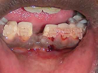
A 43 years old male reported in the OPD of Department of Prosthodontics with the complaint of missing tooth in the right upper back tooth region.On further asking he explained that he has gradually lost his teeth due to gradual decaying of teeth. He has lost his teeth 1 year back. On intraoral examination it was revealed that he had missing 16 and 36. On radiographic examination it was found that the mandibular edentulous area for an easy
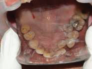
Pre op Maxillary Occlusal View

Prep Intraoral view

Pre op radiograph
Properly sterilized surgical instruments were arranged on a surgical trolley.Subject was seated in the dental chair and prepared following the standard guidelines for asepsis. Posterior superior alveolar nerve block, infraorbital nerve block and greater palatine nerve block with 2% lidocaine (epinephrine 1:80000) were administered.
After confirming the effectiveness of local anaesthesia crestal incision with relieving incision on buccal aspects were given. After elevating the mucoperiostal flap the lateral window was prepared on the buccal aspect of edentulous ridge with the help of diamond bur. The cortical bone so created after window preparation was left attached to the membrane so as toprevent its perforation. After this the Schneiderian membrane was lifted using hand instruments. While elevating the membrane caution was given to not to perforate the bony septa present 7 mm above the floor of sinus. Followed by this, sequential drilling was done through the crestal approach to prepare recipient site for implant. Blood sample was collected from ante cubital vein without any anticoagulant in 10 ml tube, which was immediately centrifuged at 3000 rpm for 10-12 min. Blood centrifugation immediately after collection resulted in fibrin clot in the middle of the tube, just between the red corpuscles at the bottom and acellular plasma at the top. The fibrin clot thus obtainedwas separated from the RBC fraction and gently pressed between two layers of sterile gauze to form a membrane. Synthetic hydroxyapatite particulate graft was placed in association with the PRF membrane in the sinus cavity obtained after lifting the Schneiderian membrane. A 10 mm implant was placed at the site equicrestally with the primary stability of 40 Ncm. The implant placement was assessed through the lateral window. Healing abutment was screwed on the top of implant which prevented accidental slippage of implant into sinus cavity. The window which was stuffed with graft was closed with thehelp of collagen membrane. The flaps were approximated and 3-0 black braided silk was used for interrupted suture. Sutures were made water tight and knot was tied away from the incision line.

Pre op preparation

Flap elevation

Window preparation

Elevating Schneiderian membrane

Implant placement

Graft placement
Post-operative instructions were given to the subject regarding diet, oral hygiene and medications were prescribed as per requirement. Patient was recalled after 24 hours for review, after 7 days for suture removal and soft tissue evaluation. Prosthesis was delivered at 6 months and regular follow up was scheduled. Soft tissue profile was well contoured around the implant supported prosthesis.

Post op occlusal view

Post op radiograph after 1 year
DISCUSSION
Pneumatised maxillary sinus has always been a challenge for the implantologists. A number of options have been explored to place a stable implant in the posterior maxilla. One can either place an angulated implant or a pterygoid implant or zygomatic implant. One of the easy options to manage a large sinus is elevating the Schneiderian membrane. However
sinus lift procedure is a complicated surgical procedure and requires a skilled implantologist. A great care is required while elevating the membrane trying to avoid membrane perforations. The direct sinus lift procedure makes access to sinus through lateral wall making it clear, easy access and longer implants can be placed when compared with indirect sinus lift procedure which is a crestal approach.[5] Tatum preferred crestal approach for achieving 1mm rise in sinus membrane but Boyne advocated lateral wall access.[1,2] Boyne,s
technique gained more popularity among the implantologists.
Whenever planning for ridge augmentation procedures a graft should be selected according to the need of the area. Hydroxyapatite particulate graft is a good option as it is osseoconductive and being porous allows for capillary action to allow for neovascularisation. Augmentation with PRF leads to release of autologous growth factors.[6] Platelet rich fibrin is a simple, natural and inexpensive entity for the production of leukocyte and growth factors. As a strong solid fibrin membrane, it is particularly easy to use in implant
dentistry. Additionally, it offers a protective effect both mechanical and biological to the grafted area. PRF is a good source of growth factors like transforming growth factor B1 (TGFB1), platelet derived growth factor AB (PDF-AB), vascular endothelial growth factor (VEGF) and matrix glycoproteins such as thrombospondin-1, insulin-like growth factor (IGF) and platelet-derived angiogenic factor. Proliferation was significantly higher on PRF membranes and PRF clot as pointed in the study conducted by Boora P, Rathee M, Bhoria
M.[7] Ali S, Bakry SA, Abd-Elhakam H also supported placement of PRF in sinus augmentation procedures.[8]
CONCLUSION
This combination of PRF with hydroxyapatite graft offers a speedy healing and bone formation while placing implants in posterior maxilla associated with direct maxillary sinus lift procedures. This case report points out the excellent bone quality obtained after healing as obtained with PRF in conjunction with hydroxyapatite.
Acknowledgement
The author declares no financial interest in any product referenced herein.
REFERENCES
- Boyne PJ, James RA. Grafting of the maxillary sinus floor with autogenousmarrow and bone. J Oral Surg. 1980; 38: 613-616.
- Tatum H, Jr. Maxillary and sinus implant reconstructions. Dent Clin North Am.986; 30: 207-229.
- Wang X et al. Behavior of Gingival Fibroblasts on Titanium Implant Surfaces inCombination with either Injectable-PRF or PRP. Int J Mol Sci. 2017;18(2).e331.
- Amo FSFD et al. Biologic Agents for Periodontal Regeneration and Implant SiteDevelopment. Biomed Res Int. 2015;2015:957518.
Disclaimer from Guident
The views and opinions expressed in articles published in Guident are solely those of the authors and do not necessarily reflect the views of the editors, the editorial board or the publisher, Ivory India. Guident does not endorse any product, technique or treatment described. Clinical content is intended for qualified dental and medical professionals and is not a substitute for individual clinical judgement. Guident, its editors and the publisher accept no liability for any consequence arising from the use of information published in the journal.
How to cite this article
Dr. Aditi Singh, Dr. Manu Rathee, Dr. Akanksha Gopal Shetye. PRF Adjunction with Direct Sinus Lift for Implant Placement. Guident. 2018 Sep;11(10). Available from: https://ivoryindia.in/articles/implantology/prf-adjunction-with-direct-sinus-lift-for-implant-placement.html