
Pyogenic Granuloma
Career Post Graduate Institute of Dental Sciences & Hospital, Lucknow
ABSTRACT
Pyogenic granuloma is a reactive hyperplasia of connective tissue in response to local irritants. It is a tumor like growth of the oral cavity, frequently located surrounding the anterior teeth or skin that is considered to be neoplastic in nature. It usually arises in response to various stimuli such as low-grade local irritation, traumatic injury, hormonal factors, or certain kinds of drugs. Histologically, the surface epithelium may be intact, or may show foci of ulcerations or even exhibiting hyperkeratosis. It overlies a mass of dense connective tissue composed of significant amounts of mature collagen. Gingiva is the most common site affected followed by buccal mucosa, tongue and lips. Pyogenic granuloma in general, does not occur when excised along with the base and its causative factors. This paper presents one case of a pyogenic granuloma managed by surgical intervention.
Key Words: Pyogenic granuloma, begin neoplasm, hyperplastic lesion
INTRODUCTION
The term “pyogenic granuloma” is a misnomer because the lesion does not contain pus and does not represent a granuloma histologically.1 Pyogenic granuloma is a relatively common, soft tissue tumor of oral cavity that is believed to be reactive and not neoplastic in nature.2 Approximately one-third of the lesions occur due to trauma and poor oral hygiene may also be one of the precipitating factors. It often presents as a painless, pedunculated, or sessile mass of gingiva. Pyogenic granuloma is the most common of all the oral tumorlike growths.3 While the terminology implies a benign neoplasm, most if not all fibromas represent reactive focal fibrous hyperplasias due to trauma or local irritation. Although the term “focal fibrous hyperplasia” more accurately describes the clinical appearance and pathogenesis of this entity, it is not commonly used.4 Pyogenic granuloma is a hyperactive benign inflammatory lesion that occurs mostly on the mucosa of females with high levels of steroid hormones. It is generally believed that female sex hormones play important roles in its pathogenesis.5 It is a tumour like growth of the oral cavity (frequently located surrounding the anterior teeth) or skin that is considered to be neoplastic in nature.4 It usually arises in response to various stimuli such as low-grade local irritation, traumatic injury, hormonal factors ,or certain kinds of drugs.6,7
CASE REPORT
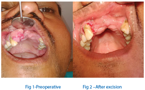
A 38 years old male patient was reported to our department with a chief complaint of growth upper right front region of jaw since three month. The lesion was whitish red in color irregular in shape, 1x2 cm in longest diameter present in relation to labial mucosa of 11,12 &13.Surface of lesion rough in appearance. Growth was pedunculated, firm in consistency. It was compressible, & does bleed on pressure and non tender on palpation.No other oral anamolies were detected. This clinical examination led to a tentative diagnosis of a pyogenic granuloma.After hematological and general medical evaluation of patient. An excisional biopsy was performed, intra-orally under local anesthesia and pressure pack was given.(Fig-2&3). Specimen sent for histopathological examination.
Postoperative instruction was given along with antibiotics Cap. Amoxicillin(500mg) and NSAID tab. Ketorolac(10mg) were given t.i.d for 5 days. After a week patient was recalled, healing was uneventful(Fig-5).
Histopathological examination of the tissue revealed Stratified squamous orthokeratinized epithelium covering cellular connective tissue(Fig-4) The epithelium shows area of ulceration below which can be seen inflammation in the connective tissue. the connective tissue shows proliferating fibroblasts and collagen fibres interposed in which can be seen lot of epithelial lined spaces with in the connective tissue can be seen patchy distribution of lymphocytes and plasma cells. There was no evidence of malignancy. The clinical and histopathological findings confirmed it to be a case of pyogenic granuloma.

DISCUSSION
Pyogenic granuloma is an inflammatory hyperplasia affecting the oral tissues. Hullihen’s description in 1844 was most likely the first pyogenic granuloma reported in the English literature. It was only in1904 that Hartzell first ever introduced the term pyogenic granuloma.8 It is now universally agreed that this lesion is formed as a result of an exaggerated localized connective tissue reaction to a minor injury or any underlying irritation.9 The irritating factor can be calculus, poor oral hygiene, nonspecific infection, over hanging restorations, cheek biting etc. Because of this irritation, the underlying fibrovascular connective tissue becomes hyperplastic and there is proliferation of granulation tissue which leads to the formation of a pyogenic granuloma.10 Pyogenic granuloma may occur at all ages but is predominantly seen in the second decade of life in young adult females, possibly because of the vascular effects of female hormones.11 The gingiva is the most commonly site affected followed by the buccal mucosa, tongue and lips.8 Pyogenic granuloma in general, does not reoccur when excised along with its base and all the causative factors are removed. Other treatment modalties includes cryosurgery, elctrodessication, sodium tetradecyl sulphate sclerotherapy, steroid injections and ultrasonic scissors. This paper presents a case of a pyogenic granuloma managed by surgical intervention.
CONCLUSION
It can be concluded that the combinations of various etiological factors might have caused the inflammatory tissue to cross the threshold from regular gingivitis to granuloma formation. The lesion was painless as nerves do not proliferate within the reactive hyperplastic tissue. Surgical excision is a successful treatment of choice in minimizing the recurrence of lesion. So, the consideration should also be given to correct diagnosis and proper treatment planning.
ACKNOW LEDGEMENT
Authors of this article acknowledge the contribution and help of the following Dentists: Prof. K.Srinivas, Prof.P.Ratnakar, Prof.Jyoti Gupta ,Dr. Parvez Alam Khan, Dr. Arti S.Sachdev, & Dr. Vasu S.Saxena.
REFERENCES
- karthikeya Patil, mahima VG, Lahari K: Exragingival pyogenic granuloma. Indian journal of dental research 2006, 17(4):199-202.
- . Ramirez. K, Bruce G: carpenter. pyogenic granuloma: case report in a 9-year-old girl. General Dentistry 2002, 50(3):280-1.
- Macleod RI, Soames JV. Epulides: a clinicopathological study of a series of 200 consecutive lesions. Br Dent J 1997; 163 : 51-3.
- Vilmann A, Vilmann P, Vilmann H. Pyogenic granuloma: evaluation of oral conditions. Br J Oral Maxillofac Surg 1986; 24(5): 376-82.
- Verma et al, Pyogenic Granuloma - Hyperplastic Lesion of the Gingiva: Case Reports” The Open Dentistry Journal, 2012, 6, 153-156.
- Mussalli NG, Hopps RM, Johnson NW. Oral pyogenic granuloma as a complication of pregnancy and the use of hormonal contraceptives. Int J Gynaecol Obstet 1976; 14(2): 187-91.
- Miller RAW, Ross JB, Martin J. Multiple granulation tissue lesions occurring in isotretinoin treatment of acne vulgaris: successful response to topical corticosteroid therapy. J Am Acad Dermatol 1985; 12 (5): 888-9.
- Jafarzadeh H, Sanatkhani M, Mohtasham N. Oral pyogenic granuloma: a review. J Oral Sci 2006; 48(4): 167-75.
- Mathur LK, Bhalodi AP, Manohar B, Bhatia A, Rai N, Mathur A.Focal fibrous hyperplasia: a case report. Int J Dent Clin 2010; 2(4):56-7.
- Kerr DA. Granuloma pyogenicum. Oral Surg Oral Med Oral Pathol 1951; 4(2): 158.
More references are available on request.
Disclaimer from Guident
The views and opinions expressed in articles published in Guident are solely those of the authors and do not necessarily reflect the views of the editors, the editorial board or the publisher, Ivory India. Guident does not endorse any product, technique or treatment described. Clinical content is intended for qualified dental and medical professionals and is not a substitute for individual clinical judgement. Guident, its editors and the publisher accept no liability for any consequence arising from the use of information published in the journal.
How to cite this article
Dr.Md.Asdullah, Dr. Akhtar Husain, Dr. Payal Tripathi Department of Medicine & Radiology. Pyogenic Granuloma. Guident. 2015 Jun;8(7). Available from: https://ivoryindia.in/articles/oral-medicine/pyogenic-granuloma.html


