Endoscope Guided Management of Condylar Fracture
Impressions Dental and Maxillofacial Centre Umrao Hospitals Mira Road, Mumbai, India
Controversy about the treatment protocol for condylar fracture has a long history in oral and maxillofacial traumatology. Reduction and miniplate osteosynthesis of condylar fractures often require a wide extraoral approach with the risk of aesthetic impairment and possible facial nerve palsy. The endoscopic-assisted approach for the treatment of condylar fracture is a less invasive alternative and overcomes the limited access to the operation field to obtain an accurate reduction and fixation avoiding other complications. In two of our cases we achieved good anatomic reduction and fixation. The treatment of condylar mandible fractures with a minimal invasive endoscopically assisted technique is reliable and may offer advantages for selected cases, particularly concerning the lower occurrence of facial nerve damage.
Introduction
Modern surgery attempts to minimize as much as possible the patient’s somatic and psychological trauma. The expanding interest in the minimal access surgery has spearheaded the development of endoscopic techniques in facial surgeries. Increased availability and interest has led maxillofacial surgeon to utilize the endoscopic techniques in maxillofacial surgeries (facial cosmetic surgeries, TMJ surgeries and trauma). Condylar fractures of the mandible are frequent, their treatment being one of the most controversial aspects in maxillofacial surgery. Whether to treat it by open reduction and fixation or by closed method. All surgical approaches for the open treatment of condylar fractures require a extra oral facial incision, and nearly all will result in a perceptible scar and chances of facial nerve injury. The use of the endoscope to treat condylar injuries was a extension of minimally invasive techniques for managing condylar fracture either by transoral or small extraoral submandibular incision. Endoscopic assistance allows the surgeon to get anatomic reduction, and to avoid the negative sequelae of condylar fracture. The endoscopic approach described here has the potential to reduce morbidity by limiting scars, reducing the risk to the facial nerve, and eliminating the need for MMF.
The objective of the article is to describe the endoscopic assisted
General requirements for endoscopic surgery include the following:
- The ability to surgically obtain and maintain an optical cavity
- The ability to insert a fiberoptic endoscope
- The ability to maintain adequate hemostasis
- The ability to apply instrumentation
Advantages of endoscopic repair include the following:
- More accurate fracture visualization
- Small external incisions
- Reduced soft tissue dissection
- The potential for visualization around corners
- The possibility of reduced duration of hospital stays
- Improved teaching opportunities (since the procedure can be visualized on a television monitor)
Disadvantages of endoscopic repair include the following:
- A current lack of dedicated instrumentation
- A moderate learning curve for the techniques
- A narrow field of view
- A limited ability for bimanual instrumentation without an assistant
CLINICAL CASES PRESENTATION
A 24 years male patient with no relevant medical history was admitted to hospital due to facial trauma. On physical examination, malocclusion and a right parasymphyseal fracture was observed. The radiographic study by CT Face confirmed the existence of a right parasymphyseal fracture and a left high subcondylar fracture. Endoscopic guided surgery was performed for subcondylar fracture through intraoral approach and parasymphseal fracture through
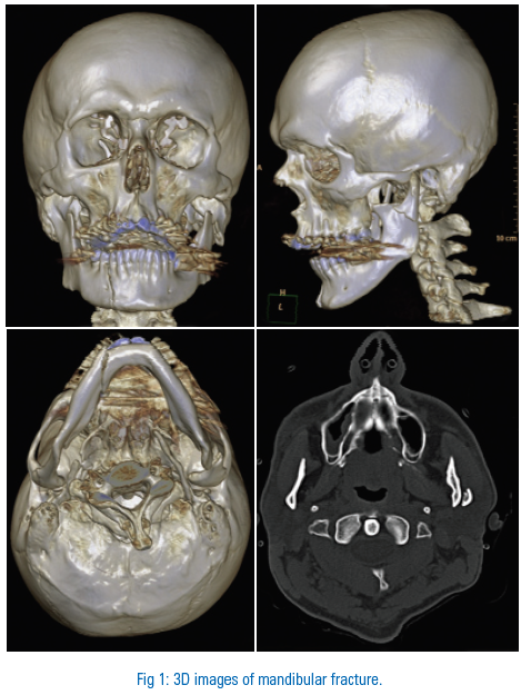

direct intraoral vestibular approach. Both the fractures were fixed with titanium miniplates. Intermaxillary fixation was not done. We experienced good visualization of the fracture and a good intraoperative reduction and osteosynthesis was achieved. Post operative radiographic evaluation showed correct anatomic reduction and restoration of the ascending ramal height with no disturbance in occlusion. After 21 days complete range of mobility was observed. At 5 months follow up the aesthetic and functional outcomes were satisfactory.
DISCUSSION
The management of facial trauma has evolved from the basic fundamentals of orthopaedic surgery. The main principles still remain the same, i.e. anatomical reduction under direct vision with rigid fixation. But the continuous need of minimal invasive procedures so as to maintain aesthetic facial appearance and function, has led to the evolution of many new techniques for example endoscopic surgeries.
Endoscopic procedures has been preferred more recently as it causes lesser soft tissue damage, minimises blood losses, fastens recovery and also gives aesthetically and functionally acceptable outcomes. Endoscopic procedures have been used for the fixation of fractures of the extremities. Its result motivated the use of similar techniques in management of maxillofacial trauma.
Condylar fractures of the mandible are very frequent, but their treatment is one of the most controversial. This controversy is based on the outcome of open versus closed treatment of fracture condyle. These Traditional treatment options of condylar fractures often results in a malreduction and significant functional and aesthetic sequelae, including facial asymmetry, decreased jaw opening, and potential for late derangements of the temporomandibular joint. The endoscopic technique will reliably restore condylar, thus obviating the consequences of malunion with no significant facial scarring or facial nerve damage has resulted.
Though we have less experience in this technique the result that has obtained was satisfactory, favourable occlusion, mouth opening approximately around 34mm with no signs of TMJ dysfunction after 5 month follow-up.
The main difficulty in this is steep learning curve, handling of endoscope and at the same time availability of required instrument.
CONCLUSIONS:
Endoscopic guided treatment of condylar fracture by transoral approach combines the positive aspects of both conventional techniques: closed and open reduction; allowing anatomic reduction and a stable fixation leaving no visible facial scars and with a minimum risk of injury to the facial nerve.
REFERENCES
- Zhang Q B, Dong Y J, Li Z B, Zhao J H. Coronal incision for treating zygomatic complex fractures. J Craniomaxillofac Surg. 2006; 34:182–185.
- Phillips J H, Gruss J S, Wells M D, Chollet A. Periosteal suspension of the lower eyelid and cheek following subciliary exposure of facial fractures. Plast Reconstr Surg. 1991; 88:145–148.
- Silvennoinen U, Iizuka T, Lindqvist C, Oikarinen K. Different patterns of condylar fractures: an analysis of 382 patients in a 3-year period. J Oral Maxillofac Surg. 1992; 50:1032–1037. [
- Silvennoinen U, Raustia A M, Lindqvist C, Oikarinen K. Occlusal and temporomandibular joint disorders in patients with unilateral condylar fracture. A prospective one-year study. Int J Oral Maxillofac Surg. 1998;27:280–285. Dahlstrom L, Kahnberg K E, Lindahl L. 15 year’s follow-up on condylar fractures. Int J Oral Maxillofac Surg. 1989; 18:18–23.
- Throckmorton G S, Ellis E, III, Hayasaki H. Masticatory motion after surgical or nonsurgical treatment for unilateral fractures of the mandibular condylar process. J Oral Maxillofac Surg. 2004; 62:127–138.
- Ellis E, III, Throckmorton G S. Bite forces after open or closed treatment of mandibular condylar process fractures. J Oral Maxillofac Surg. 2001;59:389–395.
- Ellis E, III, Simon P, Throckmorton G S. Occlusal results after open or closed treatment of fractures of the mandibular condylar process. J Oral Maxillofac Surg. 2000; 58:260–268.
- Ellis E, III, Throckmorton G S, Palmieri C. Open treatment of condylar process fractures: assessment of adequacy of repositioning and maintenance of stability. J Oral Maxillofac Surg. 2000; 58:27–34. Discussion 35.
- Ellis E, III, Palmieri C, Throckmorton G. Further displacement of condylar process fractures after closed treatment. J Oral Maxillofac Surg. 1999; 57:1307–1316. Discussion 1316–1317.
- Ellis E, III, Carlson D S. The effects of mandibular immobilization on the masticatory system. A review. Clin Plast Surg. 1989; 16:133–146.
- Mueller R V, Czerwinski M, Lee C, Kellman R M. Condylar fracture repair: use of the endoscope to advance traditional treatment philosophy. Facial Plast Surg Clin North Am. 2006; 14:1–9.
- Walker R V. Condylar fractures: nonsurgical management. J Oral Maxillofac Surg. 1994; 52:1185–1188.
- Feifel H, Albert-Deumlich J, Riediger D. Long-term follow-up of subcondylar fractures in children by electronic computer-assisted recording of condylar movements. Int J Oral Maxillofac Surg. 1992; 21:70–76.
- Guven O, Keskin A. Remodelling following condylar fractures in children. J Craniomaxillofac Surg. 2001; 29:232–237.
- Stanley R B., Jr The zygomatic arch as a guide to reconstruction of comminuted malar fractures. Arch Otolaryngol Head Neck Surg. 1989; 115:1459–1462.
- Chemin J. [Treatment of isolated fracture of the zygomatic arch by Gillies' technic.] Revue Stomatol. 1954; 55:586–589.
- Popescu V, Stanescu S, Lascar I. [Conclusions from our experiences with the Gillies-Dan Theodorecu method in fractures of the zygomatic arch.] Stomatologia (Bucur) 1968; 15:499–505.
- Chen C T, Lai J P, Chen Y R. Application of endoscope in zygomatic fracture repair. Br J Plast Surg. 2000; 53:100–105.
- Appling W D, Patrinely J R, Salzer T A. Transconjunctival approach vs subciliary skin-muscle flap approach for orbital fracture repair. Arch Otolaryngol Head Neck Surg. 1993; 119:1000–1007.
- Mullins J B, Holds J B, Branham G H, Thomas J R. Complications of the transconjunctival approach. A review of 400 cases. Arch Otolaryngol Head Neck Surg. 1997; 123:385–388.
Disclaimer from Guident
The views and opinions expressed in articles published in Guident are solely those of the authors and do not necessarily reflect the views of the editors, the editorial board or the publisher, Ivory India. Guident does not endorse any product, technique or treatment described. Clinical content is intended for qualified dental and medical professionals and is not a substitute for individual clinical judgement. Guident, its editors and the publisher accept no liability for any consequence arising from the use of information published in the journal.
How to cite this article
Dr. Abdul Hameed. Endoscope Guided Management of Condylar Fracture. Guident. 2013 Dec;7(1). Available from: https://ivoryindia.in/articles/oral-surgery/endoscope-guided-management-of-condylar-fracture.html