Soft Ti Ssue Oral Surgery the Removal of a Fibroma
Mississauga, Ontario.
Soft tissue oral surgery encompasses a large percentage of surgeries performed by a dentist on a day to day basis. It includes everything from the surgical removal of hyperplastic tissue to enucleation of cysts. Every dentist who performs surgical procedures on a routine basis should be familiar with this aspect and category of oral surgery and be prepared to perform it as the need arises.
There are numerous textbooks written that describe soft tissue surgery but the vast majority of them are complicated and written primarily for the specialist. Many of these elaborate texts ignore the everyday routine surgical procedures, and instead concentrate on major surgical techniques that are of no interest or clinical value to the general dentist.
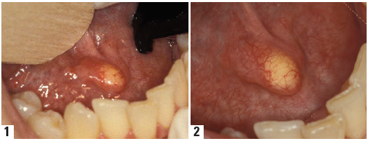
The technique for the removal of a soft tissue Fibroma on the oral mucosa of the right cheek is a simple but effective procedure that will appeal to the general dentist. It requires very few instruments and can be performed easily in the dental office. I encourage my fellow dental colleagues to study the following procedure and to utilize it when presented with a similar case.
I would also point out that any tumor of this nature that is present in a patient who is to undergo fabrication of dentures, operative dentistry, crown and bridge and implants, be performed first. This will eliminate the possibility of the fibroma interfering in any way with the proper fit of a denture, bridge, fillings and implants. Manipulation of prosthetic material may traumatize existing tumors or excess tissue and cause additional problems such as excess bleeding, ulceration and infection. It is important for those involved in all dental procedures, such as the dentist, dental technologist, denture therapist and dental hygienist to recognize that surgical tumors should be attended to first before other work is accomplished.
Technique For The Surgical Removal Of A Soft Tissue Fibroma
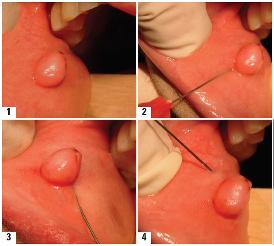
- Local anesthesia is accomplished by utilizing and performing a Field Block. A Field Block is accomplished by injecting the anesthetic around the periphery of the fibroma in the form of an enclosed circle. The anesthetic is also deposited under the base of the tumor as well. This approach not only serves to anesthetize the tumor and prepare it for removal, but the vaso-constrictor serves to reduce the amount of bleeding that accompanies the surgery. Less bleeding will allow for better access and visibility, providing a much drier field in which to work. Figures #1, 2, 3, 4.

- The fibroma, approximately 1 cm. in diameter, is held at its base using an Allis forcep #5237, Figure #5
- Dissecting scissors, Kelly #4001, are used to make a cut below the base. Holding the Allis forcep in a taut fashion, the entire tumor is removed with one or two clean cuts of the scissors. Figures #6,7,8, 9. The scissors must be extremely sharp or tearing and jagged edges of the tissue will ensue. This may leave portions of the fibroma still attached to the mucosa and a possible recurrence. The use of a scalpel and blade is discouraged as it does not have the accuracy, control and stability that the scissors possess with this type of surgical excision. In actual fact, a good pair of sharp scissors really represent two scalpel blades fixed together with a screw. In this case, two is better than one and one is insufficient.

- Following removal of the fibroma, the surgical site is sutured with 2-0 gut sutures on a cutting needle. Non-cutting needles should not be used as they may tear and lacerate the mucosa. Figures # 10,11.In this case, three sutures were used to reposition and close the surgical site.

- The fibroma is now sent for pathological examination. Removal of pathological tissue of this nature is never discarded. The specimen is always sent to an oral pathologist to determine its exact microscopic identity. Oral cancer must be confirmed or ruled out. This is to be considered an essential part of the entire surgical procedure.
- The patient is seen one week following surgery for assessment and to follow the surgical healing. Gut sutures will usually be biodegraded on this second visit.
Disclaimer from Guident
The views and opinions expressed in articles published in Guident are solely those of the authors and do not necessarily reflect the views of the editors, the editorial board or the publisher, Ivory India. Guident does not endorse any product, technique or treatment described. Clinical content is intended for qualified dental and medical professionals and is not a substitute for individual clinical judgement. Guident, its editors and the publisher accept no liability for any consequence arising from the use of information published in the journal.
How to cite this article
Dr. Lawrence I. Gaum. Soft Ti Ssue Oral Surgery the Removal of a Fibroma. Guident. 2013 Feb;6(4). Available from: https://ivoryindia.in/articles/oral-surgery/soft-ti-ssue-oral-surgery-the-removal-of-a-fibroma.html