Surgical Removal of a Stone in the Submandibular Duct
Mississauga, Ontario.
Many general dentists today are doing more and more surgical procedures. These ambitious colleagues are also providing their patients with more endodontics, periodontics, crown and bridge, implants and restorations. They are constantly and successfully taking continuing educational courses and I commend them for this effort to not only keep abreast of the latest and best that is being offered, but to also strive to go beyond the cutting edge in many ways. I witness many of these individuals attending my seminars in oral surgery and I do my utmost to teach them the “Bread and Butter” surgical procedures that will prove to be so valuable on a day-to-day basis in their offices.
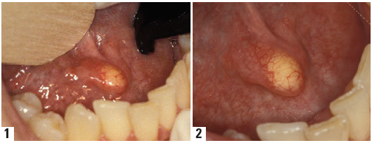
In keeping with my quest to teach oral surgery to my fellow G.P. colleagues, I will present a case here that can and should be accomplished by a dentist in his/her office. The following case illustrates a large stone in the submandibular duct, on the right hand side. As the photos illustrate, the stone can be easily visualized and is close to the opening of the duct in the floor of the mouth. Photos #1, #2
STEP #1
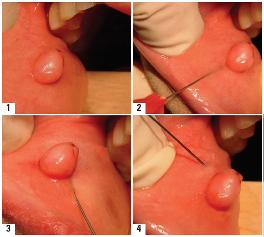
The first step in the removal of the stone is to place a 2-0 gut suture distal to the stone and securely tying the suture around the duct in a loop wise fashion. The purpose of this suture is quite obvious. Without this suture, the stone may begin to slip distally within the duct and end up deeper and closer to the Submandibular gland. If this occurs, the simple and routine removal of the stone may now require a much more involved surgical approach that may necessitate that it be completed in a hospital setting. One must be cautioned in not allowing this mishap to occur. The assistant now holds the loose ends of the suture in an upright or “ lifting fashion” to allow greater visualization and access to the stone. Photo #3

STEP #2
Using a #15 blade, an incision is made through the mucosa and the duct, on the superior portion of the stone. In this case, because the stone is very superficial and visible, the incision is very shallow in order to expose the stone and a deep incision is not required. Picture #4

STEP #3
Using a spoon curette, gently “tease” the stone from its soft tissue covering until it is completely free from the duct and mucosa. The curettage action should be performed carefully so as not to traumatize or tear the very small and fragile duct and thin mucosa. Pictures #5,#6


STEP #4
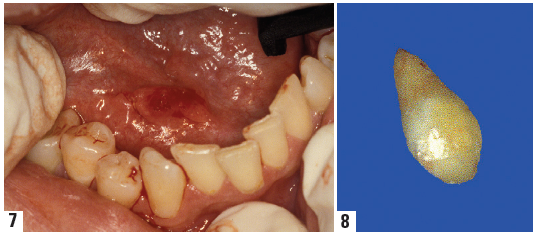
Flush and clean the surgical site with clean water to remove any debrisassociated with the stone that may still be present. Note that the stone is large and shaped like a supernumery tooth. (Pictures #7, #8)
Using a small suture such as 3-0 gut, on a reverse cutting needle, 3/8 inch, the mucosa and duct are sutured to reposition the tissues
The stone should be sent to an oral pathologist for identification.
Disclaimer from Guident
The views and opinions expressed in articles published in Guident are solely those of the authors and do not necessarily reflect the views of the editors, the editorial board or the publisher, Ivory India. Guident does not endorse any product, technique or treatment described. Clinical content is intended for qualified dental and medical professionals and is not a substitute for individual clinical judgement. Guident, its editors and the publisher accept no liability for any consequence arising from the use of information published in the journal.
How to cite this article
Dr. Lawrence I. Gaum. Surgical Removal of a Stone in the Submandibular Duct. Guident. 2013 Mar;6(5). Available from: https://ivoryindia.in/articles/oral-surgery/surgical-removal-of-a-stone-in-the-submandibular-duct.html