Management of a Maxillary Lateral Incisor Associated with Talon Cusp Using Multidetector Computed Tomography
Department of Orthodontics and Dentofacial Orthopedics, Department of Conservative Dentistry and Endodontics, Department of Prosthodontics, Manipal College of Dental Sciences, Manipal University, Manipal, Karnataka, India
Abstract
The complicated root canal system seen in special cases such as talon cusp may produce substantial diagnostic, treatment planning and procedural challenge to the dentist. Newer diagnostic aids such as computed tomography (CT) can be valuable diagnostic tool to ascertain root canal morphology in these cases. This article reports a case where interdisciplinary approach assisted with multidetector computed tomography was used to manage a talon cusp affecting the maxillary lateral incisor.Key words: Dental anomaly, diagnosis, root canal morphology, multidetector computed tomography, talon cusp, tooth displacement
INTRODUCTION
Talon cusp is a rare dental anomaly which occurs as an additional cusp-like structure from the cingulum area or cemento-enamel junction of the maxillary or mandibular anterior teeth in either primary or permanent dentition. It was first described by Mitchell in 1892 and was named as ‘Talon cusp’ due to its resemblance to an eagle’s talon.1 This anomalous structure is composed of normal enamel and dentin and either has varying extensions of pulp tissueinside it or is devoid of a pulp horn.2
The prevalence of this relatively rare odontogenic anomaly is lowand has been reported ranging from 0.06 to 7.7% in the literature.2The maxillary lateral incisors are the most commonly affected (67%) followed by the central incisors (24%) and canines (9%).3The exact etiology of talon cusps is still unknown; however, a multifactorial etiology has been suggested, involving genetic and environmental factors.4,5
A large talon cusp may be associated with clinical problems such as compromised aesthetics, occlusal interferences, displacement of tooth, caries, periodontal problems and irritation of the tongue and neighboring oral tissues during speech and mastication, breast feeding difficulties and even temporo-mandibular joint disordersresulting from traumatic occlusal forces.6,7 The presence of these accessory cusps may prevent proper bonding of brackets for orthodontic treatment and complicate the wearing of a mouth guard during sports activities.
The clinical management of this rare anomaly demands optimal treatment planning based on careful clinical judgment and radiographic examination. Conventional intraoral periapical radiographs are frequently used in endodontics for assessing the root canal morphology. However, in certain clinical situations such as taloned tooth, they may not provide sufficient information about the complicated morphology of the root canal system. In such clinical conditions, a newer advanced diagnostic aid such as computed tomography (CT) may be a valuable diagnostic tool which produces 3-dimensional (3D) images of individual teeth and the surrounding tissues. Here we present a case where interdisciplinary approach assisted with multidetector computed tomography (MDCT) was used to manage a talon cusp affecting the maxillary incisor.
CASE REPORT
A 24 year old female reported to our department with the chief complain of forwardly placed upper right lateral incisor. The patient was concerned about her facial appearance and also complained of interference during speech and mastication.
Clinical examination revealed a talon cusp on the palatal surface of the upper right lateral incisor extending more than the half way from the cementoenamel junction to the incisal edge. The talon cusp was pyramidal in shape and measured 6.89 mm in length (incisocervically) and 4.24 mm in width (mesiodistally). All the measurements were performed by using an electronic digital caliper. The cusp tip was pointed which might be reason of discomfort during speech and mastication. The talon cusp was diagnosed as type 1. Non-carious developmental grooves were present at the junction of the talon cusp and the palatal surface of the tooth (Fig 1a). The premature occlusal contact was noted between the affected teeth and the opposing lower lateral incisor. This occlusal interference was probably responsible for the labial displacement of the anomalous tooth (Fig 1b).
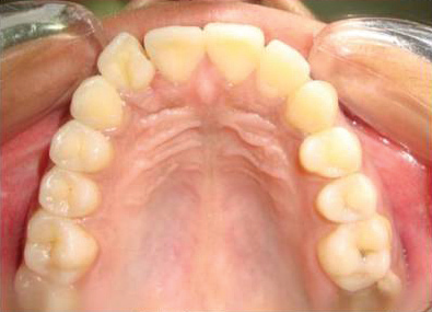
Fig 1a: Occlusal view of maxillary dental arch showing pyramidal shaped talon cusp of rigth lateral permanent incisor cusp tip extending more than half length of crown gingivo-incisally
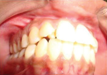
Fig 1b: premature occlusal contact between the talon cusp of maxillary rigth permanent lateral incisor an opposing lower rigth lateral incisor with labial displacement of affected maxillary incisor

Three dimensional reconstruction of CT section showing premature occlusal contact between the taloned tooth and the opposing lower lateral incisor
The taloned tooth responded normally to percussion, electric (Parkell electric pulp tester; Parkell Inc., Farmingdale, NY, USA) and thermal (ENDO-ICE; Coltene/ Whaledent Inc., Cuyahoga Falls, OH, USA) pulp tests. Localized gingival recession, marginal gingivitis and attrition were noted in relation to the opposing lower right lateralincisor. There was no history of trauma or any hereditary conditions.Medical history was noncontributory.
Periapical and upper occlusal radiograph revealed an invertedV-shaped radio opaque structure superimposed on the image of theaffected crown. The extent of pulp tissue into the talon cusp could not be determined radiographically (Fig 2a). Subsequent to obtainingan informed consent from the patient, a 3-D imaging using 64-slice multidetector computed tomography (MDCT) scanner (Brilliance CT 64-channel, Philips Healthcare, Andover, MA, USA) was plannedin order to confirm the morphology and extension of pulp horn in the talon’s cusp. A MDCT scan was performed and images were acquired using dental software program DentaScan (GE Healthcare,Milwaukee, WI, USA). Axial slice of the cervical third section of the crown of the maxillary right lateral incisor showing presence of pulp tissue in the talon cusp (Fig 2b). Sagittal view revealed two root canals (labial and palatal) joining in the middle third of the root (Fig 2c). Distance from the tip of the talon cusp to the pulp space was measured 3.6 mm in MDCT image.


Fig 2b: Axial view of MDCT image of the cervical third section of the crown of the maxillary right lateral incisor showing presence of pulp tissue in the talon cusp.

Fig 2c: Sagittal view of MDCT image showing two root canals (labial and palatal) joining in the middle third of the root of the upper right lateral incisor.
TREATMENT ALTERNATIVE
In order to attain a pleasant esthetics and functional improvement two treatment alternatives were proposed
a. A gradual reduction of the talon cusp in multiple visits along with orthodontic alignment of the affected tooth
b. Complete reduction of the talon cusp followed by root canal treatment and orthodontic alignment of the affected tooth
Patient preferred the first treatment option as it was less invasive procedure compared to second treatment alternative.
TREATMENT PROGRESS
The occlusal interference on the talon cusp was determined with the help of a articulating paper (Samit product, India) in both centric occlusion and in protrusive and lateral movements. The talon cusp was reduced using a diamond bur in a high-speed air turbine hand piece (NSK, PANA AIR ; Nakanishi INC). The cusp was ground more on the sides than the cusp tip area. No exposure of pulp tissue occurred during this procedure. After grinding, dentine was covered with an adhesive bonding agent (OptiBond FL, Kerr Corp, Orange, CA, USA). A Hawley’s appliance was placed and labial bow was activated to align the upper lateral incisor. Patient was asked towear the appliance 16-18 hours perday. (Fig 3) Six weeks later itwas found that upper lateral incisor had moved back and startedcontacting the lower incisor. Therefore, the talon cusp was furtherreduced using the same protocol followed initially and labial bowwas reactivated to continue with the alignment procedure. After a period of 12 weeks the lateral incisor was aligned. A total reduction of approximately 2 mm was performed without exposing the pulp or compromising the vitality of the tooth. The fixed retainer was placed. (Fig 4) There was no sensitivity in the affected tooth and it responded normally to thermal pulp test. The gingival inflammation in relation to the opposing lower incisor was also reduced post treatment.


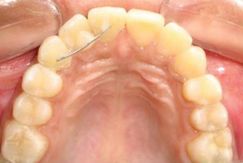
Fig 4: Realignment and retention of maxillary rigth permanent lateral incisor.
a ) Labially displaced maxillary rigth permanent lateral incisor aligned to a lateral incisor normal occlusion position. b ) Retention of aligened incisor with fixed bonded retainer
DISCUSSION
In the case presented here, talon cusp was found to be associated with the right maxillary permanent lateral incisor. The maxillary permanent lateral incisor is frequently found to be associated with various dental anomalies. The increase susceptibility of the lateral incisors to dental anomalies could partly be related to compression of the tooth germ of the lateral incisor by the adjacent central incisor and canine, which develops about 7 months earlier than the
lateral incisor. Increased localized external pressure on a tooth germ during the morphodifferentiation stage may result in outfolding of the dental lamina leading to dental abnormalities.8
The presence of a talon cusp can cause significant functional and aesthetic problems in the patients as well as substantial diagnostic, treatment planning and procedural challenge to the dentist. A talon cusp can be identified at an early stage of the formation of unerupted permanent teeth during a routine radiographic examination of the primary dentition. An unerupted talon cusp may be misinterpreted as compound odontome or a supernumerary tooth leading to unnecessary surgical intervention. This is extremely significant because approximately 90% of the supernumerary teeth occur in maxilla and half of these in incisor region.7
The presence of a talon cusp is, in itself, not indicative of dental treatment, unless it is associated with clinical problems such as compromised aesthetics, occlusal interferences, displacement of tooth, caries, periodontal problems and irritation of the tongue. In the case reported here, the occlusal trauma caused by the taloned tooth, displaced the upper right lateral incisor and produced localized gingivitis, gingival recession and attrition in the opposing lower lateral incisor. Patient also had difficulty while speaking. Traumatic occlusal interference over a period of time can cause bone loss and destruction of periodontal connective tissue. Such a tubercle is also subject to wear (attrition) or may be fractured from its base during occlusal function leading to indirect or direct pulp exposure with resultant loss of vitality.
A tooth with talon cusp is commonly thicker faciolingually and wider mesiodistally which prevents the orthodontic alignment of the displaced tooth into an ideal overbite/overjet relationship. The displaced tooth is also prone to accidental trauma. Therefore, in the case presented here, it was decided to reduce the bulk of talon cusp to prevent occlusal trauma followed by orthodontic alignment of the displaced tooth.
The treatment of talon cusp depends upon whether the cusp contains or is devoid of a pulp horn. Most talon cusps contain extensions of pulp tissue similar to pulp horns.9 Therefore, careful clinical and radiographic examination are essential for optimal treatment planning. Conventional intraoral periapical radiographs are an important diagnostic tool in endodontics for assessing the root canal configuration. However, it represents a two dimensional of a three dimensional object which has its inherent limitations. They are of rather limited value in cases with complex root canal anatomy. In case of taloned tooth, the talon cusp and its pulp extension are superimposed over the main pulp chamber; the extent of the extra pulp horn is difficult to distinguish in intra oral periapical radiograph. These problems might be overcome by using newer diagnostic methods such as spiral CT, MDCT or volume acquisition CT, which can produce 3D images of the root canal morphology and offer new perspectives for dental imaging of special clinical cases. A MDCT or volume acquisition CT can generate sub millimeter spatial resolution images of high diagnostic quality with radiation dose significantly lower than the conventional CT. However due to high cost its use should be limited to the cases with unusual tooth morphology.10
In this special case, we have used MDCT for better understanding the morphology of the root canal system. The MDCT images confirmed the presence of pulp tissue inside the talon cusp. There was sufficient dentine and enamel thickness overlaying the pulp horns. Therefore it was decided to reduce the talon cusp gradually at 6-8 week intervals to allow the deposition of reparative dentin to preserve pulpal vitality. Consideration should be given to where the majority of the odontoblasts are located and the effect on them when the tip of the cusp is ground. Because most of the odontoblasts lie along the length of the cusp, grinding should be performed on the sides of the cusp to initiate the formation of reparative dentine. For the same reason, in this case, the talon cusp was ground more on the sides than the cusp tip area. The other option could have been the complete reduction of the talon cusp followed by the root canal treatment. However this approach was not preferred by the patient as it was more radical in nature.
CONCLUSION
The presence of a talon cusp can cause significant functional and aesthetic problems in the patients as well as substantial diagnostic, treatment planning and procedural challenge to the dentist. A newer diagnostic aid such as MDCT can be a valuable diagnostic tool to ascertain root canal morphology in special cases such as talon cusp.
REFERENCES
- Schulze C. Developmental abnormalities of the teeth and jaws. In: Gorlin RJ, Goldman HM, eds. Thoma’s Oral Pathology. 6th ed. Vol. I. St Louis, Mo: CV Mosby; 1970:p.96–97.
- Chawla HS, Tewari A, Gopalakrishnan NS. Talon cusp: a prevalence Study. J Ind Soc Pedod Prev Dent 1983; 1:28-34.
- Jowharji N, Noonan RG, Tylka JA. An unusual case of dental anomaly: a facial talon cusp. J Dent Child 1992; 59:156-158.
- Hattab FN, Yassin OM. Bilateral talon cusp on primary central incisors:a case report. Int J Paediatric Dent 199;6:191-195.
- Hattab FN, Yassin OM, Al-Nimri KS. Talon cusp in the permanent dentition associated with other dental anomalies:Review of literature and reports of seven cases. J Dent Child 1996; 63:368-376.
- Sicher S, Bhasker SN, Orban S. Oral Histology and Embryology, 7th edn. St Louis, MO: C. V. Mosby Co., 1972.
- Soares AB, de Araujo JJ, de Sousa SM, Veronezi MC. Bilateral taloncusp: a case report. Quintessence Int 2001;32:283-286.
Disclaimer from Guident
The views and opinions expressed in articles published in Guident are solely those of the authors and do not necessarily reflect the views of the editors, the editorial board or the publisher, Ivory India. Guident does not endorse any product, technique or treatment described. Clinical content is intended for qualified dental and medical professionals and is not a substitute for individual clinical judgement. Guident, its editors and the publisher accept no liability for any consequence arising from the use of information published in the journal.
How to cite this article
Dr.Santosh Kumar, Dr. Sunil Gupta, Dr. Priyanka Jayaswal. Management of a Maxillary Lateral Incisor Associated with Talon Cusp Using Multidetector Computed Tomography. Guident. 2013 Mar;6(5). Available from: https://ivoryindia.in/articles/orthodontics/management-of-a-maxillary-lateral-incisor-associated-with-talon-cusp-using-multidetector-computed-tomography.html