Analysis of Mandibular Behaviour in Patients with Cleft Lip and Palate: A Cephalometric Study
Department of Orthodontics and Dentofacial Orthopaedics, Punjab University, Chandigarh, India, (Sr. Professor & H.O.D), Department of Orthodontics and Dentofacial Orthopaedics, A B Shetty Memorial Institute of Dental Sciences, Mangalore, India. Dr. Neelen Shah( P. G. Student)
ABSTRACT
Aims: The aim of the study is to evaluate the mandibular behaviour in left lip and palate patients when compared to a control group through lateral cephalometric radiographs
Methods and Material: The lateral cephalograms of Thirty eight adult patients with cleft lip and palate were taken. The lateral cephalograms of forty five adult individuals with normal occlusion and without cleft lip and palate was used as the control group. Various parameters both linear and angular were used to evaluate the changes in the anterior cranial base, morphology of the mandible (five parameters) and the altered spatial positioning of the mandible in relation to the anterior cranial fossa ( six parameters).
Statistical analysis used: Two way analysis of variance was used to observe the difference between group means. Comparisons between the cleft group and control group were undertaken with large sample test.
Results: The results showed a short anterior cranial base, shorter mandibular ramus, body and total length, a more obtuse gonial angle, a vertical growth pattern, and the spatial relation of the mandible to the cranial base showed a downward and backward mandibular rotation in cleft lip and palate patients.
Conclusions: Hence, this present study concludes that a thorough knowledge of the anatomy and physiology of the naso-oro-pharyngeal cavities supplemented with the knowledge of the growth and age changes and how they relate to the facial appearance and oro-pharyngeal function is pertinent for the rehabilitation of a cleft lip and palate patient.
Key-words: Cleft lip and palate, mandibular morphology, spatial position of mandible
Key Messages: Care for the individual with a cleft continues throughout the life time that individual and the orthodontist plays a key role in the rehabilitation of these otherwise normal individuals. With the in-depth knowledge of the oro-facial structures, the levels of clinical success can be improved a lot.
INTRODUCTION
Cleft lip and palate is one of the most common congenital anomalies of the face and an orthodontist plays an integral role in the multidisciplinary team approach required for the management of these patients. With a cleft lip and palate patient, the tendency is to concentrate on the growth of the maxilla and to forget that the mandibular growth and its direction are equally important and may enhance or hurt efforts to achieve good facial profile relationships as well as desirable occlusal relationships. The position, growth and configuration of the lower jaw are equally important to the aesthetic appearance of the face.
The objective of the present study is to evaluate the variation of mandibular morphology and spatial position in cleft lip and palate patients when compared to a control group through lateral cephalometric radiographs.
SUBJECTS AND METHODS
Materials for the study were the lateral cephalomatric radiographs of 38 patients who reported to department of orthodontics and dentofacial orthopaedics. All patients displayed cleft lip and palate, all in permanent dentition and without a history of pre-orthodontic treatment. Non growing individuals were selected to standardise the influence of growth.


Cephalometric landmarks (Figure 1)
- Sella (S)
- Nasion (N)
- Gnathion (Gn)
- Gonion (Go)
- Menton (Me)
- Anterior nasal spine (ANS)
- Pogonion (P)
- Supramentale (B)
- Condilion (Co)
Following measurements were made (Figure 2)
- Anterior Cranial Base (SN)
- Structure of Mandible.
A) Go-Gn – Length of the body of the mandible
B) Co-Go – Length/height of the ramus of the mandible
C) Co-Gn – Total length of the mandible
D) P-NB – denotes the antero-posterior position of the chin in relation to the NB line
E) Co-Go-Me – denotes the gonial angle which determines the angular relationship between the body and the ramus.

3) Spatial position of Mandible in relation to Anterior Cranial Fossa
A) S-Go – Posterior facial height
B) N-Me -- Anterior facial height
C) SNB -- Anterio-Posterior relation of mandible to cranial base
D) Sn-Go-Gn -- Angular relation of mandibular plane to anterior cranial base
E) ANS-Me -- Lower anterior facial height
F) SN-Gn -- Y-axis
Two way analysis of variance was used to observe the difference between group means. Comparisons between the cleft group and control group were undertaken with large sample test.
RESULTS


DISCUSSION
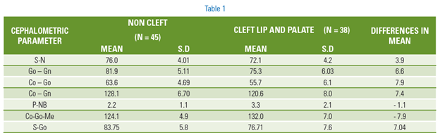
Analysis of the results shows significant difference in structure and spatial relation of the mandible in cleft lip and palate individuals when compared to the non-cleft individuals. This is in agreement with Jain1, Horowitz2 and Bishara.3
In the cleft group the mandibular dimension at the level of the ramus (S-Go, Co-Go), the body (Go-Gn) and the total length (Co-Gn) was found to be smaller. The mandible showed besides the short ramus and body length, a more vertical growth pattern. These findings can be confirmed by decreased posterior facial height (S-Go), greater anterior facial height (N-Me), more obtuse gonial angle (Co-Go-Me) and a downward and backward rotation of the mandible relative to the cranial base (Sn-Go-Gn).This is in agreement with the previous studies done by Hayashi4, Smahel5, Clarke6, Roth7 and Rantha8 and many other similar studies.
Concerning the antero-posterior spatial relationship of mandible to the cranial base (SNB), cleft group showed a significant posterior repositioning of the mandible when compared to subjects without clefts. This is due to greater downward and backward rotation of the mandible, leading to a more posterior position of landmark B. During the mixed dentition period when the maxillary segments are displaced medially, tongue cannot be accommodated in its normal position in the palate. The position acquired by the tongue becomes decisive for the pattern of further development of the maxilla as well as the mandible. Here if the nasal respiration is impeded the tongue may assume a low position to facilitate normal respiration. If the tongue in the low position does not rest under the occlusal surface of the maxillary teeth, the alveolar height will increase, resulting in a progressive lowering of the mandible, a more open gonial angle and a more retruded position of the chin. The shorter mandibular length is being attempted to be compensated by a short cranial base length. Besides the shorter ramus and body lengths, a more vertical growth pattern is seen in cleft group.
Not only is the extent or amount of lower jaw growth but also the direction of mandibular growth is important and deserves equal consideration while deciding what orthodontics can do to improve the function as well as the facial appearance of the cleft lip and palate individual. Clinically, such knowledge can be helpful in estimating the eventual mandibular position following growth and providing improved insight into treatment modalities that might be best suited for any individual patient. In formulating a treatment plan of any growing patient, it is mandatory for the operator to know about the growth and development of the craniofacial skeleton and thus predict with various methods as to what the final outcome will be without any treatment. For growth predictions in cleft lip and palate cases, one needs to know the altered pattern of growth which has taken place in all dimensions.
A cleft lip and palate patient usually show a retrognathic or concave profile and is managed with a reverse pull headgear with simultaneous palatal expansion appliances. In such cases, it is to be determined, whether the mandible should be held back during the above treatment and if so, how long to achieve an acceptable maxillo - mandibular relationship and hence an acceptable profile. Usually, in non growing patients who need an orthodontic - orthognathic approach to correct the concave profile, the morphology and spatial position of maxilla and mandible should be known to determine, how much to decompensate, which arch to do surgery, in which direction and the amount of push back or advancement to be made.
CONCLUSION
It is believed that there are many roads leading to Rome, hence it is to be accepted that many different treatment strategies and techniques may produce good results. Choosing from more than one approach may open the door to a better understanding of the various treatment strategies and their applications. This option should encourage us to explore more unorthodox ideas and approaches.
Care for the individual with a cleft continues throughout the life time of that individual. Levels of clinical success have improved a lot over the years and achieving ideal results is becoming a more frequent possibility. Orthodontics in all ages plays a valuable role in striving to achieve ideal results in the rehabilitation of the cleft lip and palate individual.
REFERENCES
- Jain, Krogman. Cleft types and sex differences in craniofacial growth in clefting from one month to ten years. Cleft Palate J 1983; 20:238-45.
- Horowitz, Graff. Factor analysis of craniofacial morphology in complete bilateral cleft lip and palate. Cleft Palate J 1980; 17:234-44.
- Bishara et al. Longitudinal changes in the dentofacial relationship of unilateral cleft lip and palate subjects. Cleft Palate J 1979; 16:391-401.
- Hayashi, Sakuda. Craniofacial growth in complete unilateral cleft lip and palate: A roentgeno-cephalometric study. Cleft Palate J 1976; 13:215-37.
- Smahel et al. Craniofacial morphology in isolated cleft palate prior to palatoplasty. Cleft Palate J 1987; 24:200-8.
- Clarke, Hepworth. Chondrodysstrophic mice with coincidental agnathia: Evidence for the tongue obstruction hypothesis in cleft palate. Teratology 1988; 38:565-70.
- Roth, Krkosk. The adaptable mandible: A product of the relative osteo-neural growth . Gegenbaurs morphol jahrb 1978; 124:765-83.
- Ranta, Altoneu. A comparative study of gonial angles and growth of the lower third molar in cleft lip and palate: An orthopantamographic study. Scand J Plast Reconstr Surg1978; 12:273-8.
Disclaimer from Guident
The views and opinions expressed in articles published in Guident are solely those of the authors and do not necessarily reflect the views of the editors, the editorial board or the publisher, Ivory India. Guident does not endorse any product, technique or treatment described. Clinical content is intended for qualified dental and medical professionals and is not a substitute for individual clinical judgement. Guident, its editors and the publisher accept no liability for any consequence arising from the use of information published in the journal.
How to cite this article
Dr. Arun Garg, Dr Harvansh Singh Judge Institute Of Dental Sciences & Hospital, Dr. U.S.Krishna Nayak Dean. Analysis of Mandibular Behaviour in Patients with Cleft Lip and Palate: A Cephalometric Study. Guident. 2013 Apr;6(6). Available from: https://ivoryindia.in/articles/orthodontics/analysis-of-mandibular-behaviour-in-patients-with-cleft-lip-and-palate-a-cephalometric-study.html