Management of Impacted Maxillary Canine
Department of Orthodontics, Department of Orthodontics, Department of Public Health Dentistry, Manipal College of Dental Sciences, Manipal, India
Abstract
The permanent canines are the foundation of an esthetic smile and functional occlusion. Factors that interfere with its development and eruption had serious consequences on esthetics, function and stability of stomatognathic system. Impacted canines are those with a delayed eruption time or that are not expected to erupt completely based on clinical and radiographic assessment. Management of the impacted canine is one of the greatest challenges for orthodontist. Success of the treatment depends upon patient cooperation, age of patient, proper diagnosis, level of canine impaction, inclination and depth of impaction, amount of root formation, type of exposure of tooth, amount of bone removal, type of attachment and method of orthodontic traction. All these parameter plays important role when managing impacted canines to achieve good canine alignment in the arch with canine guided occlusion, gingival level, and integrity of periodontium. This article briefly presents the incidence, etiology, diagnosis, general steps of mechanotherapy, treatment alternatives and complications of untreated impacted canines along with two case reports.
Key words: Impacted canine, forced eruption, flap designs.
INTRODUCTION
The ectopic eruption and impaction of maxillary permanent canines is a frequently encountered clinical problem. The diagnosis and treatment of this problem usually requires the expertise and cooperation of the general practitioner, the pediatric dentist, the oral surgeon and the periodontist, as well as the orthodontist. The aim of this article is to briefly present the incidence, etiology, diagnosis, general steps of mechanotherapy, treatment alternatives and complications of untreated impacted canines along with two
CASE REPORTS
When can a tooth considered to be impacted?
Under normal conditions, tooth erupts with a developing root and with approximately ¾ of its final root length. Hence in simple terms, a tooth can be considered impacted if its root is completed but still has not erupted. Applying this concept to maxillary and mandibular canines, they generally have their ¾ root completed by 11-12 years and 9-10 years respectively.1
According to Shafer, Hine and Levy2, impacted teeth are those which are prevented from erupting by some physical barrier in the eruption path.
Incidence
Dachi and Howell3 reported that the incidence of maxillary canine impaction is 0.92%, whereas Thilander and Myrberg4 estimated the cumulative prevalence of canine impaction in 7- to 13-year-old children to be 2.2%. Ericson and Kurol5 estimated the incidence at 1.7%. Impactions are twice as common in females (1.17%) as in males (0.51%). Of all patients with maxillary impacted canines, it is estimated that 8% have bilateral impactions. The incidence of mandibular canine impaction is 0.35%.
Etiology
In general, the causes for retarded eruption of teeth may be either generalized or localized.6 Generalized causes include endocrine deficiencies, febrile diseases, and irradiation. The most common causes for canine impactions are usually localized and are the result of any one, or combination of the following factors: (a) tooth sizearch length discrepancies, (b) prolonged retention or early loss of the deciduous canine, (c) abnormal position of the tooth bud, (d) the presence of an alveolar cleft, (e) ankylosis, (f ) cystic or neoplastic formation, (g) dilaceration of the root, (h) iatrogenic origin, and (i) idiopathic condition with no apparent cause.
Diagnosis
- Inspection: It has been suggested that the following clinical signs might be indicative of canine impaction: (1) delayed eruption of the permanent canine or prolonged retention of the deciduous canine beyond 14 to 15 years of age, (2) absence of a normal labial canine bulge, (3) presence of a palatal bulge, and (4) delayed eruption, distal tipping, or migration of the lateral incisor.
- Palpation: Bulge of permanent canine could be palpated buccally above the deciduous canine 2-3 years before its eruption; it should be palpated deep above attached gingiva in the sulcus where mucosa reflects. Palpation should be done in abnormal locations after getting clue from inspection. Deciduous canine should be checked for mobility.
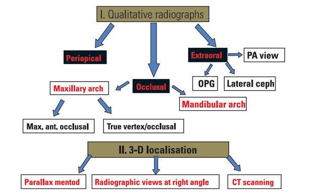
- Radiography7:
General steps of mechanotherapy
- Leveling and alignment of the erupted teeth
- Creating enough space for the impacted tooth and maintaining it
- Conversion of the arch into a rigid anchorage unit
- Surgical exposure of the crown of the impacted tooth and attachment bonding
- Application of low force (60-80g.) traction from rigid anchorage unit
- Final detailing and retention
Treatment Alternatives
- No treatment, if the patient does not desire it. Since the long term prognosis of deciduous canine is poor as its root may eventually resorb, it should be periodically evaluated.
- Auto transplantation of the canine.8
- Extraction of impacted canine and moving premolar in its position.9
- Extraction of the canine & posterior segmental osteotomy to move the buccal segment mesially to close the residual space.
- Prosthetic replacement of the canine, not amendable for juvenile cases.
- Most desirable approach is surgical exposure of the canine followed by orthodontic treatment.
- Morbidity of deciduous canine
- Cystic change
- Permanent canine crown resorption
- Resorption of roots of adjacent teeth.
Complications of untreated impacted canines 10
CASE REPORT 1
A 16-year old girl came to the Department of Orthodontics and Dentofacial Orthopedics with the chief complaint of forwardly placed upper anterior teeth. She was physically healthy and had no history of medical or dental trauma. No signs or symptoms of temporomandibular joint dysfunction or trauma were noted at the initial examination. The extraoral clinical examination showed a convex profile, without any gross asymmetry [Fig 1].
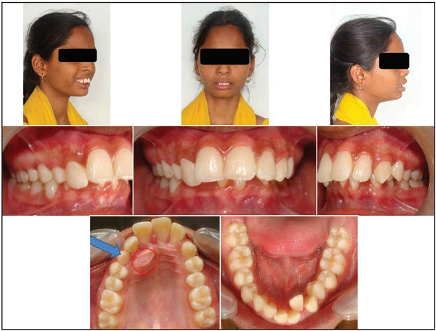
Fig1: Pre-treatment Photographs
The intraoral examination showed Angle Class II division 1 malocclusion with persisted right deciduous canine. The permanent right canine was not seen in the arch. Upper midline was shifted to the right by 3 mm and lower midline was also shifted to the right by 2 mm. Overbite was 90% with an exaggerated curve of Spee of 3 mm. Palatal bulge was identified in the maxillary right palatal region suggestive of the position of the impacted canine [Fig 1]. Cephalometrically, the patient had a Class II skeletal relationship

Fig 2: Pre-treatment OPG
with average growth pattern. The panoramic radiograph [Fig 2] showed all permanent teeth including developing third molar buds and maxillary right canine. The right canine was mesially inclined toward the midline and was apparently overlapping the distal aspect of the maxillary lateral incisor. Periapical radiographs were taken with the Clarks tube shift technique to confirm the palatal position of the right impacted canine. Occlusal X-ray was also done to co-relate with the clinical findings.
Treatment objectives
1. The treatment plan was to obtain space for the retraction of upper anterior teeth and extrusion of the impacted canine.
2. The midline had to be corrected and the arches leveled by intrusion of the anterior teeth.
3. In addition, it was planned to surgically expose the impacted right canine and bond attachment to it and erupt it carefully into the arch in its normal position and align it.
4. The final objective was to achieve a stable functional occlusion with normal overjet and overbite, Class I canine and Class II molar relationship.
5. Attaining a pleasing profile.
Treatment progress
Initially, 53 and 24 were extracted and the molars were banded and all the other remaining teeth were bonded with 0.022 × 0.028 inch pre-adjusted edgewise appliance. After initial leveling and aligning, a 0.019 × 0.025 inch stainless steel arch wire was placed in the maxillary arch. The crown of an impacted canine was exposed using wide flap, but with removal of minimal bone. An attachment with pigtail ligature was bonded and the flap is fully sutured back to its former place [Fig 3]. The maxillary right premolar had been
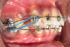
extracted which till now had also served as a space maintainer for the impacted canine and prevent any space loss. The pigtail ligature was ligated to the main 0.019 × 0.025 inch stainless steel arch wire and progressively activated to maintain a force of 60–90 g. The pull on the canine was directed distally toward the extraction space which not only caused the eruption of the canine but also corrected its mesio-distal inclination. This also pulled the canine away from the root of the lateral incisor. The duration of the traction was 7 months which elapsed between the application of the traction and the eruption of the cusp of the impacted canine. The bracket on the labial aspect of the canine was inverted to correct the torque and ligated to the auxillary 0.016 inch NiTi wire and progressively increased to 0.019 × 0.025 inch stainless steel wire. At the end of traction, the palatally impacted maxillary right canine was positioned into proper alignment, resulting in a pleasing smile [Fig 4].
DISCUSSION
According to the criteria put forth by Lindauer et al11, the present case was diagnosed with a palatally impacted right canine. It consisted of the fact that root formation had been completed for

Fig 3: Closed Eruption Technique. a) Surgically raised palatal flap., b) Attachment bonded to tooth., c) Flap sutured back

Fig 4: Treatment in progress
both right and left canines. It is also noted that left canine has erupted for more than 6 months in the oral cavity without any signs of eruption of the right canine. Power and Short12 predicted the chances of canine impaction based on calculating the angulation of the canine on an orthopantomograph; the angulation of the long axis of the canine to the midline may be monitored from the time root formation is one-third complete to 6 months after root completion, 10–13 years of age. If the tooth is angled more than 31° to the midline, its chances of eruption even after deciduous extraction are decreased. Exactly 91% of the ectopically erupting canines come into proper occlusion if the canine crown was distal to the midline of the lateral incisor at the time of removal of the deciduous canine.13 If the crown was mesial to the midline of the lateral incisor root, spontaneous incisor eruption occurred in only 64% cases.13 In the present case, the impacted maxillary canine was 25° to the midline in the pre-treatment stage which is in agreement with the criteria put forth by Power and Short12 for successful canine disimpaction; also, the canine crown was distal to the midline of the lateral incisor in agreement with the criteria of Warford et al.13

Fig 5: Pre-treatment Photographs
Correction of torque, labio-palatal root angulation in basal bone, in the impacted canine tooth is important to achieve proper functional occlusion. This has been achieved successfully by the method advocated by McLaughlin et al.14
CASE REPORT 2
A 17-year old boy came to the Department of Orthodontics and Dentofacial Orthopedics with the chief complaint of irregularly placed anterior teeth. He was physically healthy and had no history of medical or dental trauma. No signs or symptoms of temporomandibular joint dysfunction or trauma were noted at the initial examination. The extraoral clinical examination showed a mild convex profile, without any gross asymmetry [Fig 5]. Patient had Angle’s Class I malocclusion with crowding in both arches. Upper midline was coinciding and lower midline was shifted to the left by 3 mm. Complete intraoral examination of the patient revealed missing upper permanent canines following which a panoramic radiograph was advised to look for the permanent canines [Fig 5]. Also, both lateral incisors were in crossbite. The radiograph revealed bilaterally impacted maxillary canines, the right maxillary canine was located between the roots of right lateral incisor and first premolar, whereas the left maxillary canine was located high [Fig 6]. Periapical radiographs were taken with the Clarks tube shift technique to confirm the labial position of both impacted canines.Cephalometrically, the patient had a Class I skeletal relationship with average growth pattern.

Fig 6: Pre-treatment OPG
Treatment objectives
1. The treatment plan was to obtain space in the both arches by the extraction of first premolars. In upper arch space is required for the impacted canines and in lower arch space is required to relieve the crowding.
2. The midline had to be corrected and the arches leveled by intrusion of the anterior teeth.
3. Since the level of impacted right canine was not high so it was planned for natural eruption (after the extraction of first premolar).
4. In addition, it was planned to surgically expose the impacted left canine and bond attachment to it and erupt it carefully into the arch in its normal position and align it.
5. Correction of cross bite in relation to 12 and 22.
6. To achieve a stable functional occlusion with normal overjet and overbite, Class I canine and molar relationship.
7. Maintaining the pleasing profile.
Treatment progress
Initially, 14, 24, 34 and 44 were extracted and the molars were banded and all the other remaining teeth were bonded with 0.022 × 0.028 inch pre-adjusted edgewise appliance. After initial leveling and aligning, a 0.019 × 0.025 inch stainless steel arch wire was placed in the maxillary arch.
The surgical exposure by creating a window had been carried out by the oral surgeon in this stage and the attachment was bonded [Fig 7]. Attached gingival attachment was maintained on the impacted tooth for good periodontal prognosis. The attachment was ligated to the main 0.019 × 0.025 inch stainless steel arch wire and progressively activated to maintain a force of 60–90 g. The pull on the canine was directed distally toward the extraction space which not only

Fig 7: Showing Window Eruption Tecnique., a) A high buccal canine exposed by circular incision of the sulcus mucosa., b) Attachment bonded to tooth
caused the eruption of the canine but also corrected its mesio-distal inclination. This also pulled the canine away from the root of the lateral incisor. The duration of the traction was 6 months which elapsed between the application of the traction and the eruption of the cusp of the impacted canine. At the end of treatment, the buccally impacted maxillary left canine was positioned into proper alignment, resulting in a pleasing smile [Fig 8].
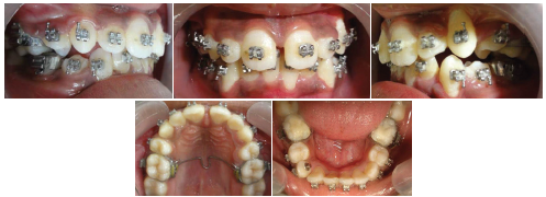
Fig 8: Treatment in progress
DISCUSSION
Maxillary permanent canines are important for an attractive smile and are also essential for a functional occlusion. Therefore, extraction of the canines should be avoided, if at all possible. In the case of maxillary impaction, surgical exposure of the related tooth and the use of fixed orthodontic appliances is the most frequently used treatment alternative as long as the tooth position is favorable. Various methods have been used for moving the canine into proper alignment.9,15-19 Fournier etal9 have proposed the use of a removable plate. Becker and Zilberman16 have recommended the use of a flexible palatal arch slotted into horizontal, soldered, palatal tubes on the molar bands of any type of fixed multibonded appliance. Jacoby17 has suggested his ballista spring to direct a palatal-occlusal force from the buccal side. In this report, the successful treatment of a buccally impacted maxillar canine using the lingual button and E-chain was presented.
Light force mechanics advocated by Becker20 and Kokich etal21 have been successfully used in the present case to bring the buccally impacted canine into the arch, while preventing any adverse effects and non-vitality of the adjacent teeth.
CONCLUSION
Orthodontic treatment of impacted tooth can be very rewarding if we are ready to accept the challenge of anticipating the changes, on the basis of a sound problem list and treatment goals.
Disclaimer from Guident
The views and opinions expressed in articles published in Guident are solely those of the authors and do not necessarily reflect the views of the editors, the editorial board or the publisher, Ivory India. Guident does not endorse any product, technique or treatment described. Clinical content is intended for qualified dental and medical professionals and is not a substitute for individual clinical judgement. Guident, its editors and the publisher accept no liability for any consequence arising from the use of information published in the journal.
How to cite this article
Dr. Ritesh Singla, Dr. Madhumitha N., Dr. Nishu Singla. Management of Impacted Maxillary Canine. Guident. 2013 May;6(7). Available from: https://ivoryindia.in/articles/orthodontics/management-of-impacted-maxillary-canine.html