
Ecto Dermal Dys Plas Ia: An Oral Genodermatosis - Overview of Literature
Faculty Of Dentistry, Jamia Milia Islamia New Delhi, India, Z.A Dental College & Hospitals A.M.U Aligarh, India, Z.A Dental College & Hospitals A.M.U Aligarh, India, Z.A Dental College & Hospitals A.M.U Aligarh, India, Department of Oral Medicine & Radiology
Abstract
The term oral genodermatosis implies to inherited diseases affecting the skin and oral mucosa. There are a large number of inherited and congenital syndromes having oral and dermatological manifestations. However, the recognition of their skin findings is important not only for the initiation of appropriate dermatologic therapy, but also for the detection of other associated abnormalities in these frequently multisystem disorders, including malignancy. (eg. Basal cell nevus syndrome, xeroderma pigmentosum, Peutz Jegher syndrome). Ectodermal dysplasia is an inheritable genodermatosis affecting the ectoderm and is characterized by abnormal development of the skin, hair, nails, sweat glands, and the stomatognathic system. There are more than 170 different types of ectodermal dysplasia reported in the literature. Early dental intervention can improve patient’s appearance,thereby minimizing the associated emotional and psychological problems in these patients. Treatment requires a team work by medical personal along with dental professionals.
Keywords: Genodermatosis, Ectodermal dyslasia, hypodontia, hypohidrosis, prosthetic rehabilitation.
INTRODUCTION
Ectodermal dysplasia is the term used to describe a group of rare, inherited disorder characterized by dysplasia of tissues of ectodermal origin- primarily nails, teeth, skin, and occasionally, dysplasia of mesodermally derived tissues.1 The condition is thought to occur in approximately1 of 100,000 live births.1,2
Clinically, ectodermal dysplasia may be divided into two broad categories: the Hypohidrotic form (X linked recessive) and the hydrotic form (autosomal inherited). Hypohydrotic ectodermal dysplasia, also termed as Christ-Siemens Tourine syndrome 3,4 is more common and is characterized by a triad of signs comprising sparse hair (hypotrichosis), abnormal or missing teeth (hypodontia or anodontia), and an inability to sweat because of the lack of sweat glands (anhidrosis or hypohidrosis). Such patients cannot perspire and consequently suffer from hyperpyrexia and patients are unable to tolerate warm temperature.
Hypodontia, in turn, causes reduced alveolar bone growth and lack of development of the alveolar ridges which often appear clinically to be extremely narrow and concave lingually. Teeth, if present, are often conical in shape, malformed and widely spaced. Other symptoms include saddle nose, prominent lips, linear wrinkles, and hyperpigmentation around the eyes, atrophy, mild facial dysmorphicfeatures and increased susceptibility to respiratory infections. The phenotype is seen in its full form only in affected males.5,6
DISCUSSION
The ectodermal dysplasias comprise a large, heterogenous group of inherited disorders that are defined by primary defects in the development of two or more tissues derived from the embryonic ectoderm. The tissues primarily involved are the skin, hair, nails, eccrine glands and teeth. The first published report of a patientwith ectodermal dysplasia was in 1848 by Thurman,7 but the term “ectodermal dysplasia” was coined by Weech8 in 1929. Freire- Maia and Pinheiro9 described 117 varieties of ectodermal dysplasia with multiple combinations of abnormal ectodermally derived structures.
The first classification system for ectodermal dysplasias was proposed by Freire-Maia and Pinheiro in 1982,10 with additional updates in 1994 and 2001.11 The patients were stratified into subgroups based on presence or absence of the following:
- Hair anomalies or trichodysplasias
- Dental abnormalities
- Nail abnormalities
- Eccrine gland dysfunction or dyshidrosis
Overall, the ectodermal dysplasias were classified either into group A disorders, which were manifested by the defects in at least two of the four classic ectodermal structures as defined above, with or without other defects, and group B disorders, which were manifested by a defect in one classic ectodermal structure. With advances in the understanding of the genetic basis of the disease, newer classifications are proposed, which are based on the defects in cell-cell communication and signaling, adhesion, transcription regulation or development; e.g., the riolo and lagana classification in 2001 and the Lamartine classification in 2003.12,13,14
Graves (1963) reported a highly literate account of the large southern Mississippi group affected with this disorder. This was also described in the Work projects administration WPA guide, 1938, wherein these individuals were referred to as “Whitaker negroes”.15
From the clinical point of view two main forms have been distinguished:
- Hypohidrotic form/Christ-Seimens- Tourian Syndrome.
- Hidrotic form/Clouston syndrome.
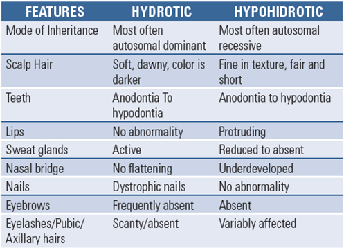
The Hypohidrotic form exhibits the classic triad-hypohidrosis, hypotrichosis and hypodontia. Usually X-linked recessive inheritance is seen. Males are affected severely, while females show only minor defects. In the hidrotic form teeth, hair and nails are affected. The sweat glands are usually spared. It is usually inherited as an autosomal dominant trait. Other inheritance modalities like autosomal recessive have also been reported.16,17
In the hypohidrotic form.the skin is soft, thin and dry. Partial or complete absence of sweat glands is responsible for the inability to perspire and accounts for intolerance to heat and frequent hyperthermia. The sebaceous glands are also defective or absent.18,19 Palms and soles are hyperkeratotic; psuedorrhagades are present around the eyes. Atopic eczema is common, especially in flexures during early childhood. In a new born, a “plastic wrap” appearance of the skin is characteristic of hypohydrotic type.
In the hydrotic type, sweat glands develop partially; ducts may be formed but secretory coils are absent and there are reduced number of sweat pores1.Scalp hair is often fine, stiff and short. Eyelashes and eyebrows are scanty and often missing18,20. The characteristic facial features are: frontal bossing, depressed nasal bridge, prominent supra orbital ridges, prominent and obliquely set ears , midface is depressed, the lower third of the face appears small due to lack of alveolar bone development, lips are protuberant.4,18 Children with hypohidrotic type of ectodermal dysplasia showed maxillary retrusion due to sagitally under developed maxilla, forward and upward displacement of the mandible and collapsed lower anterior facial height. In the oral cavity, the most striking feature is oligodontia. The teeth that are present have abnormal crown form. Teeth in the anterior region of maxilla and mandible are conical in shape.19 There is a wide midline diastema and hypoplasticlabial frenum. Cuspids are usually normal. Commonly, there is only one molar tooth in the second molar region which usually exhibits a bud crown form. Consistent variations in the number and crown forms of teeth occur which appear to be a characteristic dentalphenotype for ectodermal dysplasias with different modes of inheritance. This is demonstrated especially in autosomal recessive condition where there is a total absence of permanent teeth with or without taurodontism of primary molars.16 Cases have been reported where both primary and permanent dentition were congenitally missing.4,20
DIAGNOSTIC AIDS
RADIOGRAPHS
Orthopantomogram at an early age is to be done to rule out hypodontia and dental abnormalities.21 Xrays of the hand, feet show specific skeletal deformities
SWEAT PORE COUNT
Sweat pore count is done using yellow starch-iodine powder applied to palmar or dorsal skin. In unaffected persons, sweating turns the powder to deep purple, allowing visualization of sweat pores. Sweat pores are poorly visualized in affected children.23
BLASCHKO LINES
Streaky areas of hypohidrosis that follow blaschko lines are observed upon starch-iodine staining, which demonstrates a mosaic pattern of areas of normal number of sweat pores alternating with areas of absent pores.24
SKIN BIOPSY
Hypothenar eminence is the most reliable biopsy site to demonstrate an absence of sweat glands.22
PRENATAL DIAGNOSIS
Fetal skin biopsy helps in the diagnosis of prenatal ectodermal dysplasias.
Chorionic villus sampling at the 10th week of gestation can also be done in some cases.22
HISTOLOGIC FEATURES
Skin histopathology documents a reduction in the number of sweat glands, hair follicles and the sebaceous glands associated with the different EDs. The epidermis is thin and flattened. Eccrine sweatglands are few or poorly developed or are very rudimentary. Salivary glands may show ectasia of ducts and inflammatory changes.25
TREATMENT
Ectodermal dyspasia patients require both aesthetic and functional corrections of face. The absence of alveolar bone and teeth is a difficult reconstructive challenge for the surgeon. Complete rehabilitation of these patients is a team effort of specialists comprising of plastic surgeons and specialist dental surgeons.
Medical management is symptomatic. Patients with hyohidrosis require frequent consumption of cool liquids, wear cool clothing and should stay in a cool environment (air conditioning). Emollients for dry skin, artificial tears for patients with decreased lacrimation, saline sprays for nasal mucosa and antibiotics, if any infection occurs, may be required. Dental malalignment may require orthodontic treatment.
The typical decreased vertical facial height of the patient resulted from the congenital absence of teeth leading to underdevelopment of the alveolar ridges. In order to enable a further unrestricted development, the dentures have to be adapted to the changing intra-oral situation due to growth.26
Historically, prosthetic treatment of ectodermal dysplasia patients involved removable partial dentures, removable partial or complete overlay dentures and fixed partial dentures. The advent of dental implants and implant borne total telescopic dentures has provided an additional treatment modality for restoration of the dentition in this group of patients.2 There are aesthetic, functional and psychological reasons that make it important to start oral rehabilitation early in life. However, this is usually a difficult condition to manage prosthodontically, because of the typical oral deficiencies and the patient’s age. Numerous clinical reports have documented the importance of prosthetic dental treatment in patients of ectodermal dysplasia for physiological and psychological reasons.17
Serial sets of dentures may be required as the child matures. Shaw4 describes the need of these to restore vertical dimension and prevent the undesirable protruding lips secondary to overclosure, thereby improving the profile.
REFERENCES
- Dhanrajani PJ, Jiffy AO. Management of Ectodermal dysplasia. A literature review. Dental udate 1998; 25: 73-75
- Kupietzky K, Milton H. Hypohidrotic ectodermal dysplasia: Prosthetic management of Hypohidrotic ectodermal dysplasia. Quintessence Int 1995; 26: 285-291
- Richard AS, Karin V, Gerard K, Caries B. Placement of an endosseous implant in a growing child with ectodermal dysplasia. Oral Surg, Oral Med, Oral Path 1993; 75: 669-673
- Shaw RM. Prosthetic management of ectodermal dysplasia with anodontia. Case report: Aust Dental J 1990; 35: 113-116
- Piegno MA, Blackrnan RB, Cronin RJ, Cavazos E. Prosthetyic management of ectodermal dysplasia: A review of literature. J Prosthetic Dent 1996; 76: 541-545
- Tanner BA. Psychological aspects of Hypohidrotic ectodermal dysplasia. Birth defects 1998; 24: 263-275
- Thurman J. Two cases in which the skin, hair and teeth were very imperfectly developed. Proc RM Chir Soc 1848; 31: 71-82
- Weech AA. Hereditarty ectodermal dysplasia (congenital ectodermal defect). Am J Dis Child 1929; 37: 766-790
- Freire-Maia N, Pinheiro M, editors. Ectodermal dysplasias: A Clinical and genetic study. New York: Alan R Liss; 1984
- Pinheiro M, Freire –Maia N. The ectodermal dysplasias. Arch Dermat 1982; 118: 215-216
- Pinheiro M, Freire –Maia N. Ectodermal dysplasias. A Clinicalclassification and a casual review. Am J Med Genet 1994; 53: 153- 162
- Lamartine J. Towards a new classification of ectodermal dysplasias. Clinic Exp Dermato 2003; 28: 351-355
- Priolo M, Lagana C. Ectodermal dysplasias: A new clinical-genetic classification. J Med Genet 2001; 38: 579-585
- Itin PH, Fistarol SK. Ectodermal dysplasias: Can Med Assoc J 1929; 21: 18-31
- Srivastava AK, Durmowicz MC, Hartung AJ, Hudson J, Outzs LV,Donovan DM et al. Ectodysplasin A1 is sufficient to rescue both hair growth and sweat glands in tabby mice. Hum Mol Genet 2001; 10: 2973-2981
- Rajendran R, Shivapathasundaram B. Shafer’s text book of oral pathology. 5th edition Philadelphia: Saunders; 1983.
Disclaimer from Guident
The views and opinions expressed in articles published in Guident are solely those of the authors and do not necessarily reflect the views of the editors, the editorial board or the publisher, Ivory India. Guident does not endorse any product, technique or treatment described. Clinical content is intended for qualified dental and medical professionals and is not a substitute for individual clinical judgement. Guident, its editors and the publisher accept no liability for any consequence arising from the use of information published in the journal.
How to cite this article
Dr. Shamimul Hasan, Dr. Sarah Asif, Dr. Nabeel Ishrat Khan. Ecto Dermal Dys Plas Ia: An Oral Genodermatosis - Overview of Literature. Guident. 2013 May;6(7). Available from: https://ivoryindia.in/articles/oral-medicine/ectodermal-dysplasia-an-oral-genodermatosis-overview-of-literature.html


