Surgical Orthodontic Management of Skeletal Class II Malocclusion in an Adult Patient
Department of Orthodontics & Dentofacial Orthopaedics A.B. Shetty Memorial Institute of Dental Sciences
Abstract
In adults Class II discrepancy are treated either by orthodontic or camouflage or by surgical correction. Class II discrepancies with mandibular deficiency are treated surgically by mandibular advancement surgery. This article discusses in detail the combined orthodontic and surgical treatment carried out for an adult male with skeletal class II malocclusion. A cosmetic correction was achieved by BSSO with mandibular advancement and genioplasty and maxillary setback, along with orthodontic treatment. The patient’s facial appearance was markedly improved along with functional and stable occlusion
INTRODUCTION
In adult subjects having skeletal Class II malocclusions with mandibular deficiency, there traditionally are 2 possible treatment options. The first option is camouflage orthodontics— extracting the maxillary premolars to allow retrusion of the maxillary incisors to normalize the overjet and mask the underlying skeletal problem. The second option is orthognathic surgery to reposition the mandible anteriorly.
In the literature,a large number of surgical mandibular advancement procedures have been described for the therapy of skeletal class II malocclusions with mandibular deficiency1.However the current standard method is the retromolar mandibular sagittal split osteotomy introduced originally by Schloessmann in 1922.2 The most frequently used modifications of this method are those of Obwegeser3 and Dal Pontsup>4 and of Hunsuck5 and Epker6.
This article describes a case treated by a combination of orthodontics and surgery
CASE REPORT
An 22 year male old patient reported to the Department of Orthodontics, A B Shetty Memorial Institute of Dental Sciences with complaint of forwardly placed and irregularly placed upper and lower front teeth.
Extra oral examination
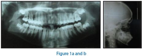
Dolichocephalic head pattern and leptoprosopic facial form. Frontal examination showed consciously competent lips. Profile was convex with posterior divergence and average mandibular plane angle. Normal breathing, deglutition and speech were diagnosed on functional examination.(Fig no.1)
Intraoral examination revealed U shaped arches with bimaxillary dento alveolar proclination of upper and lower anteriors. Upper and lower incisors showed moderate crowding with exaggerated curve of spee in lower arch.

Fig no 2: Pre-treatment intra-oral photographs

Angle’s class II molar and class II canine relationon both sides with over jet of 10 mm and over bite of 6mm.
Cephalometric examination revealed class II skeletal base due to orthognathic maxilla with retrognathic mandible.Vertical growth pattern with excess lower anterior facial ht. Dento alveolar analysis showed proclined upper and lower anteriors. Soft-tissue analysis indicates lip strain and protrusive and everted lower lip.
Diagnosis
Angle class II malocclusion on a class II skeletal base between orthognathic maxilla and retrognathic mandible with vertical growth pattern and increase lower anterior facial height, with over jet of 10 mm and over bite of 6 mm and lower anterior crowding.
Treatment objectives
To obtain class I skeletal base, to level and align the teeth, to obtain the ideal over jet and over bite, to maintain class1 molar and canine relation, improvement of soft-tissue profile.
Treatment plan
Phase I - Pre surgical orthodontics , initial levelling and alignment.
Phase II -Orthognathic surgery, maxillary set back,BSSO with mandibular advancement(previous history of orthodontic treatment, extn of all 1st PM’s)
Phase III -Post surgical stabilization.
Final finishing and detailing.
Treatment progress
The maxillary and mandibular arches were banded and bonded with 0.022 MBT [McLaughlin, Bennet and Trevisi] slot brackets. The initial levelling and aligning were done with 016 Niti, 018Niti, 16x22 Niti and 19x25 Niti. After initial alignment, upper and lower 19x25 stainless steel wires were placed.
Maxillary setback was done along with BSSO surgery. The patient was put on settling elastics post surgically.


Fig no.10 Post treatment extraoral photographs

Table 1: Comparison of pre- and post-surgical cephalometric variables

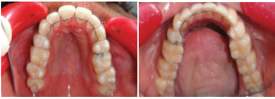
RETENTION
DISCUSSION
Combined surgical–orthodontic treatment can now be carried out successfully for patients with severedentofacial problems of any type. Interestingly,orthognathic surgery does not rate high on discomfort/morbidity scales. Often psychological evaluation of the patient before the surgery is mandatory and carefullypreparing the patient for their surgical experience would benefi t the patient to adapt to the significant facial changes.7
The most common mandibular advancement surgery done is the bilateral saggital split osteotomy.8,9 BSSO along with maxillary setback has proved to be useful method in treatment of skeletal class II malocclusion.
Based on the clinical and cephalometric findings, the patient in this case report had a normal maxilla, retrognathic mandible with a class II relation. Dentally the upper anteriors were proclined whereas the lower anteriors were proclined. The overjet in this case was found to be 10mm.
The mandibular surgery and maxillary set back surgery performed in this case. The post treatment SNB and ANB value and Witts appraisal indicated a correction of Class II discrepancy in this case by mandibular advancement (Table no.1) There was significant improvement in the soft tissue profile indicated by the position of the upper lip, lower lip and the chin. Dentally Class I molar and Class I canine relation is achieved.
CONCLUSION
Mandibular sagittal split osteotomy in combination with pre- and post surgical orthodontics is an efficient approach in the therapy of adult sleletal class II malocclusion.Saggital occlusal malrelationships are corrected and hard and soft tissue profiles straightened in a consistent way.
REFERENCES
- Proffit WR ,White RP,Sarver. Contemporary treatment of dentofacial deformity St. Louis ;2003;3 12 -344
- Hoffmann Axthelm W. Chirurgie der Zahnstellumg und Kieferanomalien. Die Geschichte der Mund-,Kiefer und Gesichtschirurgie Berlin ; 1995;121-142
- Obwegeser H. The surgical correction of mandibular prognathism and retrognathia with consideration of genioplasty. Oral surg.1957;10;677-689
- Dal Pont G Retromolar osteotomy for correction of prognathism .J Oral Surg 1961 ; 19 ;42-47
- Hunsuck EE A modified intra oral sagittal splitting technique for correction of mandibular prognathism J Oral Surg 1968; 26 ; 250 -253
- Epker BN Modifications in the sagittal osteotomy of the mandible. J Oral Surg 1977;35;157- 159
- Graber TM, Vanarsdall RL, Katherine WI Vig. Orthodontics – Currentprinciples and techniques. 3rd ed. Mosby Inc, Elsevier, 2000
- Poulton DR, Ware HW. Surgical orthodontic treat-ment of severe mandibular retrusion. Part I. Am J Orthod 1971; 59:244-65.
- Poulton DR, Ware HW. Surgical orthodontic treat-ment of severe mandibular retrusion. Part II. Am J Orthod 1973; 63:237-55.
Disclaimer from Guident
The views and opinions expressed in articles published in Guident are solely those of the authors and do not necessarily reflect the views of the editors, the editorial board or the publisher, Ivory India. Guident does not endorse any product, technique or treatment described. Clinical content is intended for qualified dental and medical professionals and is not a substitute for individual clinical judgement. Guident, its editors and the publisher accept no liability for any consequence arising from the use of information published in the journal.
How to cite this article
Dr. U. S. Krishna Nayak, Dr. Jyoti Kumari, Dr. Manmeet Kaur Alagh. Surgical Orthodontic Management of Skeletal Class II Malocclusion in an Adult Patient. Guident. 2014 Sep;7(10). Available from: https://ivoryindia.in/articles/orthodontics/surgical-orthodontic-management-of-skeletal-class-ii-malocclusion-in-an-adult-patient.html