Management of Natal Teeth: A Case Report
Department of Pediatric & Preventive Dentistry, K.D. Dental College & Hospital, Mathura, India
ABSTRACT
Natal teeth are teeth present in the oral cavity at birth, and though rare, they pose significant clinical concerns due to potential complications such as feeding difficulties, risk of aspiration, and injury to the infant or the mother. Most commonly affecting the mandibular central incisors, these teeth may be part of normal dentition or associated with developmental anomalies or syndromes. Proper clinical evaluation, including mobility assessment, is essential to determine the appropriate management approach. Conservative treatment such as smoothing sharp edges or extraction is considered based on the degree of mobility and associated risks. Extraction should be avoided in the first 10 days of life unless medically indicated, due to risk of hemorrhage from vitamin K deficiency. Parental counseling and follow-up are crucial components of management to ensure both oral health and general well-being of the neonate.
The present case report of 1-month-old infant describes the management of natal teeth present in mandibular arch with mobility. After the consent of parents, the extraction of the shell-like tooth was done immediately and hemostasis was achieved.
INTRODUCTION
The clinical age at which deciduous mandibular incisor tooth erupts is 6–7 months.1 In rare conditions, teeth might be present in the mouth at birth or might appear in the oral cavity within the first month of life. The teeth present at birth were termed as “natal teeth” by Massler and Savara 2 and those erupting within the first 30 days after birth as “neonatal teeth” and are also called “early infancy teeth.” Most commonly occurring predeciduous teeth are mandibular incisors. It has a prevalence rate of 66% occurring more commonly in girls.3 Natal tooth prevalence rate varies from 1:716 to 1:3500 live births.4 A survey was conducted with 18,155 infants; one in 716 for the 7,155 infants was the prevalence rate for those examined personally for the fetal teeth while one in 3,667 was the prevalence rate for those who were surveyed. A prospective examination for a group of another 38 infants and children was done who had a total of 61 mandibular central incisors as natal and neonatal teeth. It was determined that 95% of the natal and neonatal primary lower central incisors teeth were normal teeth, and 5% were supernumerary teeth. It has been observed that even though fetal teeth might rarely resemble the normal primary dentition in structure, they are often smaller, have a yellowish hue, and are conical in shape. They tend to have either poor, or no root formation and the crown is often formed with hypoplastic enamel and dentin.5 A dental roentgenogram is usually advised to help differentiate if the natal teeth is the premature eruption of a normal primary incisor or it is a supernumerary tooth.1 Enamel dysplasia was seen in 31% of the teeth, the probability of which is attributed to the duration of the gingival covering. Thus, it is indicated, as far as possible the natal and neonatal teeth should be left intact, and extraction is only advisable if they are extremely mobile, and chances of aspiration is likely. It was found that 61% of these infants had either natal or neonatal teeth or both5 without any exact known etiology. It is assumed that several factors like hereditary components, endocrinal abnormalities, and environmental elements may be contributory to its occurrence.6 Sometimes, developing teeth germs of congenital teeth are present in an unusual location under the alveolar bone.7 Complications of a predeciduous teeth usually includes lacerated skin of mother's breasts, aspiration of the teeth, discomfort during breast-feeding, sublingual ulceration associated with Riga–Fede syndrome;1 prevalence rate of Riga–Fede disease being six to ten percent.8 Careful evaluation of these infants is advised to prevent trauma to the tongue, to avoid aspiration of the tooth and injury to the mother’s breast as well. In 10 percent cases, trauma was noted while mobility and danger of inhalation in 94 percent cases cause complications. If the tooth is supernumerary or excessively mobile, tooth extraction is indicated. From the immunological and hematological perspective, it is indicated that the appropriate time for extraction of fetal teeth is 7-25 days after birth. Out of 50 children with dentitia praecox, extraction was carried out in 97 percent of the cases. It was seen that the neighbouring primary teeth usually move into the extraction space. In case if the tooth does not cause ulceration or pain, or interfere with breastfeeding, no treatment is necessary.1
CASE REPORT
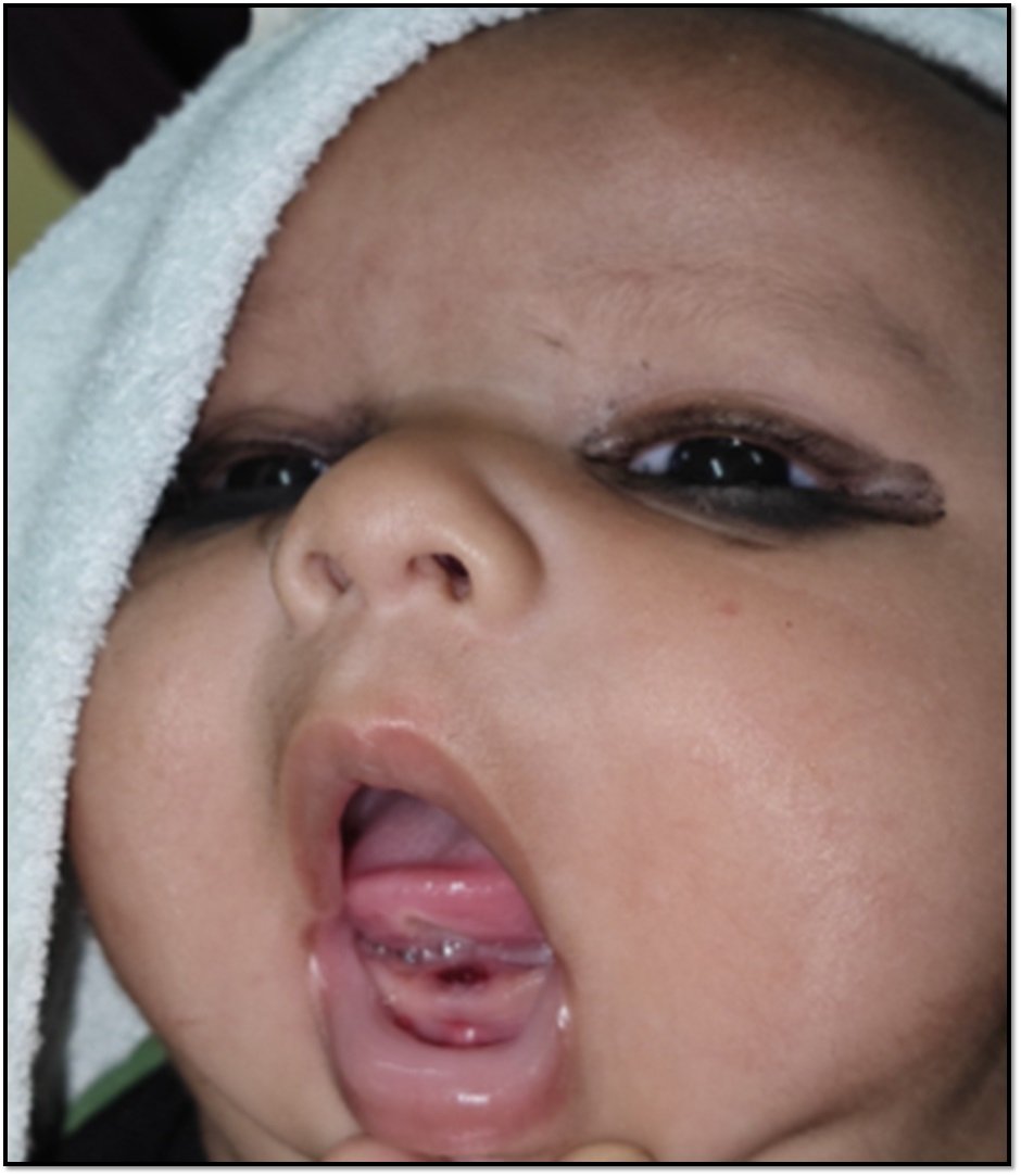
A 1 month old male infant was referred to the Department of Pediatric and Preventive Dentistry , K. D Dental College ,Mathura with complaint of two teeth in the lower jaw since birth, continuous crying, and refusal to suck milk. Oral examination revealed two crowns of the teeth in the mandibular anterior region [Figure 1], whitish opaque in color and exhibiting grade III mobility. The crown size was normal; the gingiva was of normal appearance. A diagnosis of natal tooth was made.
According to the vaccination chart the child was above 15 days therefore vitamin K prophylaxis was completed as stated by the parents.
We opted for the treatment of immediate extraction. Extraction was carried out after the topical application of local anesthesia with the help of sterile applicator (Figure 2). Post-extraction hemostasis was achieved and the patient was asked to sit in the operatory for 30 min. Post-operative instructions were also given and the patient was recalled after 1 week.
The extracted teeth had a shell-like crown ( Figure 3).

Presence of NATAL TEETH in 1 month old infant.

Extraction of natal teeth

Extracted Natal teeth
Post-extraction
DISCUSSION
Natal teeth were first documented during the Roman times by Titus Livius (59 BC) and Caius Plinius Secundus (23 BC). Massler and Savara divided tooth into two groups according to the time of eruption. According to Shafer’s, the predeciduous teeth have been described as hornified epithelial structures without roots, occurring on the gingiva over the crest of the ridge, which may be easily removed.
Natal tooth is the tooth which is being present from the time when the infant is born. Neonatal tooth is the tooth which erupts during the first 30 days of life. The prevalence of natal teeth varies from 1:7000 to 1:30000. Females are more commonly affected and also Muslim Children exhibit more natal/ neonatal tooth as compared to the Hindu children. Most common natal teeth present in infants are lower primary central incisors. Incidence of natal and neonatal teeth is mandibular incisors followed by maxillary incisors, then mandibular canine and the least found are molars and only 1% in maxillary posterior region.9
Some of the synonyms which is used to denote natal tooth are: Dentitia praecox, dens connatalis, congenital teeth, fetal teeth, infancy teeth, predeciduous teeth.10
Clinically the natal teeth are poorly developed and smaller in size and conical or normal in shape. They can be immature and can exhibit enamel hypoplasia. Sometimes there is the presence of root. They are brown- yellowish/ whitish opaque color. They are attached to a pad of soft tissue above the alveolar ridge, occasionally covered by mucosa and as a result have an exaggerated mobility with the risk of being swallowed or aspirated, in most of the cases.11
The literature reflects the association of natal teeth with reactive fibrous hyperplasia, congenital hydrocephalus associated with congenital glaucoma Walker Warburg syndrome, bilateral mandibular hamartomas, pyogenic granuloma, peripheral ossifying fibroma, eruption cyst, gingival fibrous hamartoma. Darwish et al reported a case of natal teeth in association with bifid tongue and deaf mutism.11
The radiographs of natal teeth reveal hollow calcified cap of enamel and dentin without any pulpal tissue, more like celluloid crown in appearance.
Natal and neonatal teeth were first observed by Howkins microscopically in 1932. Further investigations were carried out by Boyd and Miles. Histologically the crown of natal tooth consists of thin layer of dysplastic or hypomineralized enamel, irregular dentin and osteodentinin the cervical portions and interglobular dentin in the cervical regions. Incisal edge lacks the enamel. Hertwig’s epithelial sheath and cementum can be absent too. Sometimes there can be a greater number of blood vessels in pulpal tissue.12
If the natal teeth are not mobile for four months and is asymptomatic no intervention is required.13 Extraction should be the choice of management if the tooth is mobile as it can aspirated by the infant while suckling. If extraction is planned then the pediatric dentist should be careful to avoid unnecessary trauma to the surrounding structures. Before extraction, a thorough history of vaccination should be consulted from the parents like Vitamin k prophylaxis is utmost important as it is one of the clotting factors. If vitamin K has not been administered at birth, prophylactic administration of vitamin K (0.5e1.0 mg, IM) is advocated before and if required after extraction, as vitamin K is essential for the production of prothrombin in liver. Extraction of tooth should be followed by curettage of socket to prevent continued development of cells of dental papilla.12 Failure to curett socket might result in eruption of odontogenic remnants and necessitate future treatment.20
CONCLUSION
Natal and neonatal teeth are rare occurrence and it is not associated with any misconceptions. Therefore, the infants with erupted teeth should be thoroughly examined for further management and parent counselling should also be the part of the treatment so as to bring awareness about myths. The parents should make the visit to the dentist as soon as they notice the tooth in oral cavity of an infant. The intervention should be done immediately to avoid any complications.
REFERANCE
- KC Leung W Robson Natal teeth: A reviewJ Natl Med Assoc200698222634
- M Massler BS Savara Natal and neonatal teeth: A review of twenty-four cases reported in the literatureJ Pediatr195036334959
- J Zhu D King Natal and neonatal teethASDC J Dent Child19956212331
- MH Chow Natal and neonatal teethJ Am Dent Assoc198010021521
- GA Kates HL Needleman LB Holmes Natal and neonatal teeth: A clinical studyJ Am Dent Assoc198410934414
- RF Cunha F Boer DD Torriani W Frossard Natal and neonatal teeth: A review of the literaturePediatr Dent20012315862
- NN Nik-Hussein Natal and neonatal teethJ Pedodont19901411102
- HS Chawla Management of natal/neonatal/early infancy teethJ Indian Soc Pedod Prev Dent1993111339.
- Mhaske S, Yuwanati MB, Mhaske A, Raghavendra R, Kamath K, Saawarn S. Natal and neonatal teeth-an overview of the literature, hindawani publishing corporation. ISRN Paediatr. 2013;2013:956269.
- Seminario AL, Ivancakova R. Natal and neonatal teeth. Acta Medica. 2004;47:229-233.
- Hebling J, Zuanon ACC, Vianna DR. Dente Natal-a case of natal teeth. Odontol Clin. 1997;7:37-40.
- Delbem AC, Faraco Junior IM, Percinoto C, Delbem AC. Natal teeth: Case report. J Clin Pediatr Dent. 1996;20:325-7.
- Oliveira LB, Tamay TK, Wanderley MT, Rodrigues RM, Barboza CA, de Souza SO. Gingival fibrous hamartoma associated with natal teeth. J Clin Pediatr Dent. 2005;29:249-52.
- Darwish S, Sastry RH, Ruprecht A. Natal teeth, bifi d tongue and deaf mutism. J Oral Med. 1987;42:49-53.
Disclaimer from Guident
The views and opinions expressed in articles published in Guident are solely those of the authors and do not necessarily reflect the views of the editors, the editorial board or the publisher, Ivory India. Guident does not endorse any product, technique or treatment described. Clinical content is intended for qualified dental and medical professionals and is not a substitute for individual clinical judgement. Guident, its editors and the publisher accept no liability for any consequence arising from the use of information published in the journal.
How to cite this article
Dr. Rajeev Kumar Singh, Dr. Priyambana Pandey, Dr. Shalu Pathak. Management of Natal Teeth: A Case Report. Guident. 2025 Jun;18(7). Available from: https://ivoryindia.in/articles/pedodontics/management-of-natal-teeth-a-case-report.html