Chairside Single-Visit Feeding Plate Fabrication Using Digital Intraoral Scanning in a Newborn with Cleft Palate: A Case Report
Wisdom Dental Clinics ®, Jaipur, India
Abstract
Background: It is routine practice to document the morphology of the cleft lip and palate before undergoing palatoplasty in order to restore proper feeding. Conventional impression techniques put the airway at risk and call for extra resources and safety precautions. The initial clinical step in creating a feeding obturator for newborns with orofacial clefts is creating an intraoral impression. The aim of this clinical report is to demonstrate chairside single visit feeding plate manufacturing using a straightforward and secure digital imprint technology for maxillary impression production in a baby with cleft palate (palatoschisis) who is 7 days old. Material and method: A computerized intraoral scanner was used for prosthetic rehabilitation. Result and conclusion: The infant was delivered with a functioning feeding prosthesis to reduce parents’ concerns from multiple dental visits; it was successful in achieving the planned objectives. Thereby, opening new horizons in paediatric digital dentistry, that is minimally invasive, single visit, precise and quick in such cases.
Key words:
Cleft palate prosthesis, Feeding, Palatal, Obturator, Impression, Digital
Introduction
Documenting morphological changes in CLP (cleft lip and palate) due to growth and interventions allows assessment of treatment quality and outcome research. Orofacial defects are one of the most common anomalies of humans. [1]. New-borns with abnormal oro-nasal communication face life-threatening situations due to difficulty in feeding. [2] Hence, there is an acute demand of feeding appliance that serves as an obturator in the defect area, in view of the prevailing concerns of feeding. A prosthetic device called a feeding obturator or nasoalveolar moulding (NAM) helps to re-establish the distinction between the nasal and oral chambers. [3] Nutritional deficiency is the most important aspect in the holistic growth and development of a neonate. New-borns with CLP (cleft lip and palate) are incapable to generate the suction level and compression required for bottle/breastfeeding.[4]
In contrast to the traditional method of taking physical impressions, which carries the risk of putting the infants' airways at risk due to the impression material, digital impressions implementing intraoral scanners enable the contactless recording of the patients' oral cavity, lip, and nose morphology. [5] Despite the procedure for the fabrication of feeding obturator is well documented in studies. In light of the information deficit; this article through a case report addresses the implementation of digital impression taking with intraoral scanners in surgeons’ hands in routine clinical workflow in a 7-day-old neonate, fabrication and delivery of the feeding obturator in single visit to ward off any emergency situation while feeding.
Case report
A 7-day-old neonate (girl), weight 2.3 kg approx. referred to the clinic for fabrication of interim prosthesis for the closure of congenital cleft in soft palate. The new-born’s parents chief complaint was difficulty in feeding and nasal discharge. The baby was underweight (as normal weight of new-born is 2.7-3.1kg) which was egregiously inappropriate. Familial history was non-contributory. Intraoral examination revealed a cleft in the uvula, soft palate and secondary part of hard palate (Veau’s classification: Type II). Since palatoplasty was not attainable at this age and weight, a feeding obturator was planned as interim prosthetic management for present concerns. Parent’s counselling was done to allay their anxiety and treatment was planned for fabrication of obturator using digital impressions for which consent was obtained.
- Maxillary impression was done without any premedication or anaesthesia. The neonate was seated upright on mother's lap in dental chair with face forward. Owing to smaller mouth opening it did not permit the use of impression tray/flat wooden stick as carrier for the material, also the location of cleft was in soft palate making physical impression almost impossible. Thus, it was planned to make impression with intra-oral scanner (Omnicam, Densply Sirona). (Figure.1)
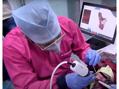
Figure.1 Intraoral scanning
- Impressions were taken in slots to avoid discomfort. In just 3 minutes, uneventful procedure was done; digital data of whole of the cleft defect was recorded along with maxilla as needed with utmost patient and parent convenience. The scanned images were redirected for digital surveying, and the appliance design was mapped out. (Figure.2 a,b)

Figure.2a Scanned image

Figure 2b. Designing of appliance
- The analysed HTML (Hypertext Mark-up Language) file is further processed for 3D models with (Excocad) software and was sent to 3D printer. The framework’s final design was 3D printed out with SG resin (surgical guide resin).
(JAMG HE, 3D printer photopolymer resin)
- Wax adaptation was done on the 3D- model using dental modelling wax (Pyrex) acting as the soft tissue, separating medium (Vaseline) was used, with this model NAM plate was fabricated using conventional acrylic dental resin (cold- cure/ self-cure) (DPI) with dough method to obtain sufficient rigidity. Though self-cure is one of the limitations but to avoid multiple visits fast setting self-cure is used. To facilitate feeding and avoid conventional impression with chances of material aspiration in infant we opted for self-cure resin.
- Any excess was trimmed and finished according to try-in on the working cast. Before the final insertion of the palatal obturator, a metal wire was connected to the prosthesis by drilling a hole on plate as a safety precaution in case of accidental swallowing. (Figure.3)

Figure.3 Master 3d model and clear feeding obturator with safety wire attached
- Then it was examined in the infant’s mouth, to our pleasant surprise no adjustments whatsoever were done during final insertion of the prosthesis.(Figure.4)

Figure.4 Feeding obturator insitu
However, during follow-up adaptation of the prosthesis seemed very good not only at the moment but also after 2 weeks. Parents were asked to feed the baby and educated for the placement, removal and cleaning of the appliance. Infant gained weight normally during the 2 months follow-up.
Discussion
There have been various techniques documented over time to improve the fabrication procedure of feeding obturators. Unfortunately, none have successfully addressed the issues of the impression taking procedure in new-borns. Given the age of newborns and the potential for aspiration and swallowing of impression material, obtaining preliminary impressions becomes extremely difficult. Clinicians have proposed customized impression tray, inverted posture of infants, and impression materials having various consistencies to ensure good detail duplication required for satisfactory fabrication of palatal prosthesis. [6] Despite all the efforts, the agitation and nervousness of neonates still remain a major factor that no technique has yet subdued.
Patients with palatoschisis and their families are confronted with series of treatment steps for cleft care. Any advancement for reducing the burden, risk and enhance treatment outcomes is exceptionally valuable, especially for infants. Therefore, alternatives to traditional impressions are required. [7] Digital impressions have completely replaced conventional impression taking. High-quality 3D intraoral scans could be reproduced without adverse events in neonates and infants while they are awake, within 100 seconds approx. Scan of lip and nose takes an additional 50 s. Acquisition of CLP allowed for low-risk data acquisition without soft tissue distortion. Integration into a digital workflow also gave the probability to digitally design and 3D print presurgical orthopedic appliances. [8]
The simplified digital impression method not only benefits the patients but also digitalization allows leveraging of technological advancements. [9] 3D imaging technique provides new as well as enhanced outcome analysis of the facial aesthetics of children with cleft. [10]
The chairside procedure might add an element of uncertainty while managing new-born patients. It is difficult to control the flow of the impression material in the cleft area and undercuts in neonates in conventional method but here no such hurdles like distortion, material handling challenges were found. To achieve optimal results, parents' education and motivation by the treating clinician’s plays a pivotal role.
Limitations: With present world scanners, like omnicam, primescan or i-Tero scanners provide with faster acquisition, high level of precision and accuracy but the only limitation can be its upscale cost to the dentist.
Conclusion
Intraoral scanner technology has advantages over conventional impression taking. In present case retention of the feeding plate was confounding in 7-day old neonate owing to the digital impressions. If the hurdles of acquisition costs and interfaces in the workflow could be overcome, conventional impressions could be replaced in clinical routine. Chairside digital impression technology enables minimally invasive, precise, and rapid fabrication of feeding obturators for neonates with cleft palate. This approach opens new horizons in pediatric digital dentistry.
Reference
- Chandna P, Adlakha VK, Singh N. Feeding obturator appliance for an infant with cleft lip and palate. J Indian Soc Pedod Prev Dent. 2011;29:71–3.
- Goldberg WB, Ferguson FS, Miles RJ. Successful use of a feeding obturator for an infant with a cleft palate. Spec Care Dentist. 1988;8:86–9.
- Rathee M, Hooda A, Tamarkar AK, Yadav SP. Role of feeding plate in cleft palate: Case report and review of literature. Internet J Otorhinolaryngol. 2010;12:1.
- Reid J, Reilly S, Kilpatrick N. Sucking performance of babies with cleft conditions Cleft Palate Craniofac J. 2007;44:312–20
- J. Radojicic. Cleft Care: intraoral 3D scanning. Cleft Palate Craniofac J, 55 (9) (2018), p. 1330, 10.1177/1055665617742806 We here successfully implemented digital impression taking with an intraoral scanner in 7-day-old neonate. Feeding plate obturator serves well to aid in optimal nourishment in new-born.
- Chandna P, Adlakha VK, Singh N. Feeding obturator appliance for an infant with cleft lip and palate. J Indian Soc Pedod Prev Dent 2011;29:71-3.
- J. Radojicic. Cleft Care: intraoral 3D scanning. Cleft Palate Craniofac J, 55 (9) (2018), p. 1330, 10.1177/1055665617742806
- Zarean P, Zarean P, Thieringer FM, Mueller AA, Kressmann S, Erismann M, Sharma N, Benitez BK. A Point-of-Care Digital Workflow for 3D Printed Passive Presurgical Orthopedic Plates in Cleft Care. Children. 2022; 9(8):1261. https://doi.org/10.3390/children9081261
- A.B. Xepapadeas, A.B. Xepapadeas, C. Weise, et al. Technical note on introducing a digital workflow for newborns with craniofacial anomalies based on intraoral scans - Part II: 3D printed Tübingen palatal plate prototype for newborns with Robin sequence BMC Oral Health, 20 (1) (2020), 10.1186/s12903-020-01159-7
- V.P. Sharma, H. Bella, M.M. Cadier, R.W. Pigott, T.E.E. Goodacre, B.M. Richard. Outcomes in facial aesthetics in cleft lip and palate surgery: a systematic review. J Plast Reconstr Aesthetic Surg, 65 (9) (2012), pp. 1233-1245, 10.1016/j.bjps.2012.04.001
Disclaimer from Guident
The views and opinions expressed in articles published in Guident are solely those of the authors and do not necessarily reflect the views of the editors, the editorial board or the publisher, Ivory India. Guident does not endorse any product, technique or treatment described. Clinical content is intended for qualified dental and medical professionals and is not a substitute for individual clinical judgement. Guident, its editors and the publisher accept no liability for any consequence arising from the use of information published in the journal.
How to cite this article
Dr. Gaurav Gupta, Dr. D.K. Gupta, Dr. Neelja Gupta. Chairside Single-Visit Feeding Plate Fabrication Using Digital Intraoral Scanning in a Newborn with Cleft Palate: A Case Report. Guident. 2025 Oct;18(11). Available from: https://ivoryindia.in/articles/pedodontics/chairside-single-visit-feeding-plate-fabrication-using-digital-intraoral-scanning-in-a-newborn-with-cleft.html