Obstructive Sleep Apnea and Prosthodontic Considerations: A Review
Department of Prosthodontics and Crown & Bridge, College of Dental Sciences, Davangere, KAR India
ABSTRACT
Sleep is a fundamental physiological process essential for maintaining hormonal balance, glycemic control, cardiovascular function, and overall well-being. However, sleep disturbances are increasingly common in modern society, often resulting in emotional instability, cognitive decline, and physical health impairments. Among various sleep disorders, sleep-disordered breathing (SDB)—particularly Obstructive Sleep Apnea (OSA)—is highly prevalent and poses significant health risks. OSA is characterized by repeated upper airway obstruction during sleep, leading to intermittent breathing cessation, oxygen desaturation, and disrupted sleep architecture. These disturbances have profound systemic consequences, contributing to cardiovascular disease, hypertension, metabolic dysfunction, and neurocognitive decline. Notably, risk factors such as obesity, aging, craniofacial anomalies, and edentulism significantly influence airway patency and neuromuscular control. In this context, prosthodontists play a critical role in the early identification and management of OSA. Through comprehensive assessment and the provision of customized oral appliances, prosthodontists contribute significantly to improving airway patency, enhancing sleep quality, and mitigating associated health risks. Their involvement underscores the importance of interdisciplinary collaboration in addressing the growing burden of sleep-disordered breathing.
KEYWORDS:
Obstructive Sleep Apnea (OSA), Apnea-Hypopnea Index(AHI), Oral appliance therapy(OAT), Continuous positive airway pressure (CPAP).
INTRODUCTION:
Sleep is an essential physiological process that plays a pivotal role in maintaining overall health, regulating hormonal balance, supporting glycemic control, and ensuring cardiovascular function. While sleep is a natural and vital part of human life, disturbances in sleep are becoming increasingly prevalent in modern society, contributing to a wide array of health issues. Sleep deprivation, whether caused by pain, stress, environmental factors, or sleep disorders, can lead to emotional instability, impaired cognitive function, and physical health problems, affecting an individual’s quality of life. Among the many sleep disorders, sleep-disordered breathing (SDB) is one of the most widespread, with obstructive sleep apnea (OSA) being the most commonly diagnosed form. These conditions disrupt the normal sleep cycle, leading to intermittent cessation of breathing during sleep, which results in a reduction in oxygen levels, increased heart rate variability, and fragmented sleep. The consequences of untreated SDB, especially OSA, are far-reaching, with associations to cardiovascular disease, hypertension, metabolic disorders, and cognitive impairments. (1)
Classification: Gastaut et al. described three types of apneas: namely obstructive, central, and mixed patterns. (6)
- Obstructive sleep apnea (OSA) - Obstructive sleep apnea, also known as occlusive apnea, is the most prevalent type, in which airflow ceases due to upper airway blockage while respiratory effort is exerted simultaneously. This respiratory effort continues despite the obstruction until the individual's sleep is disrupted. (6)
- Central sleep apnea (CSA) - simultaneous cessation of both airflow and respiratory effort is observed due to the CNS disorder. (6)
- Mixed apnea - episodes are accompanied by no respiratory effort initially, followed by respiratory muscle movement, and finally, by airflow. (6) Combination of CSA and OSA.
The most common among these, OSA, involves the partial or complete collapse of the upper airway during sleep, causing episodes of hypoxia and disrupting normal sleep stages. Apnea-Hypopnea Index (AHI) index helps to measure the severity of sleep apnea by counting the number of apnea (complete obstruction) and hypopnea (partial obstruction) episodes per hour of sleep (Table 1) (Figure 1). (2) The role of prosthodontists in managing OSA continues to expand, as oral appliances provide a crucial alternative to more invasive therapies like Continuous positive airway pressure (CPAP) and surgery. Prosthodontists’ expertise in designing and fitting these appliances helps ensure that patients receive the best possible treatment, with improved comfort and adherence. By integrating their knowledge of dental anatomy and sleep medicine, prosthodontists work closely with sleep specialists and other healthcare providers to offer personalized treatment options that address the complexities of OSA and sleep-disordered breathing.

Figure 1. Normal Breathing, Hypopnea (Partial Obstruction), And Apnea (Complete Obstruction).
This paper will explore the various types of sleep-disordered breathing, with a particular emphasis on the pathophysiology, diagnosis, and impact of obstructive sleep apnea on both adults and children. The different treatment options for OSA will be discussed, including CPAP, oral appliances, surgical interventions, and behavioral approaches, with a special focus on the critical role prosthodontists play in the management of sleep apnea through the use of oral appliances. By highlighting the advancements in the treatment of OSA and the importance of interdisciplinary collaboration, this paper aims to underscore the significance of effective management strategies in improving patient outcomes and overall quality of life.
Table 1. The severity of Obstructive Sleep Apnea (OSA) based on Apnea-Hypopnea Index (AHI).
AHI Score in Adults
Severity Level of OSA
DIAGNOSIS AND TREATMENT PLANNING:
Obstructive Sleep Apnea is a multifactorial disorder primarily associated with obesity and craniofacial anomalies. Obesity significantly increases the risk and severity of OSA, with prevalence ranging from 55% to 100% in severely obese individuals. Craniofacial features such as retrognathia, micrognathia, high-arched palate, enlarged tonsils or uvula, macroglossia, and nasal obstructions contribute to airway narrowing and obstruction during sleep. OSA is more prevalent in men, older adults, and postmenopausal women. Additional risk factors include edentulism, large neck circumference, smoking, alcohol use, hypothyroidism, acromegaly, and systemic conditions such as heart failure, stroke, type 2 diabetes, and Down syndrome. Dentists play a crucial role in early detection by screening patients for signs and symptoms of OSA using validated tools such as the Epworth Sleepiness Scale, Berlin Questionnaire, and STOP-BANG. Patients who screen positive should be referred to a physician for further evaluation.
Diagnosis involves a thorough assessment of clinical history, physical and oral examination, and sleep studies. Key symptoms include loud snoring, observed apneas, unrefreshing sleep, excessive daytime sleepiness, cognitive impairment, and mood disturbances. Physical examination may reveal obesity (BMI >30 kg/m²), enlarged neck circumference, retrognathia, and oral findings such as macroglossia, narrow palate, or TMJ disorders. Radiographic tools like lateral cephalograms, CT, and MRI help assess airway anatomy and craniofacial structure. The gold standard for diagnosis is in-laboratory polysomnography, which evaluates sleep stages, respiratory effort, airflow, and oxygen saturation. Home Sleep Apnea Testing (HSAT) is a cost-effective alternative for patients without significant comorbidities. Advanced technologies such as 3D facial scanning are emerging as non-invasive tools to assess craniofacial predictors of OSA. Early identification and appropriate referral are critical for effective management and reducing the risk of associated cardiovascular and metabolic complications.
MANAGEMENT OF OBSTRUCTIVE SLEEP APNEA:
Treatment options for sleep-disordered breathing include the following general categories:
-
Lifestyle modification: (1)
- Weight reduction
- Cessation of alcohol consumption
- Pharmacologic treatment
- Sleep position training
- Continuous positive airway pressure (CPAP).
- Oral appliances therapy: (6) Broadly classified into 3 types:
- Tongue repositioning devices (TRD), such as the tongue retaining device
- Mandibular advancement devices (MAD)
- Soft palate lifters or Palatal lift appliances (PLA)
- Upper airway surgery: (4)
- Maxillary expansion (with or without surgical assistance)
- Maxillary orthopedic protraction
- Maxillomandibular advancement
- Tracheostomy
- Nasal procedure
-
LIFESTYLE MODIFICATION FOR OBSTRUCTIVE SLEEP APNEA (OSA):
Weight Reduction: (5)
Obesity is a major risk factor for the development of OSA, with approximately 70% of OSA patients being obese (BMI ≥30 kg/m²), and 40% to 90% of morbidly obese patients (BMI ≥40 kg/m²) affected. Excess adipose tissue, particularly around the upper airway, restricts airflow and increases the risk of airway collapse during sleep. Even modest weight loss, such as a 5% reduction, can improve respiratory episodes, with a more significant weight loss (≥10%) offering substantial benefits. Weight-reduction strategies may include pharmacological options like phentermine, topiramate, or liraglutide, which have shown promise in reducing OSA severity. Bariatric surgery, such as gastric bypass, is becoming a permanent solution for many OSA patients and has shown success in achieving OSA remission in 59.2% of cases, though OSA may recur even with sustained weight loss. Regular follow-up is crucial for long-term management.
-
Cessation of Alcohol Consumption: (15)
Reducing alcohol intake, particularly avoiding alcohol at least two hours before bedtime, can be beneficial for OSA. Alcohol relaxes the throat muscles, increasing the likelihood of snoring and airway obstruction. While reducing alcohol intake may not cure OSA, it can lessen its severity, especially in moderate cases.
Pharmacologic Treatment: (28)
Several drugs have been explored for managing OSA, targeting mechanisms like REM sleep suppression, respiratory drive stimulation, and upper airway muscle tone enhancement. Tricyclic antidepressants (TCAs), particularly protriptyline, have been investigated, but there is insufficient evidence to support their use for OSA, as they may worsen REM sleep and do not improve respiratory events. Selective serotonin reuptake inhibitors (SSRIs) and clonidine, which also suppress REM sleep, have shown limited benefits in OSA patients, with no significant improvements in sleep apnea severity. Other drugs, like acetazolamide, progesterones, and theophylline, have also been studied but failed to demonstrate consistent clinical benefits in OSA treatment.
Sleep Position Training: (5)
Positional therapy is an effective treatment for patients with positional OSA (POSA), where OSA severity is significantly worse in the supine position. Changing sleep position from supine to lateral can reduce airway collapse and improve respiratory outcomes. Devices like tennis balls, position alarms, and specialized pillows are commonly used to encourage non-supine sleeping positions. While positional therapy can be effective, its long-term success is often hindered by discomfort and poor patient adherence. The "tennis-ball" method is commonly used, but more advanced position alarms with vibration cues have shown higher compliance rates. While positional therapy can significantly reduce the apnea-hypopnea index, it is generally considered a supplementary approach, not a replacement for more conventional treatments like CPAP.
-
CONTINUOUS POSITIVE AIRWAY PRESSURE (CPAP): (17)
CPAP is the first-line treatment for moderate to severe OSA, particularly for high-risk individuals such as drivers. It works by delivering continuous positive pressure, effectively acting as a pneumatic splint to keep the upper airway open during sleep. A CPAP prescription should specify the pressure setting, mask type, and necessary equipment supplies, with auto-titrating CPAP typically set at a pressure range of 5-20 cmH2O. Early patient education on equipment maintenance, therapeutic benefits, and potential issues (e.g., mask intolerance, nasal congestion, air leaks) is essential for successful therapy. Adherence to CPAP is linked to improvements in daytime sleepiness, quality of life, and blood pressure. Insurance often requires proof of adequate adherence (4+ hours per night for 70% of nights) within 30 days.
Adherence to CPAP is often determined within the first week of use. Regular follow-up visits are crucial to assess patient progress, resolve any issues, and ensure proper equipment use. Issues such as mask discomfort, nasal congestion, and pressure intolerance can often be managed with adjustments like mask selection, nasal steroids, or increased humidification. Behavioural therapies that motivate patients and educate them on OSA’s consequences, coupled with telemonitoring and automated feedback, can significantly improve adherence.
CPAP devices record usage data, which can be reviewed by physicians remotely. Regular follow-up appointments should include a review of this data to verify adherence, assess treatment efficacy, and monitor for any side effects. Long-term monitoring is necessary to ensure continued symptom management, particularly in patients who have undergone weight reduction or surgery. In cases of persistent daytime sleepiness, medications like modafinil or solriamfetol may be considered as adjuncts to CPAP therapy.
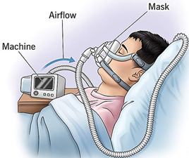
Figure 2. CPAP (Continuous Positive Airway Pressure) machine.
ORAL APPLIANCES THERAPY:
Dentists are often the first to identify signs of obstructive sleep apnea (OSA), especially mild to moderate cases, due to regular patient visits. This places them in a pivotal position to bridge the gap between patients and the broader medical community. During clinical exams, dentists assess the tonsils, tongue, uvula, soft palate, and oropharynx. The Mallampati index helps evaluate tongue volume and potential airway obstruction. Other critical observations include the mandible’s position, soft palate volume, and uvula shape—often supported by radiographic imaging.
Given the low compliance with CPAP therapy, oral appliance therapy (OAT) offers an effective alternative. OAT works by repositioning the mandible and tongue to improve airflow and reduce supine apnea episodes. As such, dentists play a key role in interdisciplinary care for sleep-related breathing disorders. The ADA has defined this role in its policy guidelines. (12,16)
The concept of oral appliances dates back to 1923 when Pierre Robin proposed a prosthesis to address airway obstruction caused by tongue displacement. Interest resurfaced with Cartwright and Samelson’s tongue-retaining device, leading to the development of mandibular advancement devices (MADs), which move the jaw forward to maintain airway patency during sleep. (4)
Fabrication of OAs for OSA: 34,35
Conventional Workflow
Maxillary and mandibular arch impressions using alginate or PVS (polyvinyl siloxane).Record protrusive bite registration (usually 60–75% of max mandibular protrusion) using materials: wax, silicone, or construction bite forks.Mount the stone casts using bite registration on an articulator.Manually design and fabricate the appliance on the models.Appliance trimmed, polished, and adjusted on the model.Try-in on patient, adjust for comfort, fit, retention, and function.Digital Workflow
Use intraoral scanner to digitally capture 3D images of maxillary and mandibular arches.Bite taken with intraoral scanner or recorded via digital construction bite device (usually 60–75% of max mandibular protrusion).Scans exported as STL files and imported into CAD software.Appliance designed virtually using dental CAD software (e.g., 3Shape, Exocad).Appliance fabricated using 3D printing (resin-based) or milling (PMMA or nylon).Cleaning, curing, polishing of 3D-printed appliance.Appliance inserted, checked for fit and comfort.Types of Oral Appliances for OSA:
Oral appliances for OSA are generally categorized into three main types: Tongue Repositioning Devices (TRDs), Mandibular Advancement Devices (MADs), and Soft Palate Lifters or Palatal Lift Appliances (PLAs). Each serves a unique function in maintaining upper airway patency during sleep and is selected based on patient anatomy, severity of OSA, and individual tolerance.
a. Tongue Repositioning Devices (TRDs) (6,9,31)
TRDs were first introduced in 1982 and are designed to hold the tongue in a forward position using negative pressure. Typically made from soft polyvinyl material, these bubble-shaped devices create a vacuum that gently retains the tongue in a protruded position, helping to reduce airway obstruction caused by tongue base collapse. TRDs are particularly useful in patients who are edentulous, have inadequate dental support, or suffer from conditions like Down syndrome. They are also prescribed for non-apneic snorers or those who cannot tolerate mandibular advancement. Common examples include Aveo TSD, SnorX, and Deepsleep Pro.
Although TRDs can reduce apneas when the patient is in the supine position, they are generally considered less effective than MADs. Side effects include excessive salivation and difficulty swallowing. Despite these drawbacks, they remain a viable alternative for specific patient populations or when other treatments fail or are contraindicated.

Figure 3. Tongue Repositioning Device.
b. Mandibular Advancement Devices (MADs)
MADs are the most widely used oral appliances for OSA. They work by advancing the mandible forward, which enlarges the upper airway and stabilizes the soft tissues, particularly in the oropharyngeal region. This forward movement stimulates muscle activity—especially the genioglossus, geniohyoid, and other pharyngeal dilators—thereby reducing snoring, apneas, and hypopneas. Cone-beam CT and other imaging studies have shown that mandibular advancement also causes anterosuperior movement of the hyoid bone and skeletal changes, which can assist in selecting suitable candidates for MAD therapy.
MADs have been shown to improve oxygenation and reduce both subjective and objective symptoms of OSA, including daytime sleepiness and snoring, across all severity levels. Additionally, modest reductions in blood pressure have been observed with long-term use. However, the effect of MADs on major cardiovascular outcomes remains unclear. (32)
MADs are classified in several ways. By design, they are either prefabricated or custom-made. Prefabricated devices are often bulkier and may lack adequate retention, which can lead to discomfort and reduced efficacy. In contrast, custom-made MADs offer better fit, patient comfort, and treatment success. These are further divided into non-adjustable (monoblock) devices, which are fixed in position, and adjustable (bi-block) devices that allow gradual mandibular advancement through titration.
The coupling mechanism—which connects the upper and lower parts—varies between devices. These may include elastic straps, rods, fins, or springs, each designed to maintain jaw position and minimize backward movement. For optimal comfort and treatment effect, advancement should be gradual (in increments ≤1 mm) to reduce side effects such as temporomandibular joint (TMJ) pain.
Vertical adjustment is another design consideration. While increasing the vertical dimension (interincisal distance) can aid some anatomical situations, excessive opening may lead to discomfort and lower patient acceptance. Therefore, minimal vertical increase is generally recommended unless otherwise clinically indicated. (32)
There are many types of MADs, each with distinct features.
The PM Positioner is an older design that uses orthodontic expanders and thermoplastic material, requiring heating before use.
The Elastic Mandibular Advancement (EMA) appliance is thin, lightweight, and utilizes elastic straps to adjust mandibular position, allowing lateral movement and reducing TMJ strain. (Figure 4)
The Klearway Appliance is fully adjustable with an orthodontic screw and is suitable for mild to moderate OSA, offering comfort through controlled advancement. (Figure 5)

Figure 4. Elastic mandibular advancement (EMA)

Figure 5. Klearway oral appliance
The Thornton Adjustable Positioner (TAP) is widely studied and features a screw mechanism for fine-tuned titration of the lower jaw. (Figure 6)
The Modified Herbst Appliance employs telescopic rods to maintain forward mandibular posture but limits side-to-side motion, offering high stability but possible TMJ discomfort. (Figure 7)

Figure 6. Thornton adjustable positioner (TAP)
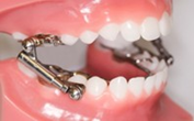
Figure 7. Modified Herbst Appliance
The DUOBLOC allows mandibular advancement via anterior attachments, emphasizing customization and patient comfort. (12,30) (Figure 8)
The Silencer System uses precision titanium hinges to permit vertical, lateral, and anteroposterior adjustments, offering unmatched control but at a higher cost.
For edentulous patients, the Simple Maxillomandibular Splint holds the jaw in a pre-recorded forward position and is easy to use but may lack sufficient retention. (Figure 9)

Figure 8. Duo-bloc MAD

Figure 9. Simple maxillomandibular splint
The Dorsal Appliance, with its two-part design and dorsal fins, allows for functional jaw movement and better comfort during speaking or yawning. (Figure 10)
The Moses Appliance adds tongue space and reduces parafunctional activity like bruxism while enabling speech and hydration. (12,30,33) (Figure 11)

Figure 10. Dorsal Appliance
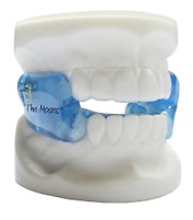
Figure 11. Moses Appliance
Digital solutions like the ProSomnus® Sleep and Snore Device utilize CAD/CAM technology to create sleek, comfortable, and precisely fitting devices. (Figure 12)
Finally, the Twin Block Appliance, originally an orthodontic device, guides the mandible forward using angled bite blocks and is particularly effective in younger patients or those undergoing orthodontic treatment. (2,30) (Figure 13)

Figure 12. ProSomnus®

Figure 13. Twin block appliance
c. Soft Palate Lifters (Palatal Lift Appliances - PLA): (6)
Palatal Lift Appliances (PLAs) are designed to reposition the soft palate upward and backward, thereby increasing the dimensions of the upper pharyngeal airway. These appliances are especially useful in patients with neuromuscular disorders that impair soft palate function, such as myasthenia gravis, cerebral palsy, cleft palate, or after surgeries like tonsillectomy or maxillary resection.
PLAs offer several benefits: they reduce gag reflex by applying consistent pressure to the soft palate, preserve tongue movement due to their superior positioning, and can be adjusted in stages for patient comfort. They are appropriate for a broad range of patients with soft palate insufficiency (Figure 14). However, limitations exist—PLAs are not suitable for patients who are uncooperative, have poor retention of the base prosthesis, or possess difficult-to-manage palatal anatomy.

Figure 14. Palatal Lift Appliances (PLAs)
ROLE OF EDENTULISM IN OSA PATHOGENESIS: (13)
Edentulism (tooth loss) is increasingly recognized as a contributing factor in the development and severity of obstructive sleep apnea (OSA). The absence of teeth leads to anatomical changes such as reduced vertical dimension of occlusion, mandibular and hyoid bone repositioning, and impaired function of oropharyngeal muscles. These changes compromise airway patency, contributing to increased airway collapsibility during sleep (Figure 15). Although a direct causal link is limited, edentulism clearly correlates with OSA through these physiological mechanisms.

Figure 15. a. Elongated uvula
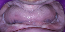
Figure 15. b.Enlarged tongue

Figure 15. c. Loss of vertical dimension in an edentulous patient.
Oral Appliance Therapy in Edentulous and Partially Edentulous Patients (13)
Oral appliances (OAs) offer a conservative, non-invasive treatment for OSA. However, edentulism presents challenges due to lack of dental retention. Mucosa-supported appliances, like monobloc acrylic splints or adjustable PM Positioners, have been developed specifically for edentulous patients. These devices advance the mandible and/or stabilize the tongue, improving airway function.
For partially edentulous patients, monobloc appliances using remaining teeth for support have shown positive outcomes in reducing AHI and snoring, while improving sleep quality.
Effect of Increased Vertical Dimension (13)
Increasing the vertical dimension with dentures can improve the retropharyngeal airway space, as noted by Gupta et al. and others. This adjustment, especially with a 2 mm increase, enhances mandibular positioning and airway patency. However, excessive vertical changes may strain the temporomandibular joint (TMJ) and require longer patient adaptation.
Disadvantages of Denture Use During Sleep (13)
Despite their benefits, sleeping with dentures poses risks, including chronic mucosal irritation, alveolar bone loss, and TMJ strain, particularly when increasing vertical dimension. Careful design, monitoring, and patient-specific adjustments are essential to mitigate these complications.
Implant-Supported Oral Appliances (13)
Implant-retained devices provide improved stability in completely edentulous patients. Hoekema et al. and Flanagan demonstrated success using implants to anchor mandibular repositioning appliances (MRAs), allowing mandibular advancement with better retention and reduced airway obstruction. These devices are especially beneficial when mucosal support alone is insufficient.
- UPPER AIRWAY SURGERY:
Upper airway surgery for OSA includes several options based on patient anatomy and severity. Maxillary expansion, either orthodontic or surgically assisted (SARME/SARPE), widens the nasal floor to reduce airflow resistance, particularly in adults with fused sutures. Maxillary orthopedic protraction is used in children with skeletal Class III malocclusion to improve airway space, though current evidence of OSA benefit is limited. Maxillomandibular advancement (MMA) surgically advances both jaws to enlarge the airway, significantly reducing AHI and improving oxygen saturation and quality of life, especially in adults with retrognathia. Tracheostomy, the most definitive treatment, bypasses the upper airway entirely but is reserved for life-threatening, treatment-resistant cases due to serious complications. Nasal procedures and uvulopalatopharyngoplasty (UPPP) can support OSA management by correcting obstructions or removing redundant soft tissue, though UPPP has modest success (~50%) and may cause complications like speech changes and nasal regurgitation, making patient selection critical. (18,19)
RECENT DEVELOPMENTS:
Complex Sleep Apnea Syndrome (CompSAS) is a condition where patients diagnosed with OSA develop central sleep apnea (CSA) after starting CPAP therapy. It affects 10–20% of OSA patients and leads to poor CPAP tolerance due to frequent central apnea episodes. Up to 50% may require treatment with adaptive servo-ventilation (ASV) instead of standard CPAP. Due to its complexity, suspected cases should be referred to specialized sleep centers. (2)
A novel device, Provent, offers an alternative treatment by using a one-way valve to maintain positive airway pressure without machines, tubing, or electricity. It is worn like a tape over the nostrils during sleep. Though FDA-approved and available in Europe, results from a recent multicenter, double-blind clinical trial are still pending. (2)
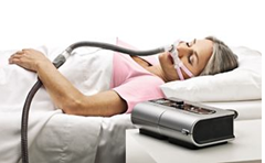
Figure 16. Adaptive Servo- ventilator

Figure 17. Provent
CONCLUSION:
Over recent decades, advancements in understanding Obstructive Sleep Apnea (OSA) have underscored its prevalence, health risks, and the importance of early, effective management. Oral appliances have emerged as a valuable non-surgical alternative to CPAP therapy, particularly for patients with mild-to-moderate OSA, offering symptom relief and long-term adherence rates of 50–70% when provided by trained dental professionals. For edentulous patients, increasing the vertical dimension of occlusion in complete dentures may improve airway patency and reduce apnea events, highlighting the potential of prosthodontic solutions in OSA care. As sleep medicine evolves, prosthodontists play a critical and expanding role in the interdisciplinary management of OSA, offering personalized interventions that enhance patient outcomes and quality of life.
REFERENCES
- Dr. Divya Chandra S, Dr. K Sounder Raj, Dr. Prema, Dr. Manisha Minz. Obstructive Sleep Apnea and Its Prosthodontic Management- An Overview. International Journal of Health Sciences & Research. 259 Vol.8; Issue: 3; March 2018.
- Mona Juneja1, Sumit Singh Phukela, Bhupender Yadav, Abhishek Nagpal, Omkar Shetty, Pankaj Ritwal. Prosthodontic Management of Obstructive Sleep Apnea. Bull. Env. Pharmacol. Life Sci., Spl Issue [2] 2022: 550-560.
- Anna Abbasil, Sushilkumar Satish Gupta, Nitin Sabharwal, Vineet Meghrajani, Shaurya Sharma, Stephan Kamholz, Yizhak Kupfer. A comprehensive review of obstructive sleep apnea. Sleep Sci. 2021;14(2):142-154.
- Annapurna K, Suganya S, Vasanth R, Ranjith Kumar P. Prosthodontic Approach to Treat Obstructive Sleep Apnea. Annals of Medical and Health Sciences Research | Jul-Aug 2014 | Vol 4 | Issue 4 |.
- Winfried Randerath, Jande Lange, Jan Hedner, Jean Pierre T.F.Ho, Marie Marklund, Sofia Schiza, Jorg Steier and JohanVerbraecken. Current and novel treatment options for obstructive sleep apnoea. ERJ Open Res 2022; 8:00126-2022.
- Dr. Pranathi Rajesh, Dr. Annie Susan Thomas, Dr. Aby Mathew T, Dr. Suja Joseph, Dr. Gopika Sali, Dr. Melwin Anna Abraham. Can Sleep Apnea be Prosthodontically Managed? –A Review. IOSR Journal of Dental and Medical Sciences (IOSR-JDMS) e-ISSN: 2279-0853, p-ISSN: 2279-0861.Volume 19, Issue 6 Ser.3 (May. 2021), PP 36-4.
- S. Savini, a Ciorba, c. Bianchini, f. Stomeo, v. Corazzi, c. Vicini, s. Pelucchi. Assessment of obstructive sleep apnoea (OSA) in children: an update. ACTA OTORHINOLARYNGOLOGICA ITALICA 2019;39:289-297; doi: 10.14639/0392-100X-N0262.
- Dr Sahil Dahale, Dr Prasad Adhapure, Dr Yeshwante Babita, Dr Sakshi Suryawanshi, Dr Kanchan Mokashi. PROSTHODONTIC MANAGEMENT IN OBSTRUCTIVE SLEEP APNEA. 2024 International Journal of Creative Research Thoughts (IJCRT) | Volume 11, Issue 6 May 2024 | ISSN: 2320-2882.
- Viranchi Oza. Dental Appliances and Mandibular Advancement Devices in Obstructive Sleep Apnea. International Journal of Head and Neck Surgery (2019): 10.5005/jp-journals-10001-1366.
- Prachi Gupta, Ram Thombare, Sameer Singhal, A J Pakhan. Obstructive sleep apnea and edentulism role of complete dentures/oral appliance from prosthodontics perspective: a review. Indian Journal of Sleep Medicine (IJSM), Vol. 5, No. 4, 2010.
- Aspasia Pachiou DDS, MSc Panagiotis Roulias DDS, MPH, MSc Paschalis Steiropoulos MD, PhD Demetrios J. Halazonetis DDS, MS Stefanos Kourtis DDS, Dr. Odont. Association of edentulism and obstructive sleep apnea: A systematic review. J. Prosthodont. 2024;1–10.
- Navya N B, Sanjayagouda B Patil, Abhilasha S B, Ankitha Thejus. OBSTRUCTIVE SLEEP APNEA AND ITS PROSTHODONTIC MANAGEMENT: A DENTAL OUTLOOK. JPID – The journal of Prosthetic and Implant Dentistry / Volume 7 Issue 1 / September–December 2023.
- Lt Gen S. Murali Mohan (Retd), Col E. Mahesh Gowda, Maj A.S. Banari. Obstructive sleep apnea (OSA): A prosthodontic perspective. medical journal armed forces india 71 (2015) S395eS399.
- Marie Marklund, Marc J.A. Braem, and Johan Verbraecken. Update on oral appliance therapy. Eur Respir Rev 2019; 28: 190083.
- NARKHEDE MB, CHINCHOLE PP, MEHETRE GD AND RATNAPARKHI NG. ANTI-SNORING DEVICE: A REVIEW. IJBPAS, June, 2022, 11(6): 2598-2608.
- CM MARYA, SONAM JAIN, RUCHI NAGPAL, PRATIBHA TANEJA, SAKSHI KATARIA, VANDANA MARYA. Obstructive Sleep Apnea: A Dental Perspective. International Healthcare Research Journal 2019;3(8):260-266.
- Sanjay R. Patel, MD, MS. IntheClinic® Obstructive Sleep Apnea. 2019 American College of Physicians ITC82 In the Clinic Annals of Internal Medicine 3December2019.
- Najla S. Alrejaye , Hamdan Al-Jahdali. Dentists’ role in obstructive sleep apnea: A more comprehensive review. Sleep Epidemiology 4 (2024) 100073.
- David Sherring, Norman Vowles, Ral Antic, Suren Krishnan, Alastair N Goss. Obstructive sleep apnoea: A review of the orofacial implications. Australian Dental Journal 2001;46:(3):154-165.
- Dr. Vijaya Laxmi Murty, Dr. Gulab Chand Baid, Dr. Saumya Sharma, Dr. Sanjeev Singh, Dr. Vivek Lath, Dr. Priyabrata Jena. Obstructive Sleep Apnea: A Prosthodontist’s Viewpoint. Vijaya Laxmi Murty., Sch J Dent Sci, May, 2023; 10(5): 102-107.
- G. J. Gibson. Obstructive sleep apnoea syndrome: underestimated and undertreated. British Medical Bulletin 2004;72.
- Amy Veenstra, RN, CCRN, CMC, Emylene Untalan, MSN, RN, CCRN, CPAN. Implications and Interventions Related to Obstructive Sleep Apnea. Adapted from Gutierrez C, Brady P. Obstructive sleep apnea: a diagnostic and treatment guide. J Fam Pract 2013;62(10):570.
- Johan Verbraecken, Jan Hedner and Thomas Penzel. Pre-operative screening for obstructive sleep apnoea. Eur Respir Rev 2017; 26: 160012.
- Bao Qian, Zhan-Jun Chen, Yong-Sheng Wang, Xiao-Yan Hu, Xiao-Biao Hu, Yong-Hua Zheng. Clinical efficacy and mechanism study of mid-frequency anti-snoring device in treating moderate obstructive sleep apnea-hypopnea syndrome. World J Clin Cases 2024 February 16; 12(5): 942-950 ISSN 2307-8960.
- S. R. Apoorva, Hiremath Shaashi, Sunantha Selvaraj. Oral appliances: An odds-on in obstructive sleep apnoea management. Journal of Academy of Dental Education • Volume 7 • Issue 2 • July-December 2021.
- Dr. Aparna S Barabde, Dr. Mradul Gupta, Dr. R. U.Thombare, Dr. Shailesh M Barabde, Dr. B.R.Thombare. Principles of Prosthodontic Management of Sleep Apnea Syndrome. Heal Talk // July-August 2015 // Vol 07 // Issue 6.
- Victor Hoffstein. Review of oral appliances for treatment of sleep-disordered breathing. Sleep Breath (2007) 11:1–22.
- Timothy G. Quinnell and Ian E. Smith. Obstructive Sleep Apnoea in the Elderly Recognition and Management Considerations. Drugs Aging 2004; 21 (5): 307-322.
- Yeongrok Lee, MD, Jae Yong Lee, MD, PhD, Ji Ho Choi, MD, PhD. Oral Appliance Therapy for Obstructive Sleep Apnea: Clinical Benefits and Limitations. Sleep Med Res 2023;14(4):175-182.
- Dr. Aushili Mahule, Dr. Jaykumar Gade, Dr. VandanaGade. MANAGING OBSTRUCTIVE SLEEP APNEA: A PROSTHODONTIST’S WAY. International Journal of Current Advanced Research Vol 9, Issue 02(B), pp 21193-21198, February 2020.
- Diane S. Lazard, M.D; Marc Blumen, M.D; Pierre Levy, M.D; Pierre Chauvin, M.D; Dorothee Fragny, M.D; Isabelle Buchet, M.D; Frederic Chabolle, M.D. The Tongue-Retaining Device: Efficacy and Side Effects in Obstructive Sleep Apnea Syndrome. Journal of Clinical Sleep Medicine, Vol.5, No. 5, 2009.
- Izabella Paola Manetta, Dominik Ettlin, Pedro Mayoral Sanz, Isabel Rocha, Miguel Meira e Cruz. Mandibular advancement devices in obstructive sleep apnea: an updated review. Sleep Sci. 2022;15(Special 2):398-40.
- Suresh Nayar, BDS, MDS, and Jeremy Knox, BDS, MScD, PhD. Management of obstructive sleep apnea in an edentulous patient with a mandibular advancement splint: A clinical report. THE JOURNAL OF PROSTHETIC DENTISTRY. AUGUST 2005 VOLUME 94 NUMBER 2.
- Shouresh Charkhandeh, DDS; David Kuhns, PhD; Sung Kim, BS. A Fully Digital Workflow and Device Manufacturing for Mandibular Repositioning Devices for the Treatment of Obstructive Sleep Apnea: A Feasibility Study. Journal of Dental Sleep Medicine. Vol. 4, No. 4, 2017.
- Banu F, Jeyapalan K, V A (July 27, 2021) Custom-Made Dual-Functional Oral Appliance for Management of Obstructive Sleep Apneic Completely Edentulous Patient. Cureus 13(7): e16656. DOI 10.7759/cureus.16656.
Disclaimer from Guident
The views and opinions expressed in articles published in Guident are solely those of the authors and do not necessarily reflect the views of the editors, the editorial board or the publisher, Ivory India. Guident does not endorse any product, technique or treatment described. Clinical content is intended for qualified dental and medical professionals and is not a substitute for individual clinical judgement. Guident, its editors and the publisher accept no liability for any consequence arising from the use of information published in the journal.
How to cite this article
Dr. Hombesh M.N, Dr. Swetha Mullapudi, Dr. Raksha Suresh Bajpe. Obstructive Sleep Apnea and Prosthodontic Considerations: A Review. Guident. 2025 Oct;18(11). Available from: https://ivoryindia.in/articles/prosthodontics/obstructive-sleep-apnea-and-prosthodontic-considerations-a-review.html