Enhancing Esthetics in Maxillary Anterior Region: A Clinical Case Report
Department of Prosthodontics and Crown & Bridge, Seema Dental College & Hospital, Rishikesh, Uttarakhand, India.
ABSTRACT
Implant therapy is now universally accepted as a predictable treatment modality for the replacement of missing teeth for both function and esthetics. This case report explains the sequential step of esthetic rehabilitation in the anterior maxilla, emphasizing the meticulous planning and judicious execution involved to attain the optimal outcome. Starting with the most comprehensive evaluation of the patient's oral health, including bone morphology and soft tissue status, a tailored treatment plan was designed as a joint effort by the implant surgeon and restorative dentist. The single tooth implant was inserted surgically with accuracy so that it could be optimally positioned and angulated to provide aesthetically pleasing outcomes while respecting the adjacent vital structures. Prosthetic rehabilitation employed proper chosen materials and techniques to harmonize fully with the rest of the natural teeth. With interdisciplinary collaboration and careful attention, anterior maxilla was aesthetically restored successfully, illustrating the restorative capacity of modern implant dentistry.
Key Words – Conventional Implant, Esthetic Zone, Abutment, Prosthesis
INTRODUCTION
Restoration of aesthetic area with dental implants is a long-standing task. In comparison to conventional means, implants provide clear benefits. Implants enable the production of restorations very similar to natural teeth and improve aesthetics as well as functionality. Formerly, insufficient bone supply, particularly in the anterior mandible, hindered the placement of implants. Presently, prosthetic needs mainly determine implant positioning, demonstrating the changing modus operandi of implant dentistry in response to esthetical demands in the anterior area.1
In the current era of digital dentistry, patient expectations for natural-looking, durable, and minimally invasive restorations—especially in the esthetic zone—have grown significantly. The esthetic zone, typically encompassing the maxillary anterior teeth, plays a crucial role not only in facial appearance but also in phonetics, self-confidence, and psychosocial well-being. As a result, the demand for restorative solutions that blend seamlessly with the surrounding dentition while maintaining long-term function has led to the evolution of both dental materials and technologies.2
Technological advances in dentistry have resulted in stable success rates, guaranteeing predictable outcomes. These developments are a hallmark of major progress in the field, typified by better techniques and materials. Patients now have confidence in undergoing implant treatment, anticipating reliable results. This advance reflects increased faith in dental implants as a trusting means of replacing teeth, a major achievement in the pursuit of excellence in modern dentistry.3
Unlike posterior implant placement, where function predominates, anterior implant rehabilitation must account for several esthetic parameters, including gingival symmetry, interdental papilla preservation, and the natural emergence profile of the prosthesis. The slightest deviation in implant position or soft tissue management can compromise the final esthetic result, making this one of the most technique-sensitive areas in implant dentistry.4
The importance of anterior maxilla is compounded with its high-profile exposure. When there is a high lip line, the smile is more exposed, enhancing the demand for beauty perfection. Some experts highlight the demand to reconcile the function and aesthetics'. Obtaining an optimal outcome here not only restores function but also significantly enhances the patient's confidence and overall well-being esthetic results: A natural look is of utmost importance.5 The patient's lip line, position of the implant, and the soft tissue surrounding it are all important considerations. When treatment fails, difficult situations may ensue that require the removal of the implant and the augmentation of tissues.
Construction and optimal emergence profile for implant supported prosthesis in esthetical zone requires use of bone regenerative materials, soft tissue augmentation and suitable abutments. This region often presents challenges such as reduced bone volume, thin gum tissue, and the need to preserve gum tissue between teeth. The bone anatomy heavily influences soft tissue contours around an implant.6 Various techniques can be used, including traditional drilling, osteotome techniques, piezoelectric bone surgery, flapless placement, and osseodensification. Undersized drilling can increase stability. Successful implant placement in the maxillary anterior region requires a comprehensive approach prioritizing esthetics, addressing anatomical and surgical considerations, and managing potential complications. Detailed planning, bone and soft tissue grafting, and meticulous techniques are essential for predictable, long-lasting results.7 This case report highlights the optimum implant positioning in the maxillary aesthetic region providing improved structural stability and prosthetic aesthetics that harmonize with the natural dentition.
CASE REPORT
A 23-year-old male patient came to the Department of Prosthodontics and crown & bridge (SDCH Rishikesh) with chief complaint of missing teeth in upper left front tooth region. On detailed intra oral examination revealed that congenitally missing teeth wrt 21,22 regions. Patient was undergone orthodontic treatment since last 1 year. [Figure 1 & 2]. Two conventional alpha bio implants w.r.t 21,22 was planned.
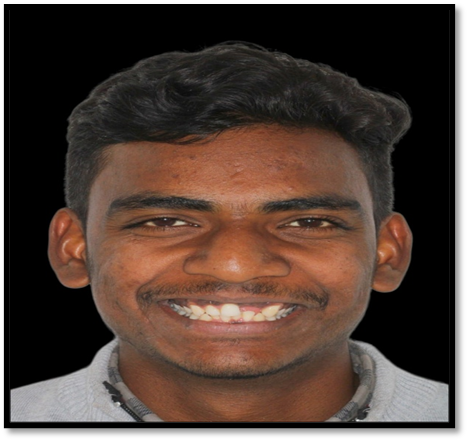
Figure 1 (Extraoral frontal view)
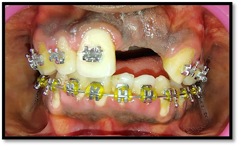
Figure 2 (Intraoral frontal view)
Then the patient was well informed about all the treatment modalities which can be performed to him and informed about the pros and cons of each treatment. He was highly concerned about his esthetics and wishes to have dental implant among all the treatment option. Subsequently, additional investigation for placing the implant was carried out using Dentsply sirona CBCT scan. Finally, after radiographic evaluation in detail and consideration of approximating landmarks, important structures and bone density two alpha bio dental implant of dimensions 3.75x13mm was chosen.
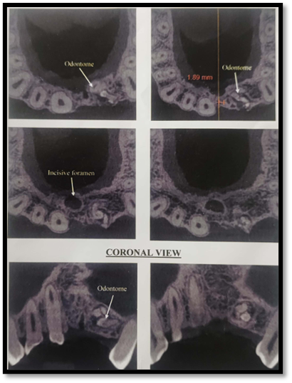
Figure 3 (CBCT investigation)
The surgical procedure was performed in sterile surgical ?eld and the armamentarium used for surgery are shown in (Figure 4).

Figure 4 (Armamentarium)
Preoperative oral cavity decontamination with chlorhexidine 0.2% mouth wash for 1 min and perioral skin decontamination with 5% povidone-iodine solution was performed. Site was anesthetized with 2% Lidocaine with 1:80000 adrenaline (Xicaine ICPA).
Mid crestal incision on the alveolar crest and two vertical releasing incisions were given and a full thickness ?ap was re?ected. A well round edge type ridge was seen on re?ection of ?ap. Meanwhile, 2 Implant alpha bio implants were placed in 21,22 of size 3.75 X 13mm ,3.75 X 13mm. A good primary stability was achieved, thereafter cover screw was placed and flap was sutured. Post operative rvg was taken as shown in (Figure 5 a-f).
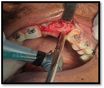
Figure: 5A
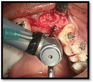
Figure: 5B

Figure: 5C
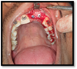
Figure: 5D
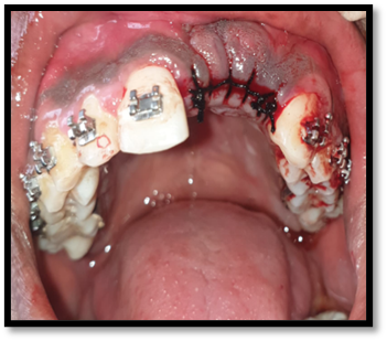
Figure: 5E
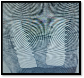
Figure: 5F
Figure 5 (Stage 1 Surgery)
Temporary, crows were given (Figure 6). Post operative instructions were given to the patient, and was asked to report after 7 days patient for suture removal.
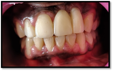
Figure 6 (Temporary crown)
The patient was reported back after 3 months for the final prosthesis. Implant stability Quotient test done again, which showed excellent biological stability with reading measuring in between 75-78. Thereby, showing excellent secondary stability or osseointegration. The cover screw was removed and a final angled abutment (Alpha Bio) was attached to the implant. (figure 7).
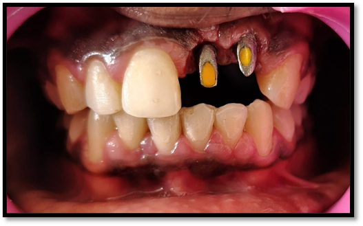
Figure 7 (Final abutment)
With a digital scanner (Omnicam Dentsply Sirona) direct abutment scanning was done and STL file was send to the laboratory for the fabrication of CAD CAM zirconia crown. (figure 8).


Figure 8 (Intra oral scans)
The ?nal CAD CAM zirconia crown, (figure 9) was seated on the ?nal abutment. Then the ?nal prosthesis was cemented with resin modified glass ionomer cement (3M ESPE RelyX U200) on the abutment (figure 10).

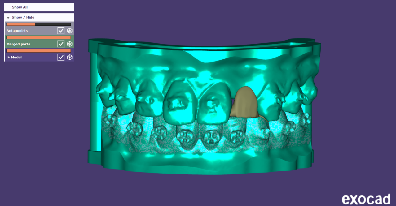
Figure 9 (3D printed model with final crown)
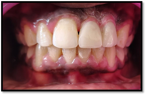
Figure 10 (final cementation of crown)
DISCUSSION
Rehabilitation of the anterior maxilla with a dental implant poses a unique clinical challenge due to the dual necessity of functional success and high esthetic demands. The anterior region, often referred to as the "esthetic zone," requires precise planning and execution to achieve harmonious integration with surrounding natural dentition in terms of color, contour, and gingival architecture.8
In the present case, the placement of a single implant in the anterior maxillary region followed by prosthetic rehabilitation with a zirconia crown demonstrated favorable functional and esthetic outcomes. The selection of zirconia as the restorative material offered significant advantages. Zirconia exhibits excellent biocompatibility, high flexural strength, and superior optical properties that closely mimic the translucency of natural enamel, making it an ideal choice for anterior restorations.9 Moreover, zirconia's minimal plaque accumulation and soft tissue response make it advantageous in areas where gingival aesthetics are critical.
Immediate implant placement or early placement protocols, when feasible, offer benefits in preserving alveolar bone and soft tissue contours. However, careful assessment of the socket morphology, periodontal status, and soft tissue volume is vital to determine the timing of implant placement. In our case, implant placement was performed following complete healing of the extraction socket, which allowed adequate osseous volume and predictable primary stability, minimizing the risk of implant failure.10
From an esthetic standpoint, monolithic zirconia offers a superior alternative to metal-ceramic restorations, especially in the anterior region. Its tooth-like color, translucency, and ability to be customized with stains and glazes make it favorable for esthetic restorations. Moreover, the absence of a metal substructure eliminates the risk of gray margins or gingival discoloration over time.11
One of the concerns with zirconia on implants is its rigidity and limited shock-absorbing capacity compared to natural teeth or other restorative materials like PEEK or PMMA. However, recent modifications such as translucent and high-strength zirconia generations have addressed the balance between strength and esthetics. The low plaque affinity of zirconia and its favorable soft tissue response also contribute to long-term peri-implant health.12
In the presented case, the use of a CAD/CAM-milled zirconia crown on an implant abutment demonstrated excellent marginal fit, esthetic integration with adjacent dentition, and patient satisfaction. The digital workflow also reduced clinical chairside time and the number of appointments, improving overall treatment efficiency.13
However, the success of CAD/CAM zirconia restorations also depends on accurate implant positioning, appropriate emergence profile design, and proper occlusal load distribution. Failures, if any, are often linked to biomechanical overload or improper design rather than material deficiencies. Therefore, comprehensive diagnosis, prosthetically driven implant planning, and interdisciplinary collaboration are critical for successful outcomes.14
One of the key considerations in anterior implant restoration is the emergence profile and the preservation or augmentation of the interdental papilla. Inadequate management can lead to black triangles and compromised esthetics. The provisionalization phase played a crucial role in shaping the peri-implant soft tissues and ensuring a natural emergence profile. Studies have emphasized the importance of a properly contoured provisional restoration in achieving optimal peri-implant mucosal architecture.15
The long-term success of the anterior implant was further supported by the use of screw-retained prosthesis, which allowed easy retrievability and eliminated the risks associated with residual cement. Additionally, a platform-switched implant design was used, which has been shown to minimize crestal bone loss and support soft tissue stability .16
While zirconia crowns present excellent esthetics and strength, they can pose challenges in terms of bonding protocols and potential wear on opposing dentition. However, in this case, careful occlusal adjustment and selection of a monolithic zirconia crown with polished occlusal surfaces minimized these risks.17
Overall, the case highlighted the importance of an interdisciplinary approach involving precise diagnostic planning, surgical execution, prosthetic design, and esthetic consideration. The use of digital technologies like CBCT and CAD/CAM further enhanced the predictability and accuracy of treatment outcomes.
CONCLUSION
The rehabilitation of anterior missing teeth with dental implants demands a meticulous approach due to the critical esthetic and functional requirements of this region. This case demonstrated that, with proper case selection, precise surgical planning, and the use of advanced prosthetic materials like zirconia, highly satisfactory esthetic and functional outcomes can be achieved. The zirconia crown provided excellent gingival harmony, durability, and natural esthetics, making it a preferred choice for anterior restorations. Moreover, a patient-specific approach, including soft tissue management and provisionalization, proved essential in achieving optimal peri-implant esthetics. Long-term success in such cases is best ensured through a multidisciplinary strategy, digital planning, and strict adherence to prosthodontic principles.
REFERENCES
- El Askary AS. Aesthetic considerations in anterior single tooth replacement. Implant Dent. 1999;8:61-67.
- Spielman HP. Influence of the implant position on the aesthetics of the restoration. Pract Periodontics Aesthet Dent. 1996; 8:897-904.
- Bashutski JD, Wang HL. Common implant esthetic complications. Implant Dent. 2007; 16(4):340-8.
- Simeone P, De Paoli C, De Paoli S, Leofreddi G, Sgrò S. Interdisciplinary treatment planning for single-tooth J Esthet Restor Dent. 2007;19(2): 79-88;
- Misch CE. The importance of dental implants. Gen Dent. 2001;49:38-45.
- Tischler M. Dental implants in the esthetic zone. Considerations for form and function. N Y State Dent J. 2004;70:22-26.
- Kopp KC, Koslow AH, Abdo OS. Predictable implant placement with a diagnostic/surgical template and advanced radiographic imaging. J Prosthet Dent. 2003; 89:611-615.
- Figliuzzi M, Mangano FG, Fortunato L, De Fazio R, Macchi A,and GI. Vertical ridge augmentation of the atrophic posterior mandible with custom-made, computer-aided design/computer-aided manufacturing porous hydroxyapatite scaffolds. JCraniofac Surg.2013;24(3):856–9.
- Mangano F, Macchi A, Shibli JA, Luongo G, Iezzi G, Piattelli A, et al. Maxillary ridge augmentation with custom-made CAD/CAM scaffolds. A 1-year prospective study on 10 patients. JOral Implantol. 2014;40(5):561–9.
- The Glossary of Prosthodontic Terms: Ninth Edition. J Prosthet Dent 2017;117(5S):e1-e105.
- Raghavendra S, Wood MC, Taylor TD. Early wound healing around endosseous implants: a review of the literature. Int J Oral Maxillofac Implants 2005;20(3):425-31.
- Oncu E et al. Positive effect of platelet rich fibrin on osseointegration. Med Oral Patol Oral Cir Bucal 2016;21(5):e601-7.
- Shah R, M G T, Thomas R, Mehta DS. An Update on the Protocols and Biologic Actions of Platelet Rich Fibrin in Dentistry. Eur J ProsthodontRestor Dent 2017;25(2):64-72.
- Fujioka-Kobayashi M et al. Optimized Platelet-Rich Fibrin With the Low-Speed Concept: Growth Factor Release, Biocompatibility, and Cellular Response. J Periodontol 2017;88(1):112-121.
- Clark D et al. Advanced platelet-rich fibrin and freeze-dried bone allograft for ridge preservation: A randomized controlled clinical trial. J Periodontol 2018;89(4):379-387.
- Margono DA et al. Comparison of Advanced Platelet Rich Fibrin (A-PRF) and Culture Media Conditioned Warton's Jelly (CMCWJ) on Fibroblast Cells Proliferation.Pesq Bras Odontoped Clin Integr 2018;18(1):e4085.
- Masuki H et al. Growth factor and pro-inflammatory cytokine contents in platelet-rich plasma (PRP), plasma rich in growth factors (PRGF), advanced platelet-rich fibrin (A-PRF), and concentrated growth factors (CGF). Int J Implant Dent 2016;2(1):19.
Disclaimer from Guident
The views and opinions expressed in articles published in Guident are solely those of the authors and do not necessarily reflect the views of the editors, the editorial board or the publisher, Ivory India. Guident does not endorse any product, technique or treatment described. Clinical content is intended for qualified dental and medical professionals and is not a substitute for individual clinical judgement. Guident, its editors and the publisher accept no liability for any consequence arising from the use of information published in the journal.
How to cite this article
Dr. Nikhil Kumar, Dr. Varun Kumar, Dr. Geeta Arya. Enhancing Esthetics in Maxillary Anterior Region: A Clinical Case Report. Guident. 2025 Oct;18(11). Available from: https://ivoryindia.in/articles/prosthodontics/enhancing-esthetics-in-maxillary-anterior-region-a-clinical-case-report.html