Lasers and Little Smiles: A Pediatric Case of Gingival Depigmentation
Department of Pediatric and Preventive Dentistry, College of Dental Sciences, Davangere, Rajiv Gandhi University of Health Sciences, Karnataka, India
ABSTRACT
Background:
Gingival pigmentation, though physiologic and benign, may cause aesthetic concern, particularly in paediatric patients. Increasing awareness and demand for cosmetic dental procedures have led to the use of various depigmentation methods. Among them, diode laser therapy has gained popularity due to its precision, patient comfort, and favourable healing outcomes.
ABSTRACT
Methods:
This case report describes the management of gingival melanin pigmentation in a 12-year-old male patient using a diode laser. The pigmentation was present in the anterior mandibular gingiva extending posteriorly to the molars. After clinical examination and hematological evaluation, laser-assisted depigmentation was performed under local anaesthesia using a 980 nm diode laser in gated pulsed mode. Standard laser safety protocols were followed. The procedure involved superficial ablation of the pigmented gingival epithelium, with no periodontal dressing applied postoperatively. Follow-ups were conducted at 7 days, 1 month, and 3 months.
Discussion:
The diode laser provided effective removal of pigmentation with minimal discomfort, excellent healing, and high patient satisfaction. No repigmentation was observed at the 3-month follow-up. Literature supports diode laser’s advantages over conventional methods, including better haemostasis, less postoperative pain, and reduced recurrence in the short term.
Conclusion:
Diode laser-assisted depigmentation is a safe, effective, and well-tolerated technique in paediatric patients. Continued long-term follow-up is essential to monitor for recurrence.
Key words : paediatric , gingival depigmentation, diode laser,
INTRODUCTION
A pleasing smile is often considered an important aspect of facial aesthetics and social interaction. While tooth alignment, shape, and colour typically dominate aesthetic concerns, the appearance of gingival tissues also plays a significant role in the perception of an attractive smile.1 Gingival health in the form of size, shape, consistency and colour are vital components for an attractive smile. The color of gingiva varies, from pale pink to deep bluish purple. Between these normal limits, a large number of colour variations are observed, which depends primarily upon the vascular supply, the thickness of the epithelium, the degree of keratinization, and the presence of pigments within the epithelium.2
Gingival pigmentation is more prevalent among individuals with darker skin tones and can vary in intensity, extent, and pattern.3 While it is widely accepted and often overlooked in adults, increasing awareness and concern about aesthetics in younger populations have led to greater demand for cosmetic procedures including gingival depigmentation in children and adolescents.4
A variety of techniques have been proposed for gingival depigmentation, including scalpel surgery, cryosurgery, chemical cauterization, abrasion, electrosurgery, and more recently, laser therapy.5 Among these, laser-assisted depigmentation has gained popularity due to its numerous advantages: precision, reduced intraoperative bleeding, sterilization of the surgical site, minimal postoperative discomfort, and faster healing.6 Various laser systems such as diode, Nd:YAG, Er:YAG, and CO2 lasers have been employed, with the diode laser being particularly favored in dental practice for soft tissue procedures due to its efficiency, affordability, and safety profile.7
This case report aims to present the clinical management of gingival hyperpigmentation in a paediatric patient using diode laser therapy, detailing the procedural steps, outcomes, and advantages observed. The report also emphasizes the importance of addressing aesthetic concerns in paediatric dentistry and supports the use of laser as a safe and patient-friendly modality in selected cases.
CASE REPORT
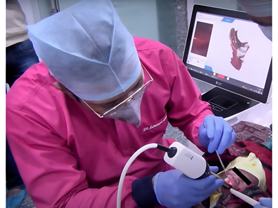
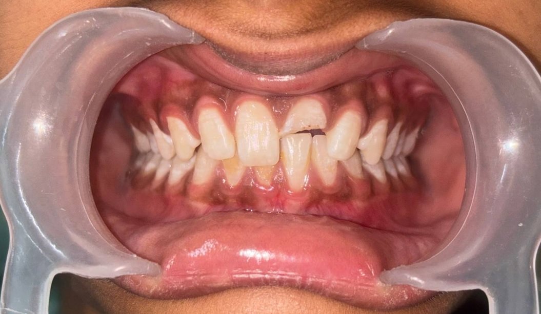
A 12-year-old male patient reported to the Department of Paediatric and Preventive Dentistry, College of Dental Sciences, Davangere, Karnataka, India. with his mother, who was concerned about the dark coloration of the gums in the front tooth region, particularly in the lower arch, due to aesthetic reasons. On further history, the mother stated that the pigmentation had been present since early childhood and had not changed significantly over time, suggesting a non-pathological origin. On examination melanin deposits was seen all over the gingiva from incisor to molar region [Figure 1]. Following blood investigations [Figure 2], Laser assisted depigmentation procedure was planned after taking informed consent of the parent. After oral prophylaxis and maintenance of oral hygiene instructions the procedure was carried out. Infiltration of local anesthesia was done using insulin syringe [Figure 3]. Before the procedure began, patient and dentist were protected from laser light by wearing protector spectacles. A diode laser unit of with a wavelength of 980 nm, in gated pulsed mode, was used. The tip was moved using interrupted brushing [Figure 4]. The saline soaked gauze was used to remove the epithelial remnants. No periodontal dressing was placed [Figure 5]. Follow up was done after 7 days [Figure 6], 1month [Figure 7], 3months [Figure 8]

Figure 1: Preoperative gingival hyperpigmentation

Figure 2: Administration of local anesthesia

Figure 3: Administration of local anesthesia

Figure 4: During the laser depigmentation

Figure 5: Immediately after the procedure

Figure 6: 1 week follow-up

Figure 7: 1-month follow-up

Figure 8: 3-Months follow-up
DISCUSSION
Gingival melanin pigmentation, though benign, often raises aesthetic concerns especially in paediatric patients, prompting demand for cosmetic correction. The diode laser has gained popularity due to its precision, haemostasis, and favourable healing profile. In our paediatric case, diode laser depigmentation yielded excellent aesthetic results with minimal discomfort and no postoperative complications, consistent with findings by Gupta et al8 who reported similar outcomes with no regimentation at 1-month follow-up. When compared to conventional scalpel techniques, diode laser treatment offered a lower early regimentation rate. Mahajan et al9 found slower recurrence in laser-treated sites at 3 months, though by 9 months both methods showed some recurrence. Long-term stability is further supported by Gupta.10 Case reports in adults, like those by Salian et al1(2024) and Esmaeili et al. (2022)11, demonstrate effective diode laser use, with high aesthetic satisfaction and manageable recurrence rates. Overall, these findings reinforce that diode laser is a safe and effective option for gingival depigmentation not just in adults, but also in paediatric patients, offering excellent aesthetic outcomes, patient comfort, and acceptable long-term results. Continued follow-up, especially regarding recurrence, remains essential.
CONCLUSION
Gingival depigmentation using a diode laser in the paediatric patient demonstrated excellent aesthetic outcomes, minimal discomfort, and no postoperative complications. At the 3-month follow-up, no signs of regimentation were observed, indicating stable short-term results. These findings support the diode laser as a safe, effective, and well-tolerated treatment modality for managing gingival pigmentation in children, with favourable healing and patient satisfaction. Continued long-term follow-up is recommended to monitor for potential recurrence.
REFERENCES
- Salian SS, Dhadse PV, Patil RT, SALIAN SS, Dhadse P. Laser-assisted gingival depigmentation: A case report. Cureus. 2024 Jan 4;16(1).
- Jalan P, Ghosh A, Zahir S, Kundu GK. Laser assisted management of hyperpigmented gingiva and eruption cyst in children–Two case reports. International Journal of Pedodontic Rehabilitation. 2020 Jan 1;5(1):10.
- Kela N, Salian S, Dhadse P, Patil R, Punse S, SALIAN SS, Patil RT, Punse SD. Laser Depigmentation: A Case Report. Cureus. 2024 Jul 20;16(7)
- Bahadure RN, Singh P, Jain E, Khurana H, Badole G. Management of pigmented gingiva in child patient: a new era to the pediatric dentistry. International Journal of Clinical Pediatric Dentistry. 2013 Oct 14;6(3):197.
- Murthy MB, Kaur J, Das R. Treatment of gingival hyperpigmentation with rotary abrasive, scalpel, and laser techniques: A case series. Journal of Indian Society of Periodontology. 2012 Oct 1;16(4):614-9.
- Prasad D, Sunil M, Mishra R, Reddy S, Reddy MJ, Ramesh A. Gingival melanin depigmentation using a diode laser: A case series. J Clin Diagn Res. 2014;8(10):ZC122-5.
- Khosravi R, Daneshparvar P, Ahmadi R. Clinical effectiveness of lasers in gingival depigmentation: A systematic review. Photobiomodul Photomed Laser Surg. 2020;38(1):1-7.
- Gupta G. Management of gingival hyperpigmentation by semiconductor diode laser. J Cutan Aesthet Surg. 2011;4(3):208–210.
- Mahajan G, Bhola RS, Shukla P, Raina A. To compare the gingival melanin repigmentation after diode laser application and surgical removal. J Indian Soc Periodontol. 2017;21(2):112–118.
- Chandra GB, Kumar NS, Mehta DS. Evaluation of surgical scalpel versus semiconductor diode laser techniques in the management of gingival melanin hyperpigmentation: A split-mouth randomized clinical comparative study. J Indian Soc Periodontol. 2020;24(1):47–53.
- Esmaeili S, Shahbazi S, Asnaashari M. Gingival melanin depigmentation using a diode 808?nm laser: a case series. J Lasers Med Sci. 2022;
Disclaimer from Guident
The views and opinions expressed in articles published in Guident are solely those of the authors and do not necessarily reflect the views of the editors, the editorial board or the publisher, Ivory India. Guident does not endorse any product, technique or treatment described. Clinical content is intended for qualified dental and medical professionals and is not a substitute for individual clinical judgement. Guident, its editors and the publisher accept no liability for any consequence arising from the use of information published in the journal.
How to cite this article
Dr. Tayaba Anjum, Dr. Neena I. E., Dr. Mallikarjuna K.. Lasers and Little Smiles: A Pediatric Case of Gingival Depigmentation. Guident. 2025 Nov;18(12). Available from: https://ivoryindia.in/articles/pedodontics/lasers-and-little-smiles-a-pediatric-case-of-gingival-depigmentation.html