Medicinal Carrier: A Treatment Modality for Atypical Gingivostomatitis
Department of Prosthodontics, Seema Dental College & Hospital, Rishikesh, Uttarakhand, India
INTRODUCTION
Atypical Gingivostomatitis is a rare benign entity, characterized by an infiltration of plasma cells in the subepithelial tissues of the gums. It is an uncommon condition of unknown etiology and has been described in the literature, also plasma cell gingivitis, idiopathic gingivostomatitis, and allergic gingivostomatitis.1 Plasma cell gingivitis (PCG), is a distinct condition, among the potpourri of gingival lesions which is often characterized by particular allergen which causes a dense infiltration of plasma cells in gingival connective tissue.1 Allergens that are thought to be the cause of Plasma cell gingivitis includes a brand of herbal tooth paste, a specific type of mint candy, peppers used for cooking, environmental and food flavoring allergens and khat leaves2 Depending upon the etiology of PCG, it has been classified as:
a. Lesions caused by an allergen
b. Neoplastic lesions, and
c. Lesions of unknown cause3
The clinical features of PCG includes a diffuse reddish and edematous swelling, with a clear demarcation along the mucogingival junction, Ulcerations are rare features and are commonly asymptomatic, although some individuals may complain of pruritus, burning, or pain sensations.4, A rapid onset of sore mouth, which is often intensified by dentifrices and hot or spicy food, the entire free and attached gingiva demonstrates a diffuse enlargement with bright erythema and loss of normal stippling2, Extensions onto the palate can occur and edentulous areas typically exhibit less intense changes, More prevalent in young women., In chewing gum cases of the early 1970s, involvement of the lips and tongue was typical. The lips were dry, atrophic, occasionally fissured, and angular cheilitis was frequent. Tongue involvement resulted in erythematous enlargement with furrows, mild crenation, and loss of the typical dorsal coating.2
The condition often affects the anterior gingiva and is usually seen in the maxilla, although direct contact with an allergen in the mandibular gingiva has reportedly produced a case of PCG.4 Histologically PCG is characterized by dense infiltration of the plasma cells separated with the strands of the collagen fibers in gingival connective tissue.5
The case reported here describes a rare case of PCG in which the identification of the etiologic agent remained inconclusive with intense gingival inflammation and enlargement predominantly in the upper and lower anterior region.
CASE REPORT
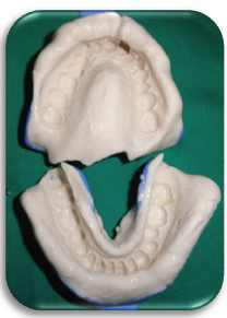
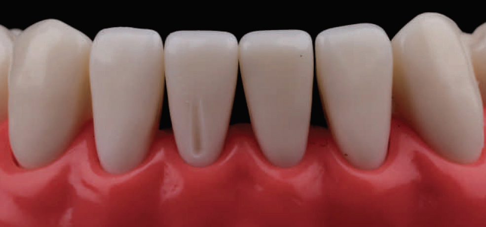
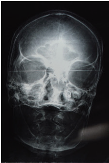
A 30-year-old female patient reported in the Department of Prosthodontics with a chief complaint of bleeding gums since one year with history of burning sensation on taking spicy food but no pain. The patient was on medication for the same but not on regular basis, since 2 months. On examination a uniform erythematous area was seen on both maxillary and mandibular arches, anteriorly as well as posteriorly extending into the attached and interdental gingiva. Figure 1 On palpation the lesion was positive for bleeding but non-tender. In addition, generalized gingival recession and severe halitosis was present. An occlusal splint was advised that would extend up to the level of gingiva and hence can be used as a medicine carrier for the patient. A diagnostic impression was taken and splint was fabricated Figure 2 & 3. Investigations done were ELISA and Plasma Protein. A complete hemogram and a routine biochemical and radiographic examination did not show any abnormality. Peripheral blood smear to rule out a typical immature cells. The results were normal. The patient was then advised topical application of Triamcinolone Acetonide thrice daily for one week with the use of splint. Figure 5 and Betamethasone (Betnosol) mouthwash twice daily for one week. Patient was also advised to avoid intake of spicy food.
On follow up after a week, the patient reported with good symptomatic relief, no bleeding and considerable reduction in the burning sensation. On examination there was 50% reduction in the erythema and gingival lesions. Figure 4 Plasma protein levels were normal.

Figure 1- Erythematous areas in both the arches

Figure 2 – Diagnostic model

Figure 3- Fabricated splint

Figure 4- 50% reduction after 1 week

Figure 5- Splint used as a medicine carrier
DISCUSSION
Atypical gingivostomatitis, is a benign condition of the gingiva with characteristics of generalized edematous and erythematic gingival tissues. The edematous tissue often extends to the mucogingival junction with a deep red appearance.
Depending upon the site of involvement, this condition could be considered a type of gingivitis (or gingival enlargement); a type of cheilitis; glossitis; or stomatitis. Sometimes the lips, the gums and the tongue can simultaneously be involved, and some authors have described this triad as a syndrome "plasma-cell gingivostomatitis".6 The mucous membranes of the genitals can also be involved by a similar condition, termed "plasma cell balanitis"7 or "plasma cell vulvitis".8
Plasma cell gingivitis and allergic gingivostomatitis are terms that are associated with allergic responses to substances. Other terms that have been used are Plasma cell gingivitis (PCG) idiopathic gingivostomatitis, allergic gingivostomatitis, hypersensitivity, and plasmacytosis. Several contact disorders of the oral tissues are known and are called orofacial disorders.
Dental materials such as crowns and restorations, food products, preservatives, and additives are reported to cause hypersensitivity as well. However, dental materials often produce a localized reaction rather than a generalized reaction of the tissue. Early publications by Silverman and Lozada, as well as Kerr et al., suggested that chewing gum was a major factor in reported cases cited within these publications. Specifically, those products with essential oils have been noted. Peppermint, wintergreen, menthol, spearmint, and cinnamon are known to cause tissue erythematic symptoms in some individuals.9
In 1968 Poswillo10 described a gingivitis, presumed to be this condition, in an article entitled "Plasmacytosis of the Gingiva." He speculated as to its cause without making any positive suggestion concerning the true nature of the process, and he did not suggest any treatment. Owings11 in September, 1969, published a report of four cases which he designated as atypical gingivostomatitis. He described the clinical and histologic findings and speculated that the disease might be allergic in nature or that it might be an autoimmune response to aerobic organisms. He reported performing full-mouth gingivectomy in two cases, with satisfactory results on short-term follow-up. In February, 1970, Vickers and Hudson12 reported on the condition under the designation of plasma-cell gingivitis and suggested that it was due to Candida infection. They were relatively unsuccessful in treating it with antifungal drugs.
Curto et al. reported that PCG is a type of plasma cell circum orificial mucositis or plasmacytosis circum orificialis. It has been reported to occur on the larynx, epiglottis, vulva, conjunctiva, and nasal aperture apart from the lips and tongue.13 Plasma cell granuloma, also known as an inflammatory pseudotumor, inflammatory myofibroblastic tumor or xanthomatous pseudotumor is an uncommon non-neoplastic lesion used to describe an enlargement confined to one specific site.14 The most common location of occurrence is the lungs, but it may also be found in the brain, kidney, heart, and rarely in the gingiva. In our case, the involvement was limited to the gingiva of the anterior segment of the jaws.
According to the literature, PCG management indicates that in many patients where no underlying cause could be discovered, topical or systemic immunosuppressive medication have been used as a non-definitive approach with variable results. Some patients do not respond to treatment, and no possible cause for the disease can be identified, in spite of all evaluations and therapeutic interventions.15 In this case, the patient’s thorough history was taken to rule out any of the probable causative factors predisposing to PCG.
However, there was no family history or no specific allergen was identified.
The therapeutic treatment includes corticosteroids, antimicrobials, topical and systemic antihistaminics, while surgical interventions like electrocoagulation, excision by laser etc. our patient responded to topical triamcinolone acetonide.16
CONCLUSION
Atypical gingivostomatitis is a rare disorder of idiopathic origin. Possible allergens should be identified by careful study of the patient’s history, appropriate diagnostic tests should be carried out to reach a definitive diagnosis to rule out similar lesions of the gingiva such as pyogenic granuloma while eliminating the probable cause. Topical and systemic steroids give good results.
REFERENCES
- Rawal SY, Rawal YB, Anderson KM, Bland PS, Stein SH. Plasma cell gingivitis associated with khat chewing. PERIO 2008;5:21-8.
- Neville oral and maxillofacial pathology, second edition.
- Marker P, Krogdahl A. Plasma cell gingivitis apparently related to the use of khat: Report of a case. Br Dent J 2002;192:311-3.
- Serio FG, Siegel MA, Slade BE. Plasma cell gingivitis of unusual origin. A case report. J Periodontol 1991;62:390-3.
- Janam, P; Nayar, BR; Mohan, R; Suchitra, A (January 2012). "Plasma cell gingivitis associated with cheilitis: A diagnostic dilemma!". Journal of Indian Society of Periodontology. 16 (1): 115–9.
- Hedin, CA; Karpe, B; Larsson, A (1994). “Plasma-cell gingivitis in children and adults. A clinical and histological description”. Swedish Dental Journal. 18 (4): 117–24.
- Hanami, Y; Motoki, Y; Yamamoto, T (Feb 15, 2011). “Successful treatment of plasma cell cheilitis with topical tacrolimus: report of two cases”.Dermatology Online Journal. 17(2): 6.
- Plasma Cell Gingivitis: Investigate products that cause gingival hypersensitivity NANCY W. BURKHART, BSDH, EdD
- Poswillo, David: Plasmacytosis of the Gingiva, Br. J. Oral Surg. 5: 194, 1968.
- Owings, 5. R.: An Atypical Gingivostomatitis; a Report of Four Cases, J. Periodontol. 40: 538, 1969.
- Vickers, R. A.,and Hudson, Carolyn D.: A Clinicopathologic Investigation of “PlasmaCell Gingivitis,” I.A.D.R. Ahstr. 755, March., 1971.
- Román CC, Yuste CM, González MA, González AP, López G. Plasma cell gingivitis. Cutis 2002;69:41-5.
- Manohar B, Bhuvaneshwari S. Plasma cell granuloma of the gingiva. J Indian Soc Periodontol 2011;15:64-6.
- Neville BW, Damn DD. Periodontal diseases. In: Neville BW, Damm DD, Allen CM, Bouquot JE, editors. Oral and Maxillofacial Pathology. 3rd ed. Philadelphia, PA: W. B. Saunders Company; 2009. p. 154-180.
- Janam P, Nayar BR, Mohan R, Suchitra A. Plasma cell gingivitis associated with cheilitis: A diagnostic dilemma! J Indian Soc Periodontol 2012;16:115-9.
- Parashis AO, Vardas E, Tosios K. Generalized aggressive periodontitis associated with plasma cell gingivitis lesion: A case report and non-surgical treatment. Clinical Advances in Periodontics 2013;84:1-12.
Disclaimer from Guident
The views and opinions expressed in articles published in Guident are solely those of the authors and do not necessarily reflect the views of the editors, the editorial board or the publisher, Ivory India. Guident does not endorse any product, technique or treatment described. Clinical content is intended for qualified dental and medical professionals and is not a substitute for individual clinical judgement. Guident, its editors and the publisher accept no liability for any consequence arising from the use of information published in the journal.
How to cite this article
Dr. Himanshu Aeran, Dr. Jyotsna Seth, Dr. Megha Sagar. Medicinal Carrier: A Treatment Modality for Atypical Gingivostomatitis. Guident. 2019 Jun;12(7). Available from: https://ivoryindia.in/articles/prosthodontics/medicinal-carrier-a-treatment-modality-for-atypical-gingivostomatitis.html