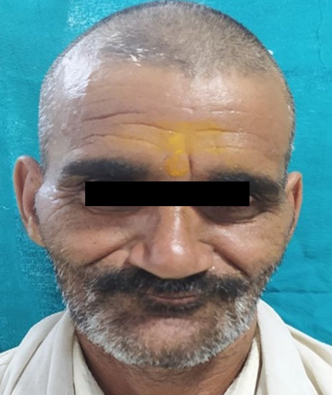
Smile Renewal: Precision and Beauty Unveiled with CAD-CAM Dental Technology
Department of Prosthodontics and Crown & Bridge, Seema Dental College & Hospital, Rishikesh, Uttarakhand, India
ABSTRACT
The evolution of materials science, bonding protocols, and innovative manufacturing techniques is driving the development of new ceramic materials, addressing the growing demand for fixed prosthodontics restorations that are both aesthetically pleasing and clinically superior. The recently launched ALD ceramic, designed for the CEREC system, stands out in this regard, offering restorations with enhanced aesthetic and flexural strength. In a detailed case report highlighting the restoration of an anterior tooth, the application of CEREC-Omnicam for tooth preparation, scanning, and design, coupled with subsequent MCXL fabrication using ALD blocks, yielded an aesthetically pleasing and functionally robust result. Notably, this approach contributed to a more efficient overall treatment time, leading to increased satisfaction for both the patient and the practitioner. This case exemplifies the impactful synergy of advanced materials and chairside technologies in meeting the contemporary demands of fixed prosthodontics.
Key words: CAD-CAM, Aesthetics, Lithium Disilicate, monolithic.
INTRODUCTION
Dental health in the contemporary era is acknowledged not only for its functional implications but also as a crucial element of aesthetics. The importance of aesthetics in dentistry has surged in recent times, with patients having heightened expectations in this regard. However, addressing patients' aesthetic preferences goes beyond surface-level considerations. Material selection, preparation procedures, cementation, and production techniques play pivotal roles in ensuring the enduring success of aesthetic dental restorations. Simultaneously, underscoring the significance of professional oral care and enhancing the patient's understanding of oral hygiene is paramount.1
Metal-supported ceramic systems have demonstrated their efficacy in crown and bridge restorations. Challenges related to biocompatibility and optical properties in metal-supported ceramics have spurred the evolution of dental ceramics. The rapid rise in aesthetic standards has boosted the popularity of dental ceramics as the preferred material in dentistry.2
The adoption of CAD (Computer-Aided Design) and CAM (Computer-Aided Manufacturing) technology in crafting restorations, utilizing ceramic blocks shaped during fabrication and reinforced with cutting-edge technology, is increasingly prevalent in dental practice. The "digital workflow" captures both sets of teeth, allowing clinicians to meticulously examine tooth preparation and design restorations according to the planned treatment. Before progressing, any necessary adjustments can be made by uploading digital files to a cloud server for swift communication with technicians. This technique is generally time-efficient, eliminating the need for traditional impression materials and often enabling the completion of the entire procedure in a single appointment.3
In recent years, there has been a growing trend towards the adoption of monolithic restorations as a preventive measure against the chipping of veneering ceramics on oxide ceramic frameworks. Lithium disilicate (LS2) ceramics stand out as the most commonly utilized materials in chairside processing, offering proven excellence in both esthetics and strength. Consequently, they are widely accepted and utilized, examples being CEREC Tessera from Dentsply Sirona in Charlotte, USA, and IPS e.max CAD from Ivoclar Vivadent in Schaan, Liechtenstein.4,5
Lithium disilicate (LS2) ceramics exhibit versatility and are well-suited for various dental applications, including single-tooth crowns, partial crowns, veneers, inlays, onlays, and even implant-supported restorations. According to information provided by manufacturers, these ceramics can be used to create 3-unit fixed partial dentures (FPDs) extending up to the first premolar, as demonstrated by products such as IPS e.max CAD from Ivoclar Vivadent in Schaan, Liechtenstein.6
The presented case report illustrates the production of monolithic single-tooth restorations using an innovative advanced lithium disilicate (ALD) ceramic, specifically the CEREC Tessera from Dentsply Sirona in Charlotte, USA, produced with the CEREC system. While the material initially appears tooth-colored, a mandatory firing process is required to achieve its final strength. This ALD ceramic possesses a unique microstructure, comprising LS2 and virgilite (lithium aluminum silicate) embedded in a glass matrix enriched with zirconia. According to the manufacturer, the firing process leads to the formation of additional virgilite crystals. The rod-shaped LS2 crystals play a crucial role in providing high tensile strength, effectively resisting crack propagation. Simultaneously, the small virgilite crystals generated during firing play a significant role in enhancing the biaxial flexural strength to over 700 MPa. This improvement is attributed to the increase in precompression stress facilitated by the presence of these crystals.
CASE REPORT
A 23-year-old male patient reported to the institution with a chief complaint of chipped maxillary left central incisor with a month old history of trauma. On clinical examination, patient had good oral hygiene and presented with Ellis’ and Davey’s Class 3 fracture with maxillary left central incisor (Fig. 1). He underwent single sitting root canal treatment (Fig. 11) for the same and reported to the department of Prosthodontics for placement of crown. An E-max lithium disilicate restoration was planned for better esthetic result.
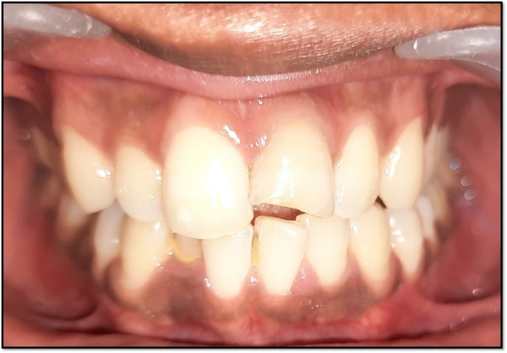
Fig. 1 Pre-operative

Fig. 2 Post-Endodontic Treatment
All the equipment for tooth preparation was prepared in advance. Three depth orientation grooves were made in each cervical, middle, and incisal third of the teeth, measuring 0.3 mm, 0.5 mm, and 0.7 mm, respectively. The finish line was established using a long-tapered diamond bur, creating a definitive shoulder margin that extended mesially, distally, and palatally. Once the finish line was in place, the land area between the depth cuts was removed. Incisal reduction was then performed to provide the necessary bulk for the porcelain. Proximal contacts were broken using a fine tapered bur, and the finishing touches were achieved with the assistance of a fine grit. Shade matching and selection were carried out using a shade guide (Vitapan classic, Vita Zahnfabrik, Bad Säckingen, Germany) (Fig. 3).

Fig. 3 Vitapan Classic Shade Guide

Fig. 4 Final Tooth Preparation
Impressions of the maxillary and mandibular arches were taken using polyvinyl siloxane impression material (Aquasil TM, DECA Regular Set, Dentsply). Subsequently, the impressions were poured with Type 2 die stone, resulting in the creation of master casts. These casts were then scanned using the CEREC Omnicam intra-oral scanner (Fig 5). A CAD model was generated, and the crown design was crafted in accordance with the specified requirements (Fig. 6).

Fig. 5 CEREC Omnicam

Fig. 6 CAD model

Fig. 7 Milling of Block
Following the completion of the design, a CEREC Tessera block of size C14 (18 x 14 x 12) was milled using CAM technology (Fig. 7). Following individual staining and glazing, the restoration was prepared for furnace firing using DS Body Stain S1, DS Incisal Stain I1, and DS Overglaze High Flu from Dentsply Sirona in Charlotte, USA. The restorations were then placed on a firing tray with a firing pad (DeguDent, Hanau-Wolfgang, Germany) and exposed to firing at 760°C (Fig. 8). Subsequent to firing, the restorations were examined to evaluate both marginal and interproximal fit.

Fig 8. CEREC SpeedFire

Fig 9. Post-Cementation
The ceramic surfaces were thoroughly cleaned with alcohol, and the inner surfaces were etched using a 5% hydrofluoric acid gel (IPS Ceramic Etching Gel, Ivoclar Vivadent, Schaan, Liechtenstein). After a 30-second etching period, the surfaces were initially rinsed in a water beaker and then with air/water spray. The dried restoration surfaces were subsequently treated with a silane coupling agent (Calibra silane, Dentsply Sirona, Charlotte, USA) for 60 seconds, with any excess removed using a strong air stream. The restoration was then cemented using adhesive resin cement (Calibra Ceram, Dentsply Sirona, Charlotte, USA). Visible excess cement was eliminated with foam pellets, and interproximal areas were cleaned with dental floss. Tack-curing was employed to facilitate the easy removal of any remaining excess cement in a gel state using a scaler. Both static and dynamic occlusions were assessed, revealing a satisfactory color match of the restoration with the adjacent teeth (Fig. 9).
DISCUSSION
Excessive loss of dental tissue due to trauma can result in a range of issues, including aesthetic, functional, and physiological concerns. While prioritizing pain relief, the discussion underscores the growing importance of addressing aesthetic considerations. Various treatment options are available for crown fractures, each with its own set of advantages and disadvantages.7
E-max crowns stand out due to several notable advantages when compared to porcelain fused to metal crowns. Recognized as an optimal match for natural teeth, E-max crowns feature a transparent color and lifelike appearance, seamlessly blending with one's own dentition. Importantly, the absence of a metal alloy base eliminates the occurrence of an unsightly grey line around the gum line, contributing to a more aesthetically pleasing result. In addition to their cosmetic benefits, E-max crowns are known for their strength and durability, surpassing many other types of crowns. Their resilience reduces the likelihood of cracks or fractures, ensuring a long-lasting solution for individuals seeking both functional and visually appealing dental restorations.8
The automation inherent in CAD/CAM (Computer-Aided Design/Computer-Aided Manufacturing) technology serves to minimize inaccuracies, ensuring precise results in the creation of dental restorations. Additionally, these systems contribute to a reduction in the risks associated with infectious cross-contamination, promoting a higher level of safety in dental procedures. Notably, machinable zirconia ceramics are particularly well-suited materials for CAD/CAM techniques. These ceramics can be effectively designed and milled in their soft pre-sintered state, allowing for intricate shaping and customization. Subsequent sintering of the milled restorations further enhances their physical properties, making machinable zirconia ceramics a versatile and reliable choice in the field of dental prosthetics.9
CONCLUSION
Advanced laboratory techniques in recent times have significantly improved the efficacy of ceramic restorations, offering a highly aesthetic and minimally invasive solution for rehabilitating fractured central incisors. This case report detailing the fractured central incisor highlight the clinical success of this approach, effectively addressing both functional and aesthetic considerations. These restorations not only exhibit long-term durability but also align with patients' aesthetic expectations, emphasizing the pivotal role of patient satisfaction in dental procedures. The positive results underscore the increasing significance of these techniques in restorative dentistry, providing practitioners with a viable and successful option for treating fractured central incisors.6
REFERENCES
- Goldstein RE, Garber DA, Schwartz CG, Goldstein CE. Patient maintenance of esthetic restorations. J Am Dent Assoc. 1992 Jan;123(1):61-7.
- Guess PC, Kulis A, Witkowski S, Wolkewitz M, Zhang Y, Strub JR. Shear bond strengths between different zirconia cores and veneering ceramics and their susceptibility to thermocycling. Dent Mater. 2008 Nov;24(11):1556-67.
- Sulaiman TA. Materials in digital dentistry-A review. J Esthet Restor Dent. 2020 Mar;32(2):171-81.
- Aziz A, El-Mowafy O, Paredes S. Clinical outcomes of lithium disilicate glass-ceramic crowns fabricated with CAD/CAM technology: A systematic review. Dent Med Probl. 2020;57(2):197–206.
- Leitão CIMB, Fernandes GVO, Azevedo LPP, Araújo FM, Donato H, Correia ARM. Clinical performance of monolithic CAD/CAM tooth-supported zirconia restorations: systematic review and meta-analysis. J Prosthodont Res. 2022 Jul 30;66(3):374-84.
- Rinke S, Pfitzenreuter T, Leha A, Roediger M, Ziebolz D. Clinical evaluation of chairside-fabricated partial crowns composed of zirconia-reinforced lithium silicate ceramics: 3-year results of a prospective practice-based study. J Esthet Restor Dent. 2020;32(2):226–35.
- Tinoco JV, Jurado CA, Sayed ME, Garcia Cortes JO, Kaleinikova Z, Hernandez A, Alshabib A, Tsujimoto A. Conservative approach for management of fractured maxillary central incisors in young adults. Clin Case Rep. 2020 Aug 23;8(12):2692-700.
- Oswal S, Nene P, Madanshetty P, Dugal R. Esthetic rehabilitation of anterior discolored teeth with lithium disilicate all-ceramic restorations. Univers. Res. J. Dent. 2016;6(2):190-1.
- Vinothkumar TS, Kandaswamy D, Chanana P. CAD/CAM fabricated single-unit all-ceramic post–core–crown restoration. J. Conserv. Dent. 2011;14(1):86–9.
Disclaimer from Guident
The views and opinions expressed in articles published in Guident are solely those of the authors and do not necessarily reflect the views of the editors, the editorial board or the publisher, Ivory India. Guident does not endorse any product, technique or treatment described. Clinical content is intended for qualified dental and medical professionals and is not a substitute for individual clinical judgement. Guident, its editors and the publisher accept no liability for any consequence arising from the use of information published in the journal.
How to cite this article
Dr. Varun Kumar, Dr. Suyashvi Gupta, Dr. Shubham Talele. Smile Renewal: Precision and Beauty Unveiled with CAD-CAM Dental Technology. Guident. 2023 Dec;17(1). Available from: https://ivoryindia.in/articles/prosthodontics/smile-renewal-precision-and-beauty-unveiled-with-cad-cam-dental-technology.html