Strengthening Dental Implant Restoration with Incorporation of Advanced-Platelet Rich Fibrin in Maxillary Anterior Zone-a Case Report
Department of Prosthodontics, Post Graduate Institute of Dental Sciences, Pt. B.D Sharma University of Health Sciences, Rohtak, Haryana, India
ABSTRACT
Dental implants have become a common approach in rehabilitating the lost natural teeth. Growth factor based products have reported to increase implant stability and enhance osseointegration. Treatment duration has reduced drastically thereby satisfying the needs of the patient. This case report presents the effect of Advanced Platelet-Rich Fibrin (A-PRF) on osseointegration of dental implant.
Keywords: Advanced Platelet-Rich Fibrin (A-PRF), dental implant, growth factors, osseointegration.
INTRODUCTION
Maxillary anterior region is the most vital aesthetic zone. Loss of natural teeth in this region impairs the patient’s normal quality of life. Nowadays, dental implants are commonly placed to replace the lost natural teeth. Implants require to be firmly osseointegrated before they are loaded. Osseointegration is interpreted as the apparent direct attachment or connection of osseous tissue to an inert, alloplastic material without intervening fibrous connective tissue.1 This process varies between 0–6 months.2 This time period can be shortened by the advent of new growth factor based materials which accelerate the osseointegration time. Platelet-based preparations produced from patient’s own blood provide an inexpensive alternative to commercially available bioactive materials. Activated platelets secrete a wide range of proteins and growth factors including bone morphogenetic protein (BMP), platelet-derived growth factor (PDGF), insulin-like growth factor (IGF), vascular endothelial growth factor (VEGF), transforming growth factor-β1 (TGF-β1), and transforming growth factor-β2 (TGF-β2), which play key roles in bone healing.3 Advanced-Platelet Rich Fibrin (A-PRF)is the new generation platelet concentrate which has shown promising results in recent times. The preparation protocol differs in centrifugation speed and time. This new formulation is thus put into effect in dental implant restoration.
CASE REPORT
A 20 year old male patient reported to the Department of Prosthodontics and Crown & Bridge with chief complaint of missing upper front tooth. Patient had a history of bike accident one year back due to which he lost his upper front tooth. Adetailed case history was recorded along with the assessment of preoperative photographs and radiographs. [Fig. 1A, B]A thorough intraoral examination was done. [Fig.1C] Patient was thereafter planned for fixed restoration using single endosseous two stage implant placement in the edentulous maxillary anterior region. All vital signs were evaluated and a complete hemogram was done to confirm the fitness of the patient for implant placement. A complete oral prophylaxis along with prescription to use 0.2% chlorhexidine gluconate mouthwash twice daily for a period of 7 days before implant placement and instructions for maintenance of oral hygienewere given to the patient.
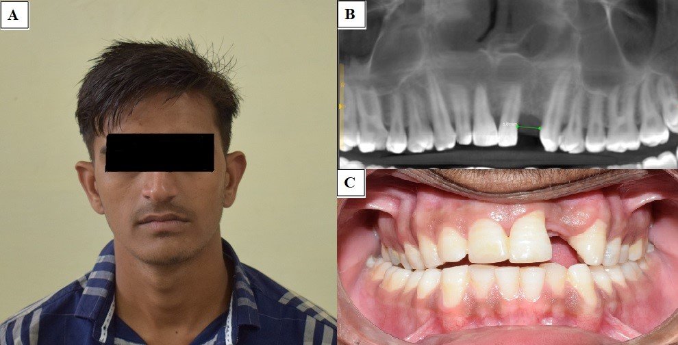
Figure 1:(A) Preoperativefull face frontal view; (B) Preoperative CBCT; (C) Preoperative teeth in occlusion
PREPARATION OF A-PRF
Patient’s blood sample was collected from antecubital vein without any anticoagulant in 10 ml tube, which was immediately centrifuged at 1500rpm for 14 minutes, in accordance with the centrifugation protocol of A-PRF.After the completion of cycle, the blood in the tube gets separated into three distinct layers; platelet poor plasma at the top, A-PRF fibrin clot in the middle and a red blood corpuscular base in the bottom. [Fig 2A]
SURGICAL PROCEDURE
Properly sterilized surgical instruments were arranged on a surgical trolley. The patient was seated on the dental chair and prepared following the standard guidelines for asepsis. Infraorbital nerve block and nasopalatine nerve block with 2% lignocaine with adrenaline 1:80000 were administered.After confirming the effectiveness of local anaesthesia, mid-crestal incision was given using surgical blade No. 11. The incision was extended to the mesial and distal teeth giving crevicular incision.Labial and palatal full mucoperiosteal flaps were reflected with the help of mucoperiosteal elevator. Labiopalatal dimensions of the alveolar ridge were assessed. Drilling was initiated with the pilot drill using surgical guide. Drilling was done at a speed of 800 to 1200rpm with continuous saline irrigation. Osteotomy preparation was completed by sequential drilling till the final length of the implant. Paralleling tool was used to check parallelism with the adjacent teeth.After determining the final length of osteotomy, the width of the osteotomy was increased with sequential drills till the final diameter of the implant. Then the A-PRF was carefully retrieved from the test tube and the RBC base was carefully removed to retain a small part of it in the clot followed by gently pressing between the two layers of sterile gauge to form a membrane.[Fig. 2B, 2C]
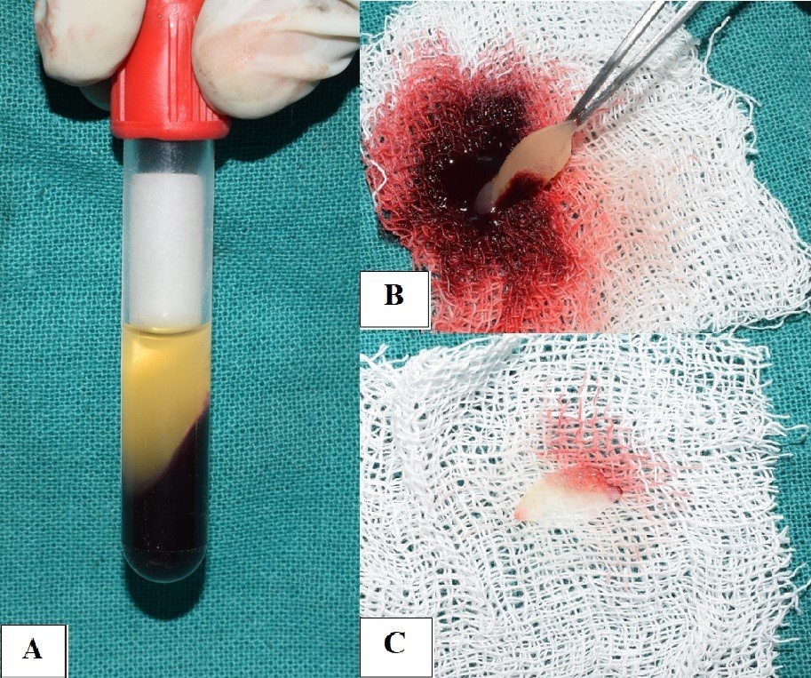
Figure 2: (A) Prepared A-PRF; (B) A-PRF separated from blood concentrate; (C) A-PRF membrane formed
The implant cavity wall was laid with A-PRF membrane all around.[Fig. 3A] A sterile manual implant connector was used to hold the selected dental implant. [Fig. 3B]With the help of dental implant connector assembly and a torque wrench, the implant was positioned in the osteotomy site flushing it with the margin of the crest. The implant was irrigated with A-PRF fluid on their surface and placed in the corresponding socket.The implant surgery was completed in one session by attaching gingival former to the implant. [Fig.4A] Gingival formers were not covered with soft tissue and mucoperiosteal flaps were sutured with 3-0 black braided silk suture material.The final surgical site was cleaned and irrigated with 0.9% saline solution. Sutures were made water tight and knot was tied away from the incision line.Postoperative instructions were given to the patient regarding diet, oral hygiene and medications were prescribed as per requirement. The patient was recommended to apply cold compresses after the surgery. Patient was recalled after one week for suture removal and soft tissue evaluation.

Figure 3: (A) A-PRF placement in the osteotomy site; (B) Dental implant before placement
Prosthesis was delivered at two weeks and regular follow up was scheduled. [Fig. 4B] Soft tissue profile was well contoured around the implant supported prosthesis.
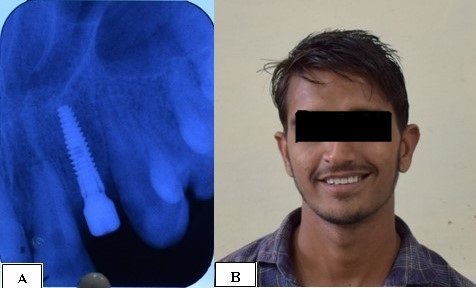
Figure 4: (A) Postoperative IOPA X-ray showing dental implant placement along with gingival former; (B) Postoperative full face frontal view after crown cementation.
DISCUSSION
Molecular biology has developed to a great extent in regards to faster bone and soft tissue regeneration. Growth factors play a vital role in bone regeneration. In the studies related to fibrin technologies, the low speed centrifugation of the autologous blood leads to the formation of a fibrin mesh existing with a three-dimensional loose mesh structure. The authors propose that such a protocol leads to enhanced B- & T-lymphocyte entrapment, more even distribution of platelets andneutrophils.4 Accordingly, this might be able to influence the differentiation of host macrophages and macrophages within the clot after implantation.5 Furthermore and in agreement with this hypothesis, it was shown that the release of several growth factors including PDGF, TGF-ß1, VEGF, as well as epidermal growth factor (EGF), and insulin-like growth factor (IGF) were significantly higher in A-PRF.5 Histomorphometric analysis done by Clark D et al demonstrated that ridge preservation using A-PRF resulted in the most vital bone formation (46±18%) across all four groups, and this bone formation was significantly greater than that observed in the freeze dried bone allograft (FDBA) treatment group which demonstrated the least amount of vital bone formation (29±14%) (P<0.05).6 12.5% A-PRF has the highest potentialability of fibroblast cells proliferation. A-PRF is multi-potent platelet derived stem cell that has agreat number of growth factors like PDGF and TGF- ß1.7 As Choukroun intended, it was observed that WBCs, as well as platelets, were highly concentrated in A-PRF preparations.8 Thus the newly formulatedadvanced-PRF stimulated significantly higher growth factor releaseover time when compared to standard PRF and may proveclinically beneficial for future regenerative procedures.
CONCLUSION
Therefore it is concluded that A-PRF would not only function as a scaffolding material but also as a reservoir to deliver certain growth factors at the site of application. Further studies arerequired to investigate the mechanism of A-PRF, and more in vivo studies are needed to identify the effect of A-PRF in bone regeneration.
REFERENCES
- The Glossary of Prosthodontic Terms: Ninth Edition. J Prosthet Dent 2017;117(5S):e1-e105.
- Raghavendra S, Wood MC, Taylor TD. Early wound healing around endosseous implants: a review of the literature. Int J Oral Maxillofac Implants 2005;20(3):425-31.
- Oncu E et al. Positive effect of platelet rich fibrin on osseointegration. Med Oral Patol Oral Cir Bucal 2016;21(5):e601-7.
- Shah R, M G T, Thomas R, Mehta DS. An Update on the Protocols and Biologic Actions of Platelet Rich Fibrin in Dentistry. Eur J ProsthodontRestor Dent 2017;25(2):64-72.
- Fujioka-Kobayashi M et al. Optimized Platelet-Rich Fibrin With the Low-Speed Concept: Growth Factor Release, Biocompatibility, and Cellular Response. J Periodontol 2017;88(1):112-121.
- Clark D et al. Advanced platelet-rich fibrin and freeze-dried bone allograft for ridge preservation: A randomized controlled clinical trial. J Periodontol 2018;89(4):379-387.
- Margono DA et al. Comparison of Advanced Platelet Rich Fibrin (A-PRF) and Culture Media Conditioned Warton's Jelly (CMCWJ) on Fibroblast Cells Proliferation.Pesq Bras Odontoped Clin Integr 2018;18(1):e4085.
- Masuki H et al. Growth factor and pro-inflammatory cytokine contents in platelet-rich plasma (PRP), plasma rich in growth factors (PRGF), advanced platelet-rich fibrin (A-PRF), and concentrated growth factors (CGF). Int J Implant Dent 2016;2(1):19.
Disclaimer from Guident
The views and opinions expressed in articles published in Guident are solely those of the authors and do not necessarily reflect the views of the editors, the editorial board or the publisher, Ivory India. Guident does not endorse any product, technique or treatment described. Clinical content is intended for qualified dental and medical professionals and is not a substitute for individual clinical judgement. Guident, its editors and the publisher accept no liability for any consequence arising from the use of information published in the journal.
How to cite this article
Dr. Akanksha Gopal Shetye, Dr. Manu Rathee, Dr. Prachi Jain. Strengthening Dental Implant Restoration with Incorporation of Advanced-Platelet Rich Fibrin in Maxillary Anterior Zone-a Case Report. Guident. 2020 Dec;14(1). Available from: https://ivoryindia.in/articles/prosthodontics/strengthening-dental-implant-restoration-with-incorporation-of-advanced-platelet-rich-fibrin-in-maxillary.html