Taming the Flabby Ridge-a Comparative Case Report of Two Impression Techniques: A Case Series
Department of Prosthodontics and Crown & Bridge, Srinivas Institute of Dental Sciences, Karnataka, India
ABSTRACT
A flabby or fibrous ridge is a frequently encountered clinical finding in long-standing complete denture wearers, in which the alveolar bone is progressively replaced by mobile, hyperplastic fibrous connective tissue. As this tissue displaces readily under the forces generated during conventional impression procedures, it poses a considerable challenge to denture support, retention and stability. A wide range of impression strategies have been proposed to record such ridges without distortion, but no single technique is clearly superior, and management continues to be guided largely by clinical judgement and material availability. This report describes the prosthodontic rehabilitation of two edentulous patients presenting with an anterior maxillary flabby ridge, each managed with a different impression philosophy. In the first case, a window (open-tray) technique was used, in which the flabby tissue was recorded in a relaxed, undisplaced state using impression plaster after the remaining supported ridge had been recorded under selective pressure. In the second case, a two-stage 'mishmash' technique was employed, combining a mucocompressive impression of the firm, well-supported ridge with a mucostatic impression of the flabby segment recorded using putty and light-body addition silicone. Both approaches allowed fabrication of complete dentures with satisfactory retention, stability and patient comfort, confirming that either technique, when selected appropriately, can produce a clinically acceptable outcome.
Keywords: Flabby ridge, fibrous ridge, complete denture, window impression technique, mishmash impression technique, impression plaster, putty–light body impression.
INTRODUCTION
A flabby or displaceable ridge refers to an area of hypermobile fibrous connective tissue that replaces the normal alveolar bone in an edentulous jaw. It is one of the more common sequelae of prolonged denture wear, and is encountered far more often in the maxilla than in the mandible, being reported in as many as 24% of edentulous maxillae compared with roughly 5% of edentulous mandibles.1,20 The anterior maxillary region is particularly susceptible, and the condition is seen most frequently in patients who have worn complete dentures for extended periods.
Several factors have been implicated in the aetiology of flabby ridges, including chronic bone resorption, unfavourable occlusal and functional loading, parafunctional habits and nutritional deficiency.1 A well-recognized contributing condition is Combination Syndrome, first described by Kelly, in which a maxillary complete denture opposes natural mandibular anterior teeth together with a distal-extension removable partial denture, leading to accelerated resorption of the anterior maxillary ridge.2 More recent reviews, however, have questioned whether the presence of anterior mandibular teeth is truly responsible for this pattern of resorption, and have found no convincing evidence that a mandibular partial denture prevents it.3 Histologically, the flabby ridge is composed of hyperplastic mucosa overlying loosely organised fibrous connective tissue, which itself sits above a more normal, denser collagenous layer, and may contain variable amounts of metaplastic cartilage or bone.1
Clinically, the mobility of this tissue makes it prone to distortion under the compressive forces generated during conventional impression making. An impression recorded without regard for this mobility tends to displace the flabby tissue apically, and the resulting cast reproduces a form that does not correspond to the tissue's resting anatomy. Dentures fabricated on such casts characteristically lose retention and stability once the tissue rebounds intraorally, and patients often describe the denture as becoming progressively looser during function.12
Management options for the flabby ridge broadly fall into three categories: surgical removal of the redundant fibrous tissue prior to denture fabrication, implant-retained prostheses (fixed or removable), and conventional prosthodontic management without surgical intervention.11 Surgical excision, while sometimes indicated, reduces the bulk of stress-absorbing soft tissue and can leave an atrophic ridge with poor resistance to lateral forces, so it is generally reserved for cases where the patient's age, systemic health, dental history and motivation favour surgery.1 Implant-supported rehabilitation may not be feasible or affordable for every patient. Consequently, conventional impression techniques that selectively record the flabby tissues while avoiding excessive compression, yet adequately capture the remaining supporting ridge, continue to be the cornerstone of management for most patients.
Selective-pressure impression philosophy is central to nearly all of these techniques. A mucocompressive impression, recorded under load, tends to produce excellent retention immediately after insertion, because the vascular contents of the soft tissue are displaced into the interstitial spaces during recording; however, once the tissue rehydrates and the blood re-enters it at rest, the denture base no longer fits as intimately, and retention deteriorates.1,12,18 A mucostatic impression, by contrast, records the tissue in its relaxed, undisplaced state and avoids this problem, but is technically more demanding to achieve when firm and flabby tissue coexist in the same arch.9,15 Various two-part and modified special-tray techniques have therefore been described to record firm ridge tissue under selective pressure while simultaneously recording the flabby segment with little or no pressure, an approach that was first popularised by Osborne for the mandible in 1964 and later adapted for the maxilla by several authors.8,9
Materials traditionally used for the low-pressure recording of flabby tissue include impression plaster, impression compound and zinc oxide–eugenol paste. Impression plaster in particular has a long history in this application, having first been advocated by Liddelow in 196424, and is still favoured because it flows passively over the tissue with minimal pressure and reproduces fine surface detail once set. It does, however, have a limited working time, cannot record undercuts, and tends to drag or fracture at the borders unless it is confined within a rigid framework such as a window in the special tray.23,25 Rubber-base (silicone or polysulfide) impression materials, available as putty and light-body viscosities in the same chemistry, offer an alternative that is more widely stocked in general dental practice, dimensionally more stable, and easier to remove without undercut-related tearing, while still allowing a comparable two-stage philosophy to be applied.19
This report presents two clinical cases of complete denture fabrication in patients with an anterior maxillary flabby ridge, each managed by a different impression strategy: a window impression technique using impression plaster in the first case, and a two-stage ‘mishmash’ impression technique using putty and light-body addition silicone in the second.
CASE REPORT 1: WINDOW IMPRESSION TECHNIQUE USING IMPRESSION PLASTER
A 59-year-old completely edentulous female patient was referred to the Department of Prosthodontics at Srinivas Institute of Dental Sciences, Mangalore after undergoing extraction of root stumps in relation to teeth 21, 22, 33, 34, 43, 44, 45, and 46. The patient's medical history was significant for diabetes mellitus and hypertension, both of which were well controlled with prescribed medications.
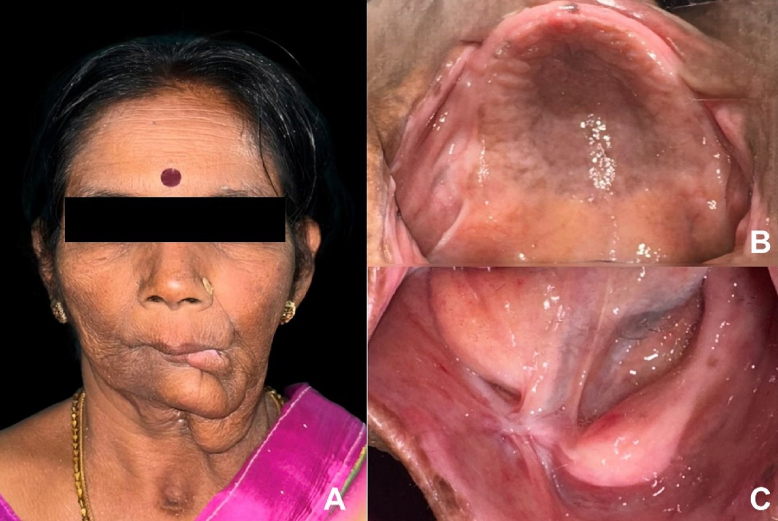
On examination, the patient presented with an area of hypermobile, displaceable tissue confined to the anterior maxillary ridge [Figure 1]. The treatment options, including surgical recontouring, implant-supported prostheses, and conventional complete dentures using a modified impression technique, were explained to the patient, and a conventional approach without surgical intervention was selected as the patient was not willing to undergo any surgical procedure.

Fig 1: Flabby anterior maxillary ridge extending from right second premolar region to left premolar region
Preliminary impressions of both arches [Figure 2] were recorded in stock trays using impression compound. On the primary maxillary impression, the boundaries of the displaceable tissue were outlined and transferred to the primary cast to guide fabrication of the custom tray10,21 [Figure 3]. A custom tray was fabricated in autopolymerising acrylic resin over the primary cast using a selective-pressure design, with the tray borders kept short of the depth of the sulcus to allow for subsequent border moulding [Figure 4].

Fig 2: Preliminary Impression of the Maxillary and Mandibular Arches

Fig 3: Marking the boundaries of displaceable tissues

Fig 4: Maxillary and Mandibular Custom tray (tray handle placed in the maxillary arch for easy handling of the tray while recording the anterior flabby ridge)
Border moulding of the custom tray was carried out using a low-fusing impression compound [Figure 5], after which the definitive impression of the firm, well-supported areas of the ridge were recorded with zinc oxide–eugenol impression paste [Figure 6]. Before the secondary impression was made, a window was cut into the tray directly over the flabby tissue using a round and fissured bur, deliberately leaving this region of the tray open.8,10 The tray was reseated to confirm that the flabby tissue was clearly visible and accessible through the window before proceeding further.

Fig 5: custom tray following border moulding with the window created in the anterior region of maxilla

Fig 6: Definitive impression of the maxillary and mandibular arches
Impression plaster was then applied over the exposed flabby tissue through the window using a soft brush [Figure 7], and the tray was reinserted into the patient's mouth. Because the window leaves this portion of the tray open rather than enclosed, the plaster is not confined and the flabby tissue is free to rebound to its resting position as the material sets, so that it is captured in an undisplaced, relaxed state rather than under compression.9,23

Fig 7: Impression plaster applied over exposed flabby ridge
Once the plaster had set, the completed impression combining a mucocompressive recording of the supported ridge with a mucostatic recording of the flabby ridge through the open window was removed intact, disinfected, and poured in dental stone without delay to obtain the master cast [Figure 9]. Denture fabrication then proceeded through conventional laboratory stages, and the completed maxillary and mandibular complete dentures were inserted with routine post-insertion instructions [Figure 10 and 11].

Fig 8: Final impression with Impression Plaster
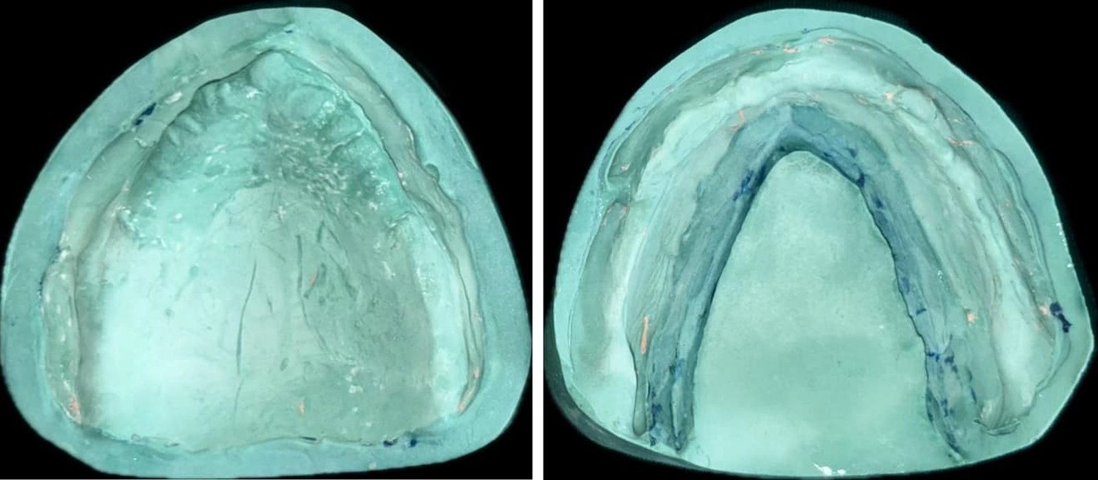
Fig 9: Master cast poured in dental stone
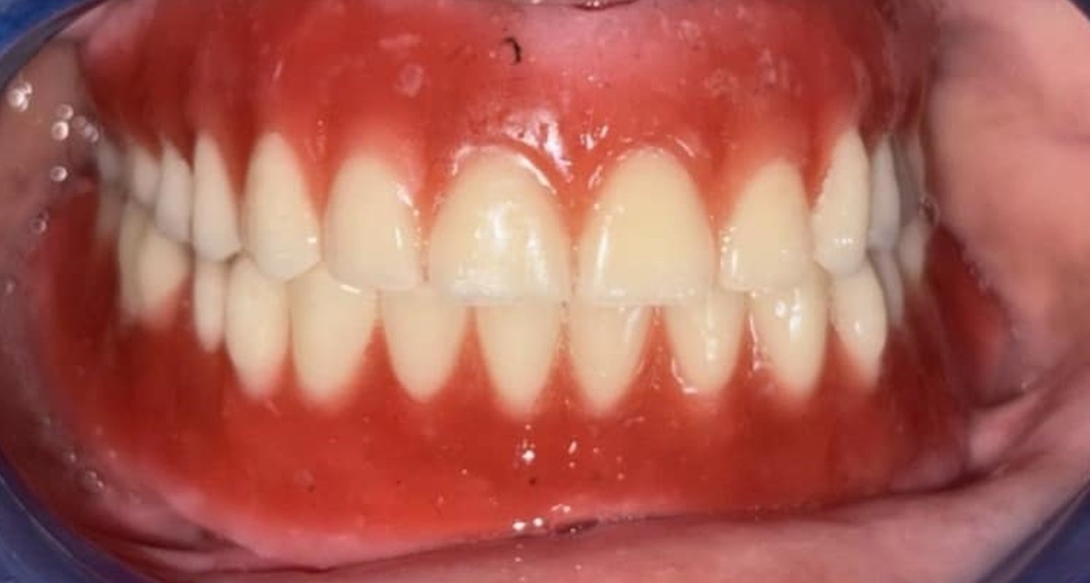
Fig 10: Denture try in

Fig 11: Pre-op and Post-op pictures
At the time of denture insertion, the retention of both maxillary and mandibular dentures was satisfactory. The patient reported no discomfort immediately following denture insertion. A follow-up appointment was scheduled one week later, during which the patient was evaluated for adaptation to the dentures. Necessary denture adjustments were carried out in areas where the patient reported pain or soreness.
CASE REPORT 2: MISHMASH IMPRESSION TECHNIQUE USING PUTTY AND LIGHT-BODY ADDITION SILICONE
A 60-year-old female patient reported to the Department of Prosthodontics, Srinivas Institute of Dental Sciences, Mangalore for the replacement of teeth in the maxillary and mandibular completely edentulous arches. The patient was a denture wearer for the last 4 years and described the existing dentures as “loose”. The patient's medical history revealed diabetes mellitus and hypertension for the past 10 years, for which she was receiving regular medication. She also had undergone coronary angioplasty 7 years previously and remained on maintenance medication.

Fig 1: Pre-op frontal and lateral view of the patient
Intraoral examination revealed a displaceable, flabby segment of the anterior maxillary ridge extending between the canine regions, with the remainder of the ridge providing firm support [Figure 2]. As the patient was unwilling to undergo surgical recontouring, it was decided to proceed with conventional complete dentures fabricated using a modified two-stage rubber-base impression technique.19
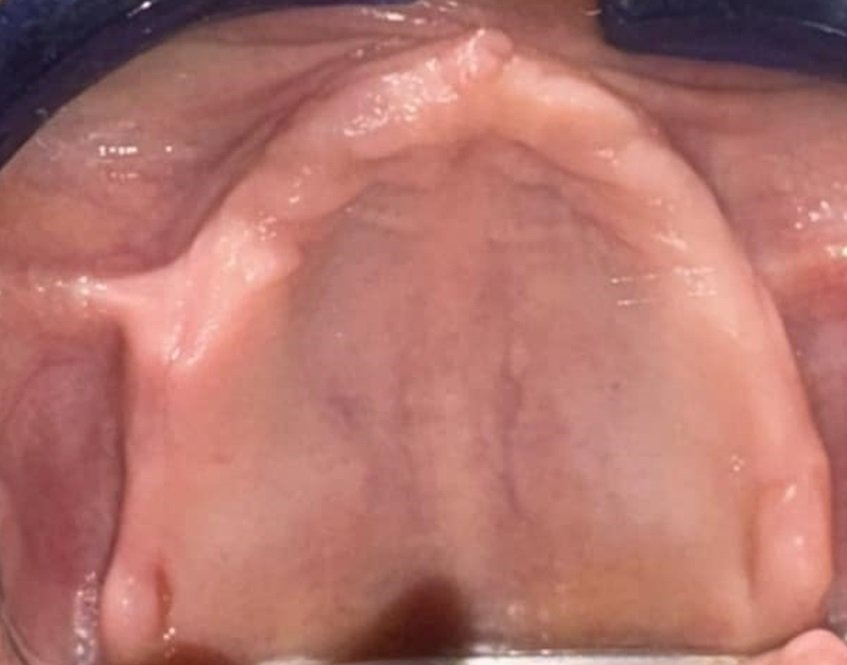
Fig 2: Intraoral view of the flabby maxillary anterior ridge
Preliminary impressions [Figure 3] were made in edentulous stock trays using impression compound, and the displaceable tissue was marked on the impression and transferred to the primary cast. A maxillary special tray [Figure 4] was then fabricated in clear autopolymerising acrylic resin, deliberately leaving the region overlying the flabby ridge uncovered and a rim-type handle was incorporated into the tray design to prevent unset impression material from flowing back toward the oropharynx while the tray was seated with the patient in a supine or semi-supine position.19 [Figure 4 and 5].

Fig 3: Preliminary Impression

Fig 4: Maxillary and mandibular special tray

Fig 5: Anterior open clear acrylic custom trays with rim handle design
The tray was tried in, adjusted, and border moulded over the firm, well-supported segments of the ridge. The definitive impression of these areas was then recorded with a zinc oxide impression paste, producing a mucocompressive recording of the supported tissue.19 [ Figure 6 and 7].

Fig 6: Border moulded tray of the firm supported areas

Fig 7: Final mucodisplacive impression of the firm supported areas
For the second stage, tray adhesive was applied around the border of this first impression in the region of the flabby ridge, and the tray was reseated. Putty-consistency addition silicone impression material was placed directly over the flabby ridge and moulded around the labial border to record this segment in an undisplaced state19 [Figure 8 and 9]. After removal, the set putty overlying the flabby region was trimmed back uniformly with a putty knife to create a uniform space, and a wash impression of this area was then recorded using light-body addition silicone, without displacing the underlying flabby tissue.19 [Figure 10]. This combined 'mishmash' of putty and light body over the flabby segment, recorded within a tray that had already captured the firm ridge under selective pressure, allowed the mobile tissue to be reproduced accurately without distortion [Figure 11].

Fig 8: Putty body impression of flabby anterior ridge by mucostatic technique

Fig 9: Mishmash impression technique for second stage impression
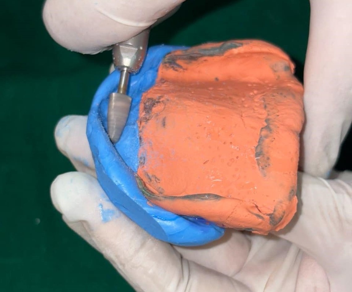
Fig 10: Set putty trimmed to create space for light body polyvinyl siloxane impression material

Fig 11: Mucostatic & mucodisplacive mishmash final impression
The completed impression was inspected, disinfected and poured immediately in dental stone, and the maxillary and mandibular complete dentures were fabricated through conventional laboratory procedures and delivered to the patient with standard post-insertion guidance [Figure 12,13,14,15 and 16]. At the time of denture insertion, both the maxillary and mandibular dentures demonstrated satisfactory retention and good stability. The patient reported no immediate discomfort following denture delivery. The patient was advised to return after one week for follow-up however, she did not attend the scheduled appointment and instead presented after two weeks. At the review visit, selective denture adjustments were performed to relieve areas of pain and soreness reported by the patient during adaptation to the new dentures.

Fig 12: Beading and boxing of the definitive impression

Fig 13: master casts

Fig 14: Jaw relation Recorded

Fig 15: try in of the maxillary and mandibular dentures

Fig 16: post op fontal and lateral view of the patient
DISCUSSION
Both cases in this report illustrate the same underlying principle applied through two different sets of materials. The firm, well-supported ridge is recorded under selective (mucocompressive) pressure, while the flabby segment is recorded separately, with little or no pressure, so that it is captured in its relaxed, undisplaced state.6,9,19 This selective-pressure philosophy addresses the central problem in flabby-ridge management. A single, uniformly compressive impression of the whole arch tends to displace the mobile tissue apically, producing a denture base that fits well only until the tissue rebounds, after which retention and stability are lost.1,12
The window technique demonstrated in the first case relies on modifying the special tray itself, leaving the region over the flabby ridge open so that a slow-setting, passively flowing material such as impression plaster can record the tissue without confinement. This approach has the advantage of being simple to execute and requires no additional appointments, although impression plaster has a limited working time, cannot engage undercuts, and needs a rigid enclosure or window to prevent it from dragging at its margins.23,25 Reports evaluating different special-tray designs have suggested that a window design produces the least distortion of simulated flabby tissue among the configurations tested.25
The mishmash technique demonstrated in the second case achieves a broadly similar mucostatic recording of the flabby segment, but substitutes putty and light-body addition silicone for impression plaster. Because these rubber-base materials are already stocked in most general dental practices for crown-and-bridge work, the technique avoids reliance on less commonly used materials, and the two viscosities used in sequence (a bulk putty stage followed by a light-body wash) allow the clinician to first create space and then refine the definitive surface detail of the flabby tissue without displacing it.19 Addition silicones also offer favourable dimensional stability and tear strength in comparison with impression plaster, which can be advantageous when the impression must be stored briefly before casting.
Neither approach has been shown in the literature to be categorically superior to the other, and the choice between them, as with most flabby-ridge techniques, is largely governed by operator preference, material availability, and the specific site and extent of the displaceable tissue.4,25 Both techniques described here share several common features that appear to be important for the success of the prosthesis. Successful management relies on using a transparent tray to evaluate tissue pressure during impression making, selectively recording the firm and flabby portions of the ridge in separate stages, and using a low-viscosity, minimally displacing impression material for the final registration of the flabby tissue.
An important advantage of both impression techniques is that they do not necessitate additional patient visits, despite requiring extra chair-side time during the impression appointment for tray modification and the sequential recording of the firm and flabby ridge tissues. This modest increase in clinical time is justified by the enhanced retention and stability of the final prosthesis. Conversely, impressions that compress the flabby tissue may produce distorted tissue records, compromising denture fit and adversely affecting long-term treatment outcomes.
CONCLUSION
The flabby or fibrous ridge remains a common and clinically significant challenge in complete denture prosthodontics, particularly in the anterior maxilla of long-term denture wearers. Surgical removal of the redundant tissue is not always necessary or desirable, and conventional prosthodontic management using a selective-pressure impression philosophy continues to offer a predictable, minimally invasive route to a stable and retentive denture. The two cases presented in this report demonstrate that this philosophy can be realised equally well using traditional materials such as impression plaster in a window-tray technique, or using contemporary putty and light-body addition silicone in a two-stage mishmash technique. The choice of material and technique should ultimately be individualised according to the site and extent of the flabby tissue, the materials available, and the clinician's experience, since the existing literature does not establish the clear superiority of any single approach.
REFERENCES
- Desjardins RP, Tolman DE. Etiology and management of hypermobile mucosa overlying the residual alveolar ridge. J Prosthet Dent. 1974;32:619-38.
- Kelly E. Changes caused by a mandibular removable partial denture opposing a maxillary complete denture. J Prosthet Dent. 1972;27:140-150.
- Palmqvist S, Carlsson GE, Öwall B. The combination syndrome: A literature review. J Prosthet Dent. 2003;90:270-275.
- Lamb DJ. Problems and solutions in complete denture prosthodontics. London: Quintessence; 1993. p.57-60.
- McCord JF, Grant AA. A clinical guide to complete denture prosthodontics. London: British Dental Association; 2000. p.10-21.
- Grant AA, Heath JR, McCord JF. Complete prosthodontics: problems, diagnosis and management. London: Wolfe; 1994. p.90-92.
- Allen PF, McCarthy S. Complete dentures: from planning to problem solving. London: Quintessence; 2003. p.48-51.
- Devlin H. A method for recording an impression for a patient with a fibrous maxillary alveolar ridge. Quint Int. 1985:395-397.
- Osborne J. Two impression methods for mobile fibrous ridges. Br Dent J. 1964;117:392-394.
- Hobkirk JA. Complete dentures — a dental practitioner handbook. Bristol: Wright; 1986. p.44-45.
- Lynch CD, Allen PF. Management of the flabby ridge: Using contemporary materials to solve an old problem. Br Dent J. 2006;200:258-61.
- MacEntee MI. Identifying the Problem. The Complete Denture. A Clinical Pathway. Chicago: Quintessence; 1996. p.1-7.
- Devan MM. Basic principles of impression making. J Prosthet Dent. 1952;2:26-35.
- Appleby RC. Mandibular impression technique for displaceable tissues. J Prosthet Dent. 1954;4:335-45.
- Porter CG. Mucostatics – Panacea or propaganda. J Prosthet Dent. 1953;3:464-6.
- Collette HA. Complete denture impressions. J Prosthet Dent. 1965;15:603-14.
- Klein IE, Broner AS. Complete denture secondary impression technique to minimize distortion of ridge and border tissues. J Prosthet Dent. 1985;54:660-4.
- Crawford RWI, Walmsley AD. A review of prosthodontic management of fibrous ridges. Br Dent J. 2006;199:715-719.
- Kulkarni P, Kulkarni RS, Shah RJ, Tomar B. Mishmash impression technique for managing maxillary anterior fibrous ridge. J Dent Mater Tech. 2018;7:63-8.
- Carlsson GE. Clinical morbidity and sequelae of treatment with complete dentures. J Prosthet Dent. 1998;79:17-23.
- Pai UY, Reddy VS, Hosi RN. A single step impression technique of flabby ridges using monophase polyvinylsiloxane material: a case report. Case Rep Dent. 2014;2014:104541.
- Mattoo KA, Kumar L, Rehman SU. Flabby ridge management using paint on technique-meticulous review. J Med Sci Clin Res. 2019;7:518-21.
- Watson RM. Impression technique for maxillary fibrous ridge. Br Dent J. 1970;128:552.
- Liddelow KP. The prosthetic treatment of the elderly. Br Dent J. 1964;117:392-4.
- Shin JO, Ko KH, Huh YH, Cho LR, Park CJ. Displacement of simulated flabby tissue by different tray designs and impression materials. J Prosthodont. 2019;28:444-51.
Disclaimer from Guident
The views and opinions expressed in articles published in Guident are solely those of the authors and do not necessarily reflect the views of the editors, the editorial board or the publisher, Ivory India. Guident does not endorse any product, technique or treatment described. Clinical content is intended for qualified dental and medical professionals and is not a substitute for individual clinical judgement. Guident, its editors and the publisher accept no liability for any consequence arising from the use of information published in the journal.
How to cite this article
Dr. Rithika, Dr. Nitesh Shetty, Dr. Vedanth. Taming the Flabby Ridge-a Comparative Case Report of Two Impression Techniques: A Case Series. Guident. 2026 Sep;19(10). Available from: https://ivoryindia.in/articles/prosthodontics/taming-the-flabby-ridge-a-comparative-case-report-of-two-impression-techniques-a-case-series.html