Evaluation of Platelet Rich Fibrin Matrix in Treatment of Periodontal Defect
The Oxford Dental College, Bangalore, India
ABSTRACT
The first generation platelet concentrate Platelet rich plasma (PRP) has treated various forms of periodontal defects. But due to its certain disadvantages, a modification of PRP was made. The platelet rich fibrin matrix (PRFM) due to its simpler preparation and robust release of growth factors which further at molecular and cellular levels leads to adhesion, proliferation, differentiation and maturation. This process further precedes the reconstruction of soft and hard periodontal tissues. The present article describes the benefit of PRFM in extraction socket and horizontal bony defect.
INTRODUCTION
In the recent era the greatest challenge faced in the medical and dental fraternity is the development of bioactive additives that will regulate inflammation and enhance healing process.1 The healing of soft and hard tissues are mediated by intra and extra cellular events mainly through the signaling proteins. From the enormous material present in the market for the treatment of periodontal defects; the platelet concentrates has emerged successfully with diversified properties of its own.
Platelets is one of the predominant cell of the blood component. They are 2-3 μm in diameter and are derived from bone marrow megakaryocytes. The α granules are spherical or oval structures with diameters ranging from 200 to 500 nm and forms an intracellular storage pool of proteins vital to wound healing. The α granules fuse with the platelet cell membrane after activation. In this process some secretory proteins are transformed to a bio-active state. The active proteins are then secreted, allowing them to bind to transmembrane receptors of the target cells. Once bound, intracellular signal proteins are activated which directs cellular proliferation, collagen synthesis, osteoid production, and so on.2
The first generation platelet concentrate Platelet-rich plasma (PRP) was introduced by Marx et al in 1998. It was used in the clinical field as liquid or gel formulation to promote tissue repair. The conventional PRP when used in clinical settings was difficult to handle and required secure implantation in a specific site and when released growth factors could not be placed in the desired location due to its poor mechanical properties. But due to its robust release of growth factors evaluated by O’Connell et al in 20064 the physical properties of PRP was further stimulated by the addition of CaCl2 and bovine thrombin, to produce a fibrin network. This too led to a weak gel along with certain disadvantages due to the addition of bovine thrombin5 To simplify the process and utilize the regenerative potential of the platelet concentrates along with the growth factors led to the introduction of Platelet rich fibrin matrix (PRFM). Various factors like thrombin-fibrinogen concentration ratios and factors such as protein and ion concentrations (Chung and Shu, 1990; Okada et al 1983; Okada et al 1985) including calcium were responsible for structural changes in the fibrin gel of the platelet concentrate6.The PRFM preparation uses calcium without the use of exogenous thrombin and with a proper centrifugation force it appears physically stronger than conventional PRPs.7
Thus two case reports have been described with the use of Platelet rich fibrin matrix (PRFm) in horizontal bone defect and extraction socket preservation.
CASE 1
A 42 years old male patient came to the department of Periodontics with chief complaint of bleeding gums since 3 months. The patient did not have any systemic disease neither had habit of smoking. On intraoral soft tissue examination, the patient had erythematous gingiva with soft and edematous consistency with scalloped and round margins and generalized bleeding on slight provocation. The patient exhibited fair plaque and moderate gingival index score of 1.8 respectively. On periodontal examination, the patient had generalized pocket depth ranging from 5-8mm and clinical attachment loss from 4 -7mm. On oropantographic examination the patient presented generalized horizontal bone defects and was diagnosed with chronic generalized periodontitis.
A thorough non-surgical periodontal therapy was given to the patient. Following it periodontal flap surgery was opted for the periodontal pockets after phase 1 periodontal therapy. An open flap debridement was performed in all the quadrants. After thorough debridement of the defect area, bleeding points were created. The PRFM were prepared as per Meresis protocol at a centrifugation of 3000 rpm for 10 minutes and was placed into the defect area as seen in figure 1. The defect areas were sutured with direct interrupted suture and periodontal pack was placed.
Every 3 months oral prophylaxis was performed and at 9 months, radio Visiography (RVG) was taken. Figure 2 demonstrates the surgical procedure and post-operative bone fill at 9 months period. Figure 3 illustrates the bone fill.
ase 2
A 38 year old male patient came to the clinic with grade 3 mobility in relation lower right back teeth region since 3 weeks. The patient complained of difficulty in mastication while eating food. The oropantogram revealed sever bone loss till the apical one third of the defect with mal aligned 3rd molar which was aligned towards the mobile teeth. The patient was systemically healthy and did not have deleterious habit of smoking.
Due to hopeless prognosis in relation to mobile teeth, a treatment plan for preservation of extraction socket was planned. Atraumatic extraction was performed from premolar to 2nd molars. A through inoculation was performed for the freshly extracted socket. As per the protocol of prepration of Meresis PRFM at a centrifugation of 3000 rpm for 10 minutes. The PRFM gel was then placed into the extraction sockets and hidden X suture was placed for complete coverage and to maintain the width of attached gingiva as illustrated in figure 4.
On 9th month post-operative a considerable amount of new formed bone can be appreciated in the cone beam computed topography as depicted in figure 5. The sockets were filled with bone and the sites did not exhibit discernable coronal invagination.

Figure 1: Preparation of PRFM gel

Figure 2: Placement of PRFM gel in horizontal bony defect
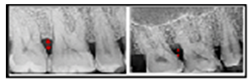
Figure 3: Pre and post operative view of horizontal defects

Figure 4: Placement of PRFM gel in extraction socket
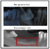
Figure 5: Pre and post operative view of extraction socket
DISCUSSION
The ideal goal of periodontal therapy is to promote healing through regeneration of lost form, function, aesthetics and comfort.8 The magnificent role of these platelet concentrates significantly releases the most important growth factors which plays a pivotal role in the wound healing process for a period of one month as mentioned in the literature. The growth factors platelet derived growth factor (PDGF), Transforming growth factor (TGF) and Insulin growth factor (IGF) has a strong mitogenic and anabolic activities further stimulating the proliferation of fibroblast and periodontal ligament cells, enhancing collagen synthesis.9,10 PDGF enhances osteoblastic cell proliferation leading to new bone formation.11 The dynamic VEGF is critical for neo angiogenesis during the wound healing and also facilitate in maintaining the integrity of endothelial cell lining of the blood vessel.12 The FGF and EGF are known to play an important role in the regulation of ectodermal and mesenchymal derived cell along being a potent chemotactic and mitogenic actions for periodontal ligament fibroblast cells.13 Thus the diverse action of the growth factors forms a key player in wound healing and regeneration.14
The PRFM gel is known to release the growth factors immediately after placement of it in the defect area in a larger quantity in the initial stages which further remains stagnant for a period of 23 days. This observation was evaluated by measuring the release of growth factors PDGF, EGF and VEGF by Carroll et al in 2009 which showed maximum release for a week period.15 The release of rapid and faster delivery of growth factor has also been evaluated for 7 days from PRFM by Lucarelli et al 201016 where the concentration of PDGF-AA, PDGF-AB, VEGF, TGF1, EGF and bFGF in the PRFM were greater at day 1 compared to following days. Gassling et al. showed that osteoblasts and fibroblasts cultured with PRP or PRF demonstrated varying expression of various growth factors, but with those cultured with PRP favoring significantly higher levels of PDGF-AB and TGFB1 expression.2
Lekovic et al17 recorded a 1.31-mm (17.79%) mean net loss of alveolar width and a 0.38-mm(11.59%) net loss of height after 4 to 6 months of healing when a polygalactide/ polylactide membrane was used for ridge preservation in intact extraction sockets (Class I sockets in this study). A more recent investigation by Iasellaet al18 found that extraction sites treated with DFDBA and a collagen membrane incurred a mean net loss of 1.2 mm (13.04%) of preoperative width. Simon et al in 2011 treated extraction sockets using PRFM. The sites treated with PRFM revealed rapid healing and excellent bone density.7 The horizontal bone though considered to be the most prevalent and common clinical entity that can come across as per Jaykumar et al in 2010; a little research has focused on regeneration in this type defect. As per thorough literature search, use of platelet rich fibrin matrix in zero walled defect is the first of its own kind which has shown an appreciable amount of bone fill.19
PRFM is a viable and biocompatible autologous biologic material that can be used alone to maintain ridge dimension during preservation procedures and in the horizontal bony defect stimulating rapid osseous fill. The cross-linkinge enable clot stabilization, prevent retraction, creates a consistency that resists displacement, and maintains space. In addition, the fibrin acts as scaffolding for migrating endothelial cells, osteoblasts, and other cells during tissue repair.20 Having an organized fibrin matrix at the start of healing increases the speed of vascular ingress into the wound compared to non-accelerated healing, which requires a longer time for fibrin formation and the development of vascularity. The earlier vascularity is established, the faster bone-forming cells will be able to enter the wound and bone formation can begin.
CONCLUSION
Based on the clinical and radiographical results obtained fromextraction socket and horizontal bone defect at 9 months period had shown a satisfactory result which could further direct our thoughts to carry out further research work with the miraculous bio active material.
REFERENCES
- Dohan DM, Choukroun J, Diss A, Dohan SL, Dohan AJ, Mouhyi J, et al. Platelet-rich fibrin (PRF): a second-generation platelet concentrate. Part I: technological concepts and evolution. Oral Surg Oral Med Oral Pathol Oral RadiolEndod. 2006;101:e37–e44. doi: 10.1016/j.tripleo.2005.07.008. [PubMed][Cross Ref]
- Gasling VLW, Acil Y, Springer IN, Hubert N, Wiltfag J. Platelet-rich plasma and platelet-rich fibrin in human cell culture. Oral Surg Oral Med Oral Pathol Oral RadiolEndod. 2009;108:45–48. doi: 10.1016/j.tripleo.2009.02.007.
- Dohan Ehrenfest DM, Rasmusson L, Albrektsson T. Classification of platelet concentrates: from pure platelet-rich plasma (P-PRP) to leucocyte- and platelet-rich fibrin (L-PRF). Trends Biotechnol. 2009 Mar;27(3):158-67.
- O’Connell SM, Impeduglia T, Hessler K, Wang X, Carroll RJ, Dardik H (2006) Autologous platelet-rich fibrin matrix as a stimulator of healing of chronic lower extremity ulcers. Wound Rep Regen 14: A76.
- Marx RE. Platelet rich plasma: evidence to support its use. J Oral Maxillofac Surg. 2004;64:489–496. doi: 10.1016/j.joms.2003.12.003.
- Roy S, Driggs J, Elgharably H, Biswas S, Findley M, Khanna S, Gnyawali U, Bergdall VK, Sen CK. Platelet-rich fibrin matrix improves wound angiogenesis via inducing endothelial cell proliferation. Wound Repair Regen. 2011 Nov;19(6): 753-66
- Simon BI, Zatcoff1 AL, Kong JW, O’Connell. Clinical and Histological Comparison of Extraction Socket Healing Following the Use of Autologous Platelet-Rich Fibrin Matrix (PRFM) to Ridge Preservation Procedures Employing Demineralized Freeze Dried Bone Allograft Material and Membrane. The Open Dentistry Journal. 2009; 3: 92-99
- Choukroun J, Diss A, Simonpieri A, et al. Platelet-rich fibrin (PRF): A second-generation platelet concentrate. Part V: Histologic evaluations of PRF effects on bone allograft maturation in sinus lift. Oral Surg Oral Med Oral Pathol Oral RadiolEndod. 2006; 101:299-303.
- Marx RE, Carlson ER, Eichstaedt RM, Schimmele SR, Strauss JE, Georgeff KR. Platelet-rich plasma: Growth factor enhancement for bone grafts.Oral Surg Oral Med Oral Pathol Oral RadiolEndod. 1998 Jun; 85(6):638-46.10.
- Myron Nevins, Marcelo Camelo, Marc L. Nevins, Robert K. Schenk, and Samuel E. Lynch. Periodontal Regeneration in Humans Using Recombinant Human PlateletDerived Growth Factor-BB (rhPDGF-BB) and Allogenic Bone. J Periodontol. 2003 Sep;74(9):1282-92.
- Geiger F, Bertram H, Berger I, et al. Vascular endothelial growth factor gene-activated matrix (VEGF165- GAM) enhances osteogenesis and angiogenesis in large segmental bone defects. J Bone Miner Res 2005;20: 2028-2035
- Terranova VP, Odziemiec C, Tweden KS, Spadone DP. Repopulation of dentin surfaces by periodontal ligament cells and endothelial cells. Effect of basic fibroblast growth factor. J Periodontol 1989;60:293-301.
- Raja S, Byakod G, PudakalkattiP..Growth factors in periodontal regeneration. Int J Dent Hyg. 2009 May;7(2):82-9
- Carroll RJ, Amoczky SP, Graham S, O’Connell SM. Characterization of autologous growth factors in Cascade platelet rich fibrin matrix (PRFM). Edison, NJ: Musculoskelatal Transplant Foundation 2005.
- Lucarelli E, Beretta R,.Dozza B, tazzaripl, S.M. O’Connell4, Ricci F,. PieriniM,Squarzoni S, Pagliaro P, Oprita E, and Donati D. A recently developed bifacial platelet-rich fibrin matrixEuropeon cells and Materials. 2010; 20: 13-23
- Lekovic V, Camargo P, Klokkevold PR, et al. Preservation of alveolar bone in extraction sockets using bioresorbable membranes. J Periodontol 1998;69:1044–1049
- Iasella JM, Greenwell H, Miller RL, et al. Ridge preservation with freeze-dried bone allograft and a collagen membrane compared to extraction alone for implant site development: A clinical and histologic study in humans. J Periodontol 2003; 74:990–999.
- van Hinsbergh VW, Collen A, Koolwijk P. Role of fibrin matrix in angiogenesis. Ann N Y AcadSci 2001;936:426–437.
Disclaimer from Guident
The views and opinions expressed in articles published in Guident are solely those of the authors and do not necessarily reflect the views of the editors, the editorial board or the publisher, Ivory India. Guident does not endorse any product, technique or treatment described. Clinical content is intended for qualified dental and medical professionals and is not a substitute for individual clinical judgement. Guident, its editors and the publisher accept no liability for any consequence arising from the use of information published in the journal.
How to cite this article
Dr. Koel Debnath Dr.Anirban Chatterjee Dr. Trisha Sircar Department of Periodontology. Evaluation of Platelet Rich Fibrin Matrix in Treatment of Periodontal Defect. Guident. 2018 Feb;11(3). Available from: https://ivoryindia.in/articles/research/evaluation-of-platelet-rich-fibrin-matrix-in-treatment-of-periodontal-defect.html