Evaluation of Two Facebow Transfer Systems in Measuring Occlusal Cant
Department of Prosthetic Dentistry Govt. Dental College & Hospital, Ahmedabad, India
Abstract
Introduction:
An angle formed between the occlusal plane of the individual and the Frankfort horizontal plane should be more or less the same when occlusal plane is transferred onto the articulator. Aim: This study was to be carried out to evaluate the accuracy of occlusal plane inclination of maxillary models with two different systems (Hanau Wide Vue and Bioart articulators) for face bow transfer by comparison with a cephalometric standard. Method: Facebow transfer of thirty dentate individuals was done on the Hanau Wide Vue and Bioart articulators and casts were mounted as per the manufacturer’s instructions. Lateral Cephalogram was used as a gold standard to compare the occlusal angle in realtion to the Frankfort horizontal plane. Results: The results showed that there was statistically significant difference between angles as measured by the 3 methods. Turkey’s HSD test showed that there was a statistically significant difference of -0.567o between mean occlusal cant as measured on Hanau Wide Vue articulator and lateral cephalogram, 4.67o between mean occlusal cant as measured on Bioart articulator and lateral cephalogram and 4.633o between mean occlusal cant of Hanau Wide Vue and Bioart articulators. The mean values for the Bioart, Hanau and cephalogram were 13.2, 8.57 and 9.13 degress respectively. The repeated measures of ANOVA test show P value <.0001 thus making the results significant. Conclusion: The values obtained with the Bioart articulator were steeper compared to the values obtained through the Lateral Cephalogram while the Hanau Wide Vue semi-adjustable articulator was more accurate.
Keywords: Occlusal Cant, Hanau, Bioart, Lateral Cephalogram.
Introduction
Facebows were developed in conjunction with articulators to relate the maxillary arch to the axis of the condylar hinge in three planes of space. A facebows a mechanical device which uses tripod localization by two posterior references approximating each of the temporomandibular joints, and an anterior reference point to relate the maxillary cast vertically to the selected horizontal reference plane[1].
The maxillary cast in the articulator is the baseline from which all occlusal relationships start and it should be positioned in space by identifying three points which cannot be on the same line. This plane is formed by two points located posterior to the maxilla and one point anterior to it[2]..
The method of orientation of the maxillary cast on the articulator has been a controversial subject. The aesthetics and the occlusal scheme developed on dentures can be affected by an erroneous orientation. Orienting the maxillary cast to a plane on the articulator influences the subsequent mountings and settings to be made on the instrument. Several investigators have recognised the significance of accurately establishing the sagittal inclination of the maxillary cast on the articulator[3-7] .The mounted maxillary cast helps position anterior teeth on a denture as they will appear when placed in the patient’s mouth , assuming the patient is standing or sitting with the Frankfort horizontal plane parallel to the floor and also helps selection and orientation of the proper plane of occlusion. Furthermore, the posterior occlusal plane will not have the correct relationship in the patient’s mouth preventing the masticatory forces from acting at right angles to the basal seat to help stabilize the denture[8].
According to Weinberg[9], steep inclination of the occlusal plane will cause an increase in the setting of the protrusive condylar inclination on the articulator to become greater than that present in the patient. In this manner, the occlusion developed on the articulator may produce an error on the balancing side in the patient’s mouth[10].
The upper member of the articulator is taken as corresponding to the Frankfort plane of the patient. Therefore, an angle formed by the occlusal plane of the individual with the Frankfort horizontal plane i.e. occlusal cant should more or less be the same when occlusal plane is transferred onto the articulator[11]. Several varieties of articulator and facebows are available in the market today. Facebows could be either the facia type or the ear piece type and tha articulators could be the arcon or non- arcon type. Each of these has distinct differences. For example, there is a difference in the principle of facebow transfer in the hanau articulator and the Bioart articulator.
Clinically, the Hanau 159-4 ear facebow, which is designed to be used with a Hanau 158-H2 articulator, relates the maxilla to the Frankfort plane by employing the external auditory meatii and the orbitale as reference points. When transferring to the articulator the ear pieces of the face-bow are seated on the auditory pins. The end of the orbital pointer attached to the face-bow is then elevated until it contacts the lower surface of the orbital plate which lies 7 mm above the condylar axis plane of the articulator. Studies of Gonzales et al showed that the position of the orbital plate is 7 mm above the condylar axis level. This position permits the orientation of the maxillary cast in a vertical plane relative to the upper member of an articulator in the same way that the maxilla is related to the Frankfort plane. Thus, the sagittal inclination of the occlusal plane, when viewed on the articulator, will be similar to that of the patient sitting in an upright position looking at the horizon[12].
The Quick-mount face-bow which is designed to be used with the Whip-mix articulator uses the nasion as the anterior point of reference[13]. The cross bar of the face-bow is located 23 mm below the midpoint of the nasion guide which is approximately the position of the orbitale. During face-bow transfer the cross bar of the face-bow supports the upper arm of the articulator, and forms the mounting unit. The cross bar serves as the anterior point of reference and lies at the level of the condylar axis of the articulator instead of 7 mm above it[14]. Since the cross bar and axis of the articulator are on a plane which is parallel to the horizontal reference plane, tine maxillary cast is positioned interiorly and as a result of this, the occlusal plane assumes a steep cant.
Thus, in order to evaluate and compare the accuracy of facebow transfer in the two articulator facebow systems described above, the following study was carried out by referring to the cephalometric occlusal cant of individuals as a gold standard.
Aim
The aim of this study was to evaluate the efficacy of two types of facebows/ articulator system (Hanau Wide Vue and Bioart) in determining the occlusal plane accurately by comparing it with occlusal plane determined by lateral cephalogram.

Materials and method
This study compared the accuracy of facebow transfer of Hanau Wide Vue Articulator (Fig. 1) with that of Bioart articulator (Fig. 2) using cephalometric record as gold standard. For this study institutional ethical clearance was obtained. A total of thirty subjects comprising of both males and females between age of 18 to 30 years were selected. The following were inclusion criteria:
- Complete permanent dentition including the second molars (presence or absence of third molars was not considered).
- Bilateral Angle’s class I molar relation.
- Minimum age of 18 years.
The following were exclusion criteria:
- Presence of any Temporomandibular joint or craniocervical disorder.
- Presence of any extensive restorations, cast restorations or cuspal coverage.
- History of previous or current orthodontic treatment.
- Presence of any pathologic and periodontal condition.
- Presence of any History of craniofacial trauma or surgery.
- Rotation of central incisors.
Two sets of irreversible hydrocolloid impressions for the maxillary arch were taken for each subject and poured in type III dental stone. The articulators evaluated in this study were Hanau Wide Vue and Bioart (standard) articulator.
Two facebow transfers were done for each of the thirty subjects, one on each articulator, with their respective face systems following manufacturers instructions. The third point of reference for Hanau Wide Vue articulator is the orbitale and for the Bioart articulator the Nasion is taken as Third point of reference. Standard mounting procedure was carried on both the articulators.
Marks were made on the mesiobuccal cusp tip of the right maxillary first molar (M) and mesioincisal edge of the right central incisor (A). The occlusal plane was thus represented from point A on right central incisor through point M on cusp tip of the molar. The plane passing through M & A represented the occlusal plane.
Two marks were marked on the Hanau and Bioart articulators. These were stationary reference points from which all measurements were recorded. These were:
- Point C- a point taken at the centre of the condylar axis. This point was determined by sticking surgical tape on the area and marking two lines intersection of which was taken as Point C
- Point I- a point determined on the incisal pin at the level of condylar axis. The markings were done in the way similar to as described for point C.
A Boley’s gauge was used to measure linear distances between several points:
(1) I and M (point on incisal pin and tip of mesio-buccal cusp);
(2) C and M (condylar axis and tip of mesio-buccal cusp);
(3) C and A (condylar axis and mesio-incisal angle of maxillary right central incisor);
(4) I and A (point on the incisal pin and mesio-incisal angle of maxillary right central incisor).
These distances were drawn on a graph paper. The angle formed by the lines CI and MA, which represented from the horizontal plane and occlusal plane respectively, was then measured to the nearest degree with a protractor.
For cephalometric evaluation, the lateral cephalograms of all the subjects were taken with Frankfort Horizontal plane parallel to the ground. Cephalometric tracing (Fig. 3) was done on fine acetate sheets by the same operator who did the clinical study. The angle on the cephalogram that reproduced the angle measured on the graph paper(Fig. 4) was Frankfort plane to maxillary occlusal plane. The Frankfort plane is a line between the Porion (Po- superiorly positioned point on external auditory meatus) and orbitale (Or- lowest point on inferior rim of orbit). Maxillary occlusal plane is between the maxillary first molar Mesiobuccal cusp and incisal edge.

Statistical analysis
The angle of occlusal plane resulting from graph were compared with the angle of occlusal plane as obtained from ‘cephalometric tracing’, which was used as ‘gold standard’ in the study. The data thus obtained was statistically analysed using Analysis Of Variance (ANOVA) test.
The difference between pair of groups was analysed using Tukey’s Honestly Significant Difference test (HSD). “P” Value of less than 0.5 was taken as significant.
RESULTS
For all the 30 subjects included in the study the sagittal inclination of mounted maxillary casts on Hanau & Bioart articulators and the lateral cephalogram were tabulated as shown in table 1.
Table 1 shows the sagittal inclination angle values of mounted maxillary casts using facebow transfer on Bioart and Hanau Wide Vue articulator and the occlusal cant as measured on Lateral Cephalogram of all 30 patients.
|
Patient No |
Bioart (angle in degrees) |
Hanau (angle in degrees) |
Cephalogram (angle in degrees) |
|
1 |
5 |
4 |
3 |
|
2 |
8 |
8 |
9 |
|
3 |
9 |
4 |
8 |
|
4 |
11 |
14 |
10 |
|
5 |
16 |
8 |
11 |
|
6 |
8 |
3 |
3 |
|
7 |
13 |
8 |
8 |
|
8 |
13 |
8 |
6 |
|
9 |
10 |
4 |
4 |
|
10 |
22 |
15 |
17 |
|
11 |
9 |
6 |
4 |
|
12 |
9 |
6 |
7 |
|
13 |
16 |
11 |
12 |
|
14 |
12 |
8 |
8 |
|
15 |
19 |
14 |
15 |
|
16 |
14 |
9 |
10 |
|
17 |
17 |
11 |
11 |
|
18 |
12 |
6 |
8 |
|
19 |
13 |
8 |
9 |
|
20 |
19 |
13 |
15 |
|
21 |
15 |
10 |
10 |
|
22 |
16 |
11 |
12 |
|
23 |
9 |
3 |
3 |
|
24 |
17 |
12 |
13 |
|
25 |
9 |
5 |
5 |
|
26 |
17 |
11 |
12 |
|
27 |
13 |
8 |
9 |
|
28 |
12 |
6 |
7 |
|
29 |
18 |
13 |
14 |
|
30 |
15 |
10 |
11 |
Table 2 shows that the minimum angle values for the Bioart articulator, Hanau articulator and cephalogram were 5, 3 and 3 respectively. The maximum angle values for the Bioart articulator, Hanau articulator and cephalogram were 22, 15 and 17 respectively. The median values for the Bioart articulator, Hanau articulator and cephalogram were 13, 8 and 8 respectively. The mean values for the Bioart articulator, Hanau articulator and cephalogram were 13.2, 8.57 and 9.13 respectively. The standard deviation values for the Bioart articulator, Hanau articulator and cephalogram were 4.029, 3.451 and 3.776 respectively.
Table 2: Repeated Measures ANOVA Results
|
Group |
N |
Mean |
Std. Deviation |
Median |
Minimum |
Maximum |
F value |
P value |
Results |
|
Bioart |
30 |
13.2 |
4.029 |
13 |
5 |
22 |
180.149 |
<.0001 |
Significant |
|
Hanau |
30 |
8.57 |
3.451 |
8 |
3 |
15 |
|||
|
Cephalogram |
30 |
9.13 |
3.776 |
9 |
3 |
17 |
TABLE 2 shows the mean, standard deviation, median, minimum, maximum, F value, P value and the results of repeated measures of ANOVA test. Results show P value <.0001 thus making the results Significant.
Graph
The graph shows the mean values of the occlusal plane inclination as measured on Bioart and Hanau articulator and Cephalogram.
It can be seen from the graph and table 2 that the Hanau Articulator came closer to the cephalometric occlusal plane and positioned the maxillary occlusal plane almost half degree less steeper than cephalometric cant. On the other hand the Bioart articulator positioned the maxillary occlusal plane almost five degrees steeper than cephalometric cant.
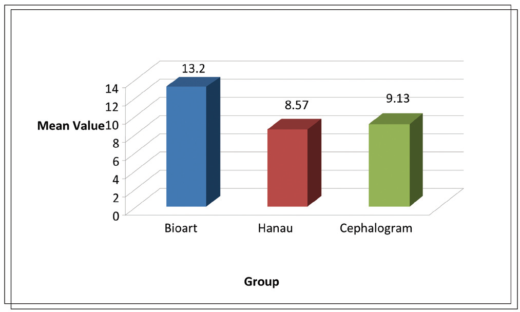
Table 3 shows that there was statistically significant difference between angles as measured by the 3 methods.
TABLE 3 shows Pairwise comparision between Bioart and Hanau, Bioart and Cephalogram and Hanau and Cephalogram by Tukey’s HSD Test.
Turkey’s HSD test showed that there was a statistically significant difference of -0.567o between mean occlusal cant as measured on Hanau Wide Vue articulator and lateral cephalogram. There was a statistically significant difference of 4.67o between mean occlusal cant as measured on Bioart articulator and lateral cephalogram. A statistically significant difference of 4.633o was found between mean occlusal cant of Hanau Wide Vue and Bioart articulators.
|
Group |
Mean Difference |
Std. Error |
p value |
95% Confidence Interval for Difference |
Results |
||
|
1 |
2 |
Lower Bound |
Upper Bound |
||||
|
Bioart |
Hanau |
4.633 |
0.388 |
<.0001 |
3.84 |
5.427 |
Significant |
|
Bioart |
Cephalogram |
4.67 |
0.303 |
<.0001 |
3.447 |
4686 |
Significant |
|
Hanau |
Cephalogram |
-0.567 |
0.274 |
0.0480 |
-1.127 |
-0.006 |
Significant |
DISCUSSION
Occlusal plane determination is an important step in complete denture construction. Maxillary cast orientation is important for this purpose which can be achieved by accurate positioning of the cast on the articulator. Failure to do this results in significant error in final occlusion of prosthesis. The position of the occlusal plane of orientation forms the basis of teeth arrangement, fulfilling the necessary mechanical, aesthetic and phonetic requirements.
The semi adjustable articulators used in our study were Hanau Widevue arcon type and Bioart Standard type with their respective facebow. Sagittal inclination of the mounted maxillary cast determined by Hanau Widevue arcon type and Bioart Standard type articulator when compared to thess gold standard technique of determining the occlusal plane i.e. lateral Cephalogram, showed that the Hanau widevue arcon type articulator was much closer to the lateral cephalogram values and less steeper than lateral cephalogram values. The Bioart Standard type articulator showed much steeper values.
The mean angle for cephalogram was 9.13 and for Hanau and Bioart articulators were 8.57 and 13.2 respectively.
Hanau articulator uses Orbitale as anterior reference point while the Bioart articulator, uses the nasion as the anterior reference point. According to Weinberg(1961)[9] various anterior point of reference used in different facebows may raise or lower the occlusalplane by +16mm and has no effect on centric occlusion, however have effect on eccentric condylar readings. Also in accordance with Gonzalez and Kingery(1968)[12], the axis orbital plane is least variable and most reliable and considered best for facebow registration as it is easy to reproduce. Bailey and Nowlin(1984)[15] evaluated the third point of refrence for mounting the maxillary cast on Hanau articulator and concluded that the Frankfort plane-maxillary occlusal plane relationship that exists in a subject is not transferred to hanau articulator with the two third points of reference.according to them , the use of middle groove on the incisal guide pin as a third point of reference positions the maxillary cast on the hanau articulator as accurately as the orbital pointer does.
According to a study conducted by Nazir et al(2012)[2] the Frankfort plane to maxillary plane relationship that exists in subjects was transferred better on Hanau widevue semiadjustable articulator as compared to the Girrbach articulator.
Mohammad Abdullah and Sherfuddin(1994)[10] conducted a comparative study of facebow transfer on Hanau and Whipmix articulators and concluded that the sagittal inclination of the occlusal plane was greater in Whip-Mix articulator as compared to Hanau-158-H2 articulator using Hanau ear piece facebow.
Malley and Milosevic(2000)[1] performed the comparison of three face-bow / semi-adjustable articulator systems for planning orthognathic surgery and concluded that the Whipmix was close to the gold standard as it flattened the occlusal plane by only 20 while Denar and dentatus differed significantly from those of the ceph as they flattened the occlusal plane by 50-6.50 respectively.
Gateno, Forres and Braincamp (2001)[16] performed a comparative study of three method sof facebow transfer recording for orthognathic surgery and concluded that the technique which eliminates the use of Nasion Relator and uses the sharpened tip of Boley’s Gauge was more accurate in determining the occlusal plane inclination as compared to the SAM Anatomical Facebow and Erickson facebow.
Lt. Col M Kumar and D’Souza(2010)[17] conducted a comparative evaluation of two techniques in achieving balanced occlusion in complete dentures and concluded that the technique that avoids the facebow presented better results in regards to time, aesthetics , comfort and stability in complete dentures.
Nascimento, Patto et al(2004)[18] conducted a double blind study for evaluating the complete dentures made by two techniques, i.e. with and without facebow and concluded that the balanced occlusion can even be provided without using facebow and is better alternative to obtain a balanced complete denture.
Gold and Setchell(1983)[19] compared the cast positions using three articulator and facebow system (Dentatus, Whipmix and Whipmix quick mount) and showed small variations. Among these the largest superior inferior variation range was seen with whipmix ear bow. Mukesh Kumar Goyal and Shelly Goyal(2011)[20] conducted a comparative study to evaluate the discrepancy in condylar guideance values between two commericially availavle arcon and non arcon articulators and concluded that the mean difference between the sagittal condylar guidance values obtained from arcon and nonarcon articualtors was highly significant in indicating a low level of reproducibility. Maxillary movements and the autorotation of the mandible affects the correct replication of the sagittal inclination on the articulator, however this study determines the accuracy required for the mounting of the maxillary cast with some limitations like the elongation of lateral cephalograms, expansion of the plaster of paris used for mounting even though the standard measurements of the weight , volume , and temperature were strictly maintained. The study showed that the replication of the sagittal inclination of the maxillary cant was much more accurate using Hanau Wide Vue articulator as compared to the bioart articulator.
CONCLUSION
The present study that was aimed at assessing the efficacy of two types of facebows/articulator system (Hanau Wide Vue and Bioart) in determining the occlusal plane accurately by comparing it with occlusal plane determined by lateral cephalogram, showed that the the values obtained with the Bioart articulator were steeper compared to the values obtained through the Lateral Cephalogram while the Hanau Wide Vue semi-adjustable articulator was more accurate.
REFERENCES
- Malley and Milosevic. Comparison of three facebow/ semi-adjustable articulator systems for planning orthognathic surgery. Br J. of Oral & Maxillofacial Surgery 2000; 38: 185-190.
- Nazia Nazir, Sujesh M, Ravikumar, Srinivas P. Accuracy of two facebow/ semiadjustable articulator systems in transferring the maxillary occlusal cant. Indian J of Dental Research 2012; 23(4): 437-442.
- Boucher C.O. The current status of prosthodontics. J Prosth Dent 1960; 10:411.
- Christiansen R. L. Rationale of the facebow in maxillary cast mounting. J Prosth Dent 1959; 9: 388-93.
- Collett H. A. Movements of the temporomandibular joint and their relation to the problems of occlusion. J Prosth Dent 1955; 5: 486-496.
- Granger E.R. Practical procedures in oral rehabilitation. Philadelphia, J.B. Lippincott Co. 1962: 63.
- McCollum, B.B., and Stuart, C.E. A research report, South Pasadena, California, Scientific Press 1955: 54-58.
- Okane H, Yamshena T, Nagasawa T, Tsuru H. The effect of antero-posterior inclination of the occlusal plane on biting force. J Prosth Dent 1979; 42: 497-501.
- Winberg L A. An evaluation of facebow mounting. J. Prosth. Dent. 1961; 11: 32-42.
- Mohammed A. Abdullah, Haneef Sherfudhin. A comparative study of facebow transfer on hanau and whipmix articulators. The Saudi Dental J 1994; 6 (1): 8-12.
- Jacobson A, Jacobson RL. Radiographic cephalometry: from basics to 3D imaging. 2nd ed. Chicago: Quintessence 2006: 308
- Gonzalez and Kingrey. Evaluation of planes of reference for orienting maxillary casts on articulator. JADA 1968; 76: 329-336.
- Stade EH, Hanson JG, Baber CL. Aesthetic considerations in the use of facebow. J. Prosth Dent. 1982; 48: 253-256.
- Pitchford JH. A re evaluation of the axis- orbital plane and the use of orbitale in the facebow transfer record. J. Prosth Dent. 1991; 66: 349-355.
- Bailey and nowlin. Evaluation of the third point of reference for mounting maxillary casts on the hanau articulator. J. prosthet. Dent. 1984; 51: 199-201.
- Gateno, Forres and Braincamp. A comparative study of three methods of facebow transfer recordings for orthognathic surgery. J. Oral and Maxillofacial Surg. 2001; 59: 635-640.
- Lt. Col Kumar M and D’Souza. Comparative evaluation of two techniques in achieving balanced occlusion in complete denture. MJAFI 2010; 66: 362-366.
- Nascimento, Patto et al. A double blind study for evaluating the complete denture made by two techniques with and without facebow. Brazilion J. Oral Science 2004; 3: 439-445.
- Gold BR, Setchell DJ. An investigation of the reproducibility of facebow transfers. J. Oral rehab 1983;10 :495-503.
- Goyal and Goyal. A comparative study to evaluate the discrepancy in condylar guidance values between two commercially available arcon and non- arcon articulators: A clinical study. Indian J. of Dental Research 2011; 22: 881-882.
Disclaimer from Guident
The views and opinions expressed in articles published in Guident are solely those of the authors and do not necessarily reflect the views of the editors, the editorial board or the publisher, Ivory India. Guident does not endorse any product, technique or treatment described. Clinical content is intended for qualified dental and medical professionals and is not a substitute for individual clinical judgement. Guident, its editors and the publisher accept no liability for any consequence arising from the use of information published in the journal.
How to cite this article
Dr. Rupal J Shah, Dr. Shilpi Sharma, Dr. Preeti Agrawal. Evaluation of Two Facebow Transfer Systems in Measuring Occlusal Cant. Guident. 2013 Oct;6(12). Available from: https://ivoryindia.in/articles/research/evaluation-of-two-facebow-transfer-systems-in-measuring-occlusal-cant.html