A Comparative Evaluation of Immediate and Early Implant Placement: A Prospective Study
Department of Oral and Maxillofacial Surgery, Swargiya Dadasaheb Kalmegh Smruti Dental College & Hospital, Nagpur, India, Department of Prosthodontics, VSPM Dental College, Nagpur, Maharashtra, India., India, Department of Oral and Maxillofacial Surgery, Swargiya Dadasaheb Kalmegh Smruti Dental College & Hospital, Nagpur, India
INTRODUCTION
The use of implant supported restoration is a predictable treatment modality. Nowadays, patients are demanding and their heightened awareness regarding advances in clinical techniques places a challenge in front of the clinician to restore a tooth in a short time span. When Branemark introduced the concept of osseointegration, in 1969(1) a waiting period of 6-8 months was advocated after extraction for healing of extraction socket before implant placement. This is the late placement technique. Implants placed by this technique has a high success rate but with main disadvantages of prolonged edentulism time and loss of alveolar bone(vertical & horizontal).Other concerns include migratory movement of teeth in edentulous space, gingival collapse and additional surgical procedure. To overcome these disadvantages clinicians introduced new techniques.
To assist the clinician in properly evaluating patients for dental implant placement, several investigators developed classification systems for the timing of implant placement following tooth extraction . Terms such as immediate, recent, delayed, late, and mature have been used in the literature in describing timing for implant placement following an extraction. Wilson and Weber’s description concerns soft tissue healing and the predictability of guided bone regeneration(2). Mayfield’s classification focuses on intervals expressed as time before installation of an implant(3). The ITI treatment guide in 2008(4)presented a new classification system according to implant placement time after extraction: type 1 (immediate) placement refers to placement of an implant at the same time as the tooth is extracted; type (early) 2 (4–8 weeks after tooth extraction) placement occurs when the implant is placed after soft tissue healing, but before any clinically significant bone fill occurs within the socket; type 3 (implant placement with partial bone healing) placement is performed after significant bone healing; and type 4 (late placement after more than 6 months of healing) placement is performed in fully healed and mature bone( branemarks technique).
Many studies have evaluated the different types of implant placement but there is paucity of studies comparing type I & type II. So, the main aim of this study was to evaluate immediate and early placement implants.The null hypothesis of this study was that type II (early) implant placement has a better outcome compared to type I (immediate) implant placement.
MATERIALS AND METHODS
This study was conducted at Swargiya Dadasaheb Kalmegh Smruti Dental College and Hospital, Nagpur. 20 patients (12 female and 8 male) were included as two separate groups in this prospective study. Group I consisted of 12 WaneCore(TM) implants
performed with immediate placement (type 1) in 10 patients and, group II included 11 WaneCore(TM) implants performed with early placement (type 2) in 10 patients. These 23 dental implants (3.5– 4.3 mm _ 11.5–15 mm) were performed by the same surgeon. The mean age was 31.5 years (42 - 19 and 44 - 21 years for groups I and II, respectively).

INCLUSION CRITERIA
All patients (ages 21 to 65) were willing to be present for examination 2 times a year for at least 2 years, as well as maintenance cleanings twice a yearimmediately after the data collection visit. All patients were free of uncontrolled diabetes (any type), existing malignancy, and were not receiving any therapy that suppresses their immune system, such as radiation, chemotherapy, or chronic steroid usage. Patients with advanced cardiovascular, pulmonary, renal, liver disease that place them in an ASA III or IV rating were excluded. Postmenopausal women with known osteoporosis as determined by their medical internist were excluded. Patients with known alcohol abuse were excluded. Patients needed the extraction of a single rooted maxillary central or lateral incisor, canine, or premolar. Patients had intact first molar occlusion to control occlusal forces on the implant restoration.If the support of all residual alveolar bone walls was adequate for immediate implant placement following the careful extraction of the unirooted teeth(Fig 1), implants were inserted immediately for group I. If there was acute or chronic periapical infection, implants were placed after the elimination of granulation tissue, epithelium and Sharpey’s fibres from the extraction socket. In the buccolingual direction minimally 2 mm of alveolar bone thickness and adequate proximal bone level for

Fig 1: Atraumatic extraction of tooth
complete interdental papilla formation should be preserved for immediate placement. 7 implants were placed in the maxilla and 5 in the mandible in group I. In group II, implants were placed 4-6 weeks after tooth removal for substantial soft tissue healing. If there was an acute endodontic or periapical infection without bone defect, lack of gingival tissue, thin periodontal phenotype or compromised mucosal blood supply, implants were placed after the 4-week healing period. 6 implants were inserted in the maxilla and 5 in the mandible in group II. When the gap between the implant surface and the surrounding bone walls was >2 mm, particulate bone harvested autogenously from the mandibular symphysis region were used for both groups I and II. Antibiotics, anti-inflammatory agents and chlorhexidine mouth-rinse were prescribed to all patients following implant surgery. Intraoral periapical radiograph was taken immediately after all implant placement procedures to determine the initial crestal bone level around the implants and at the last appointment to evaluate the vertical bone loss. Mandibular implants were loaded 3 months and maxillary implants were loaded 6 months after implant placement in both in groups. The definition of implant success and survival were determined by the implant quality scale from Misch et al.(5) If there is no pain or tenderness on use, no mobility, no history of exudates and radiographic bone loss is less than 2 mm from initial surgery implant the implant is considered successful. If bone loss is 2–4 mm, the implant is considered to have satisfactory survival. If the radiographic bone loss is less than 4 mm (less than half of the implant body) without mobility and the probing depth is less than 7 mm with a history of exudates, the implant is considered to have compromised survival. If there is pain on use, mobility, radiographic bone loss more than half the length of the implant or uncontrolled exudates it is considered a clinical failure. Periodontal pocket depth evaluation was performed at five different points around the implant. The deepest pocket depth was chosen as the periodontal pocket depth for each implant. Mesial and distal marginal bone loss was evaluated on intraoral periapical radiograph. All radiographs were taken with the same device. Initially, a transverse line was observed at the junction of the cover screw and the neck of the implant on the first radiograph and mesial and distal vertical distances between the transverse line and the crestal bone levels were documented. An additional transverse line was observed at the junction of the prosthetic restoration and the neck of the implant and the same measurements were performed on control radiographs which were taken at the last appointment. The mesial and distal vertical distances between the transverse line and the deepest marginal bone level were evaluated. Initial and former mesial and distal crestal bone levels were compared and the highest difference was chosen to determine the mean vertical bone loss.
RESULTS
The WaneCore(TM) implants were evaluated using the implant quality scale proposed by international congress of oral implantologist, consensus conference, pisa , Italy 2007(5). Pain or tenderness on

palpation, percussion or function, vertical or horizontal mobility, bone loss radiographically, any history of exudates was calculated. Probing depth was included only in compromised survival criteria.
Group I consisted of 12 implants performed with immediate placement (type 1) in 10 patients and, group II included 11 implants performed with early placement (type 2) in 10 patients. 7 implants were placed in the maxilla(58.33%) and 5 in the mandible in group I(41.66%). 6 implants were inserted in the maxilla(54.54%) and 5 in the mandible in group II(45.45%).
While placing implants in immediate & early extraction sockets an incongruity is present between the shape of socket and implant. The stability is obtained mainly from the apical part of the socket. So the discrepency present in occlusal part of the socket was filled using

Fig 4: bone graft harvested from mandibular symphysis region

Fig 5: bone graft placed in extraction socket

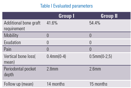
autologous bone graft from mandibular symphysis region(Fig 4,5). If discrepancy was less than 2mm bone graft was not used. In group I , the additional requirement for bone graft was 41.6% and in group II it was 54.54%. The need for bone graft was less in mandibular implants compared to maxillary implants.
The vertical bone loss (mean) was 0.4mm (0-4mm) in group I and 0.5mm(0-2, 5mm) in group II. Previously documented exact implant lengths and calculated implant lengths on radiographs were compared for each radiograph and a calibration ratio was found. This calibration ratio was used for elimination of the magnification of radiographs, which found the exact marginal bone loss.
The periodontal pocket depth( mean ) was 2.8mm in group I and 2.6mm in group II. In group I and group II the implants on compromised survival scale were 1 & 2 respectively. In both groups the periodontal pocket depth increased after occlusal loading.
On implant quality scale there were 75% success (optimum health) implants in group I and 81.8% in group II. There were no clinical failure or loss of implant. The satisfactory health implants were 8.3% & 0% in group I and Group II respectively. The survival ( compromised health) implants were 8.3% and 18.18% respectively. In this study the vertical bone loss (mean) was 0.4mm (0-4mm) in group I and 0.5 mm (0-2, 5mm) in group II. The difference in vertical bone loss in type I & type II was not clinically significant. The buccal and lingual bone loss is not evident on radiograph.
DISCUSSION
The demand for early replacement for extracted tooth leads the clinicians to develop new protocols of immediate and early implant placement. The first reported case of immediate implant was described by Schulte in 1976 using a polycrystalline aluminum surface.(6)
Hammerle CH et all(7), defined Type I as Implant placement immediately following tooth extraction and as part of the same surgical procedure. The advantages are reduced number of surgical procedures, reduced overall treatment time & optimal availability of existing bone. The disadvantages are Site morphology may complicate optimal placement and anchorage, thin tissue biotype may compromise optimal outcome, potential lack of keratinized mucosa for flap adaptation, adjunctive surgical procedures may be required and the procedure is technique-sensitive.
This disadvantage of potential lack of keratinized mucosa for flap adaptation is overcome in type II implant placement. Type II is defined implant placement after Complete soft tissue coverage of the socket (typically 4–8 wks)(7). The advantages are increased soft tissue area and volume facilitates soft tissue flap management and resolution of local pathology can be assessed. The disadvantages are treatment time is increased, adjunctive surgical procedures may be required and socket walls exhibit varying amounts of resorption.
It is well known that during the first 4 months of healing, according to observations and measurements, the buccal-lingual ridge undergoes a reduction of approximately 5 to 7 mm with a 2- to 4.5-mm loss of vertical bone height(8,9). Both techniques have the advantage of alveolar ridge preservation. But the clinician cannot predict the morphology of bone resorption which may compromise the aesthetic outcome. Covani et al.(10)reported that the vertical distance between the implant shoulder and bone crest ranged from 0 to 2 mm (mean 0.8 mm) 6 months after immediate placement. The mean distance between buccal and lingual or palatinal bone decreases from 10.5 to 6.8 mm 6 months after immediate implant placement. In the present study the vertical bone loss (mean) was 0.4mm (0-4mm) in group I and 0.5mm (0-2, 5mm) in group II. This difference of vertical bone loss between the two groups was not statistically significant.
The mesial and distal bone loss can be calculated on radiographs but buccal and lingual bone loss is cannot be evaluated on radiographs. The clinician must be aware of this and so probing should be carried out at all sides of implants in control appointments (5). The “correct pressure” for probing has not been defined for implants, but may be less important than with teeth, because there is no connective tissue attachment zone next to an implant. The potential for damage to the fragile attachment or marring of the implant surface may exist during probing(11). On the other hand, there is no clinical or experimental evidence supporting this hypothesis.
7 implants were placed in the maxilla and 5 in the mandible in group I. 6 implants were inserted in the maxilla and 5 in the mandible in group II.
The type of prosthetic restoration changes the functional loading around
the implants and affects the clinical outcome(12). The prosthetic restoration was done 3 months & 6 months after implant placement in mandibular and maxillary implants respectively. Two implants were restored using bridge and rest were restored as single crown prosthesis.
Immediate and early implant placement are equally technique sensitive. In case of incisors the root tapers evenly from the cervical line to a point approximately two thirds of its length apically(16) while the diameter of endosseous implant is same through its length. So due incongruity between socket shape and size of endosseous implant there is requirement of bone graft.(Fig 3) Sufficient bone implant contact into a healed and mature bone can be observed without bone grafting or barrier membrane usage in circumferential peri-implant bone defects less than 2 mm(13).Bone graft requirement rates were similar for both of the implant placement techniques in this study. Also the results of immediate & early implants are more predictable when bone grafts are used. So, on the basis of results of this study we recommend using bone grafts in cases of immediate & early implant placement.The 4-8 week waiting period for early implant placement did not reduce the need for bone graft. No mobility, exudation or pains were observed during clinical evaluation around any implants in groups I and II. The mean follow-up period was 14 months (range 12-16 months) in the immediate implant placement group and 15 months (range 13–17 months) in the early placement group.
There are very few studies comparing immediate and early implants.
Palattella et al.(14) evaluated implants placed immediately after extraction and those placed after 8 weeks with parameters of marginal bone resorption, papilla index, and position of the mucosal margin at a 2-year follow-up visit in 16 implants. They found no statistically significant differences in any of the studied parameters between the two groups.
Anibali et al.(15) compared the success rates of immediate, early and late placed single implants with a small sample size. They concluded that the success rates in the early period using the three techniques were similar (91.7–100%) and early placement should be considered an appropriate alternative to immediate placement when unfavourable conditions at the time of extraction could affect the clinical outcome of type 1 implant placement.
CONCLUSION
The results of this study showed that the hypothesis that the early implant placement protocol is more successful than the immediate placement was not valid. When implant success rate, bone graft requirement, cervical bone loss, implant mobility, peri-implant periodontal pocket depth, exudation and pain are considered, there was no remarkable difference between the success rates of the immediate and early placement (4 weeks waiting for soft tissue healing) techniques in long term follow up.
Also we conclude from this study that when bone grafts are used in immediate and early implant placement the success rate of osseointegration is very high which ultimately leads to more predictable results.
REFERENCES
- Branemark PI, Hansson BO, Adell R, Breine U, Hansson BO, Lindstrom J, Ohlsson A. Intra-osseous anchorage of dental prostheses. I. Experimental studies. Scand J Plast Reconstr Surg Suppl 1969;3:81–100.
- Wilson TG, Weber HP. Classification of and therapy for areas of deficient bony housing prior to dental implant placement. Int J Periodontics Restorative Dent 1993;13:451–9.
- Mayfield LJA. Immediate, delayed and late submerged and transmucosal implants. In: Lang NP, Karring T, Lindhe J, editors. Proceedings of the 3rd European workshop on periodontology implant dentistry1999:520–34.
- Chen S, Buser D. Implants in post-extraction sites: a literature update. In: Buser D, Belser U, Wismeijer D, editors. ITI Treatment Guide, vol. 3: Implants in extraction sockets. Berlin: Quintessence; 2008.
- Misch CE, Perel ML, Wang HL, Sammartino G, Galindo-Moreno P, Trisi P, et al. Implant success, survival, and failure: The International Congress of Oral Implantologists (ICOI) Pisa Consensus Conference. Implant Dent 2008;17:5–15.
- Schulte W, Kleineikenscheidt H, Lindner K, et al. The Tu¨ bingen immediate implant in clinical studies. Dtsch Zahnarztl Z 1978;33(5):348–59.
- Hammerle CH, Chen ST, Wilson TG, et al. Consensus statements and recommended clinical procedures regarding the placement of implants in extraction sockets. Int J Oral Maxillofac Implants 2004:19(Suppl):27.
- Lam RV. Contour changes of the alveolar process following extraction. Aust Dent J 1969;14:241–4.
- Johnson K. A study of the dimensional changes occurring in the maxilla following closed face immediate denture treatment. Aust Dent J 1969;14(6):370–6.
- Covani U, Cornelini R, Barone A. Vertical crestal bone changes around implants placed into fresh extraction sockets. J Periodontol 2007;78:810–5.
- McKinney RV, Koth DC, Steflik DE. Clinical standards for dental implants. In: Clark JW, ed. Clinical Dentistry. Harperstown, PA: Harper & Row; 1984:27-41
- Misch CE, Bidez MW, Sharawy M. A bioengineered implant for an ideal bone cellular response to loading forces: a literature review and case report. J Periodontol 2001;72:1276–86.
- Becker BE, Becker W, Ricci A, Geurs N. A prospective clinical trial of endosseous screw-shaped implants placed at the time of tooth extraction without augmentation. J Periodonto1 1998;69:920–6.
- Palattella P, Torsello F, Cordaro L. Two-year prospective clinical comparison of immediate replacement vs. immediate restoration of single tooth in the esthetic zone. Clin Oral Implants Res 2008;19:1148–53
- Anibali S, Bignozzi I, Iaccovazzi L, La Monaca G, Cristalli MP. Immediate, early and late implant placement in first molar sites: a retrospective case series. Int J Oral Maxillofac Implants 2011;26:1108–22.
- Ash, Nelson . Wheelers Dental Anatomy, Physiology and Occlusion. 8th Edition
Disclaimer from Guident
The views and opinions expressed in articles published in Guident are solely those of the authors and do not necessarily reflect the views of the editors, the editorial board or the publisher, Ivory India. Guident does not endorse any product, technique or treatment described. Clinical content is intended for qualified dental and medical professionals and is not a substitute for individual clinical judgement. Guident, its editors and the publisher accept no liability for any consequence arising from the use of information published in the journal.
How to cite this article
Dr. Pravin Lambade, Dr. Deepti Lambade, Dr. Dilpreet Rajkhokar. A Comparative Evaluation of Immediate and Early Implant Placement: A Prospective Study. Guident. 2013 Jul;6(9). Available from: https://ivoryindia.in/articles/research/a-comparative-evaluation-of-immediate-and-early-implant-placement-a-prospective-study.html