A Compund Odontoma in Relation to Impacted Canine
Department of Oral & Maxillofacial Surgery SDM College of Dental Science, Dharwad, India
Introduction
Odontomas are the most common odontogenic tumor. They are considered to be hamartomas rather than neoplasms, and are composed of the tissues native to teeth: enamel, dentin, and cementum and pulp tissue. Odontomas are further sub-classified based upon their gross and radiographic features into compound (small tooth like structures) or complex (a conglomeration of dentin, enamel and cementum.
CASE REPORT
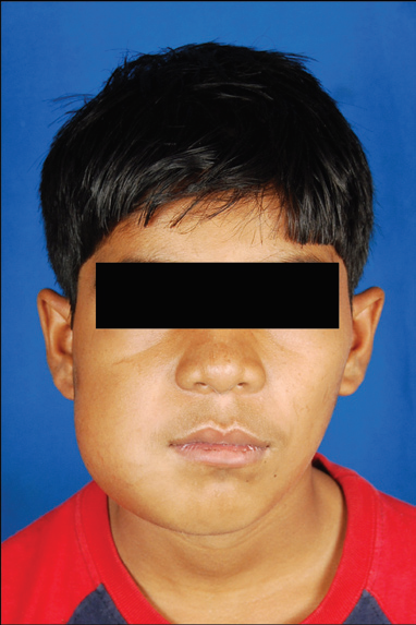
A 14 year old male patient complained of slow growing enlargement of the buccal cortical plate with missing teeth. On radio graphical assessment revealed a presence of radio opaque masses resembling odontome. Surgically impacted teeth and masses excised, Histopathological report confirmed compound odontome. Patient reviewed after 1 month. 6 months. No history of recurrence and wound healing satisfactory.
Surgical management
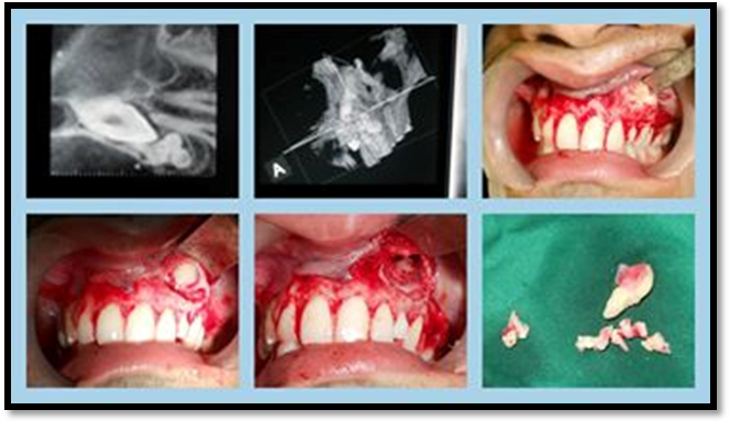
EXCISION OF LESION AND SURGICAL DISIMPACTION OF CANINE
DISCUSSION
Odontomas are the most common odontogenic tumor. They are considered to be hamartomas rather than neoplasms, and are composed of the tissues native to teeth: enamel, dentin, and cementum and pulp tissue.
It was in 1867 that Paul Broca first used the term “Odontoma.” Broca defined the term as “tumors formed by the overgrowth of transitory or complete dental tissues”.[
Classification
According to W.H.O. classification, 1 Odontomas can be divided into three groups:
1. Complex odontome - when the calcified dental tissues are simply arranged in an irregular mass bearing no morphologic similarity to rudimentary teeth.
2. Compound odontome - composed of all odontogenic tissues in an orderly pattern that results in many teeth-like structures, but without morphologic resemblance to normal teeth.
3. Ameloblastic fibro-odontome - consists of varying amounts of calcified dental tissue and dental papilla like tissue, the later component resembling an ameloblastic fibroma. The ameloblastic fibro-odontome is considered as an immature precursor of complex odontome. According to Thoma and Goldman:5
• Geminated composite odontomes - two or more, more-or-less well-developed teeth fused together.
• Compound composite odontomes - made up of more-or-less rudimentary teeth.
• Complex composite odontomes - calcified structure, which bears no great resemblance to the normal anatomical arrangement of dental tissues.
• Dilated odontomes - the crown or root part of tooth show marked enlargement.
• Cystic odontomes - an odontome that is normally en-capsulated by fibrous connective tissue in a cyst or in the wall of the cyst.
TYPES
There are essentially two types of odontomes:
1. Complex composite odontome;
2. Compound composite odontome. A new type known as Hybrid odontome is also reported by some authors “Complex Composite Odontome”
ETIOLOGY AND INDICATION FOR REMOVAL OF THE TUMOUR
The etiology of odontoma is not known but environmental causes such as infection, trauma, family history and genetic mutation are hypothesized.
Although the Odontomas erupting in the oral cavity is controversial, the reason is attributed to the eruptive forces of the apparently impacted teeth and in cases of absence of teeth, the reasons could possibly be the resorption of alveolar ridge exposing the odontoma, sequestration of overlying bone, alveolar bone remodeling in young adults, reactive growth of the capsule surrounding odontoma in elderly patients. Odontomas have been associated with trauma during primary dentition as well as with inflammatory and infectious processes, hereditary anomalies (multiple Odontomas in Gardner syndrome, Hermann's syndrome), odontoblastic hyperactivity and alterations in the genetic components responsible for controlling dental development Association of this lesion with the unerupted tooth is high and three quarters of impacted teeth related to Odontomas can erupt after removal of the odontoma. Another reason for removing Odontomas is their association with cysts in a significant number of cases (27.6%).
CLINICAL FEATURES
The complex odontoma constitute 5-30% of all odontogenic tumors and are mostly found in the posterior mandible and compound odontoma in anterior maxilla. They are seen with unerupted teeth in 10-44% and about 17% of them are associated with impacted maxillary lateral incisors. Females are affected marginally more than male (1.5:1). Majority of cases (84%) are seen below 30 years but are seen in any age group with peak incidence in second decade. Less than 10% are found in patients above 40 years of age. and most lesions are detected on routine radiographs.
Odontomas are generally asymptomatic, often associated with delayed eruption or impaction of permanent teeth and retained primary teeth. In some occasional cases, pain, infection, regional adenopathies, alveolar bone expansion and tooth displacement may be present.
Odontomas can also manifest as part of syndromes, such as Gardner syndrome, basal cell nevus syndrome, familial colonic adenomatosis, Tangier disease or Hermann syndrome
In the present case patient was asymptomatic few month back , but later complain of swelling on the left side which on radiographic diagnosis (OPG) ruled out to be multiple harmatomatous radio opaque body around the tooth in glomerulated aggregates
MANAGEMENT
Odontomas are circumscribed, encapsulated tumors that can be removed successfully by conservative surgery. Spontaneous eruption of the impacted tooth after removal of the obstruction like odontoma has been reported by many authors.
A less conservative approach is advocated by others with exposure of the unerupted tooth at the time of surgery and placement of bonded attachment and ligature/e-chain for orthodontic traction, to facilitate rapid eruption.
This approach, however, may result in poor gingival margin, inadequate gingival tissue attachment and a discrepancy of gingival level between the exposed tooth and its neighboring teeth.
Hence, in the present case, we advocated a more conservative approach of removal of odontoma and its fibrous capsule. Along with that, we removed bone unerupted canine , replaced the flap back in position.
RECURRENCE RATE AND ASSOCIATION WITH TUMORS
A clinical case of CCOT associated with odontoma and a missing deciduous tooth in a 3-year-old female patient. Described by Gorlin et al. in 1962, the calcifying cystic odontogenic tumor (CCOT) may be associated with unerupted teeth, ameloblastomas, adenomatoid odontogenic tumors, and, in many cases, with odontomas.
Prognosis is very favourable because it has no to minimal recurrence
REFERENCE
- Hidalgo-Sánchez O, Leco-Berrocal MI, Martínez-González JM. Metaanalysis of the epidemiology and clinical manifestations of odontomas. Med Oral Patol Oral Cir Bucal. 2008;13:E730–42.
- Shafer's text book of oral pathology. 5th ed. Amsterdam: Elsever; 2006. Cysts and tumors of odontogenic origin.Disturbances of development and growth. Shafer, Hine, Levy; pp. 357–432.
- Batra P, Duggal R, Kharbanda OP, Parkash H. Orthodontic treatment of impacted anterior teeth due to odontomas: A report of two cases. J Clin Pediatr Dent. 2004;28:289–94.
- Prætorius F, Piattelli A. Odontoma, Odontogenic tumors. In: Barnes L, Eveson JW, Reichart P, Sidransky D, editors. WHO Classification of tumors. Pathology and Genetics of Head and Neck Tumors.Lyon France: IARC Press; 2005. pp. 310–11.
- Yeung KH, Cheung RC, Tsang MM. Compound odontoma associated with an unerupted and dilacerated maxillary primary central incisor in a young patient. Int J Paediatr Dent. 2003;13:208–12.
- Jafarzadeh H, Abbott PV. Dilaceration: Review of an endodontic challenge. J Endod. 2007;33:1025–30
Disclaimer from Guident
The views and opinions expressed in articles published in Guident are solely those of the authors and do not necessarily reflect the views of the editors, the editorial board or the publisher, Ivory India. Guident does not endorse any product, technique or treatment described. Clinical content is intended for qualified dental and medical professionals and is not a substitute for individual clinical judgement. Guident, its editors and the publisher accept no liability for any consequence arising from the use of information published in the journal.
How to cite this article
Dr. Prit Shah. A Compund Odontoma in Relation to Impacted Canine. Guident. 2013 Jul;6(9). Available from: https://ivoryindia.in/articles/oral-surgery/a-compund-odontoma-in-relation-to-impacted-canine.html