An in-Vitro Comparison of the Antimicrobial Effects of Sodium Dichloroisocyanurate, Chlorhexidine and Sodium Hypochlorite Against Enterococcus Faecalis and Candida Albicans
Department of Conservative Dentistry and Endodontics, Yenepoya Dental College, Mangalore, India, Department of Oral Pathology, Yenepoya Dental College, Mangalore, India, Department of Conservative Dentistry and Endodontics, SIBAR Dental College, Guntur, India
Abstract
Aim: The aim of this In-vitro study is to investigate the antimicrobial action of Sodium Dichloro isocyanurate 0.6% (NaDCC) (Ecolabs, Garforth, LEEDS); Chlorhexidine 2% (CHX) (Dentochlor , Ammdent , INDIA); and sodium hypochlorite 2% (NaOCl) (Vensons,Bangalore) and a combination of 0.6% of Sodium dichloro isocyanurate and 0.2% of Chlorhexidine against Enterococcus faecalis and Candida albicans obtained by microbiological sampling procedure.
Materials & Methods: 45 patients were selected for endodontic retreatment and the microorganisms Enterococcus faecalis and Candida albicans were obtained from the dentinal shavings of the root canals. These dentinal shavings were immersed in phosphate buffered solution and sent for microbial sampling procedure. Out of these, 20 positive cultures for Enterococcus faecalis, 17 positive cultures for Candida albicans and the remaining 8 positive cultures were a mixed combination of both E.faecalis and C.albicans were obtained. Blood agar/ brain heart infusion agar were used for the culture of E.faecalis. Brain heart infusion agar/ sabouraud’s dextrose agar culture medium was used for the culture of C.albicans.
The culture plates were randomly distributed for respective microbiological sensitivity of the isolates. The isolates containing E.faecalis were incubated in candle jar culture plates, a partial anaerobic incubator and C.albicans with aerobic incubator for 48 hrs at 37°C. The diameter zones of microbial inhibition were measured in millimeters and recorded. Statistical analysis and pair wise comparison was performed with Mann Whitney U test.
Results: Sodium dichloro isocyanurate 0.6% and Chlorhexidine 2% demonstrated significantly bigger zones of microbial inhibition against E.faecalis. when Sodium Dichloro isocyanurate 0.6% and Chlorhexidine were compared Sodium Dichloro isocyanurate 0.6% showed bigger zones of inhibition than Chlorhexidine 2% and sodium hypochlorite 2% against Candida albicans. The control group of normal saline showed no inhibition.
KEY WORDS: Irrigants, Anaerobic culture, Enterococcus faecalis, Candida albicans, Sodium Dichloro isocyanurate, Chlorhexidine
INTRODUCTION
Complete debridement, disinfection and the three dimensional hermetic seal are the major objectives for predictable long term success in endodontic treatment of complex root canal system. Persistence of intracanal infection is one of the major reasons for endodontic treatment failure. Microorganisms associated with failed endodontic treatment differ from that associated with primary root canal infection. Secondary root canal infections are composed of fewer species which are dominated by Streptococcus mutans, Actinomyces israeli, Proprionibacterium granulosum, Fissurum nucleates, Porphyromas gingivalis, Yeasts and principally Enterococcus faecalis1.
Prominence of E. faecalis in root canal treated teeth with apical periodontitis has made it a focus of attention, as an etiological factor of post treatment disease2. Frequency of E.faecalis was found to be nine times more in failed root canal treatment cases than in primary endodontic infections. The prevalence range was found to be 24% to 77%, by culturing method and 67% to 77%, when a polymerase chain reaction detection method was used3. E. faecalis was found to possess various virulence factors which have the ability to maintain and have capability to invade dentinal tubules and adhere to collagen in the presence of human serum4,5
Candida albicans is another common species of yeast that has been cultured from root canals of teeth with failed endodontic treatment5. Prevalence of Candida species reported in infected root canals varies between 1% and 17%. Both C.albicans and E.faecalis are able to invade dentinal tubules to variable depth6,7. Ohara et, al. evaluated the antimicrobial effects of Sodium hypochlorite 5.25%, Hydrogen Peroxide 3%, Saline sterile NaCl 0.9% ,REDTA 17%, Chlorhexidine 0.2%, saturated solution of Calcium hydroxide against anaerobic bacteria and reported that Chlorhexidine was the most effective8,9.
Antimicrobial substantivity of 2% Chlorhexidine used as root canal irrigating solution in teeth with pulp necrosis and radiographically visible chronic Periapical lesions, reported that antimicrobial activity of Chlorhexidine with residual effects in the root canal system was for up to 48 hrs after application10 and therefore Chlorhexidine is an effective antifungal agent but its efficacy is significantly less than Sodium hypochlorite.
Sodium hypochlorite (NaOCl) is a chlorine releasing agent; it is commonly used as an irrigant for the elimination of microorganisms and necrotic tissue from the root canal system10. Another agent, Sodium Dichloro isocyanurate (NaDCC) may provide better antimicrobial action than sodium hypochlorite at similar or lower toxicity levels10,11. Minimal bacterial concentration values for E.faecalis were found to be 0.625% for NaDCC. Both NaOCl and NaDCC were lethal to fibroblasts in tissue culture; NaOCl was toxic in lower concentrations than NaDCC12. The purpose of the study was to examine the in vitro susceptibility of E.faecalis and C.albicans to Sodium Dichloro isocyanurate and Chlorhexidine with three different concentrations and the combination.
MATERIALS AND METHODS:
MICROBIOLOGICAL PROCEDURE:
45 patients were selected for endodontic re-treatment of either asymptomatic or symptomatic teeth and showed radiographic evidence of endodontic failure with or without Periapical bone lesions. Rubber dam was placed, and an aseptic technique was used for sampling procedure as laid out by Richard E Walton13. The tooth, rubber dam clamp, and surrounding parts of the rubber dam were cleaned with 30% hydrogen peroxide and then swabbed with 5% iodine tincture. After drying the tincture, the tooth surface was swabbed with 5% sodium thiosulphate solution to inactivate the iodine tincture so that remnants of iodine would not influence the bacteriologic sampling. The access of root filling was removed by instrumentation with burs, files and without the use of chemical solvents and irrigants. Radiographs were taken to ensure that all gutta-percha had been removed, and the samples of canal contents were taken with sterile forceps using sterile paper points inserted into the canal to the approximate apex region. Immediately after sampling the paper points were placed into the phosphate buffered saline solution. The canal was then instrumented so that material could be obtained from the walls. Second samples were taken with sterile reamers from the most apical dentin in the canal. Alternatively, a small volume of sterile physiological saline was applied into the canal, the canal walls were filed with a Hedstorm file, and the sample was collected with a sterile paper point.
The collected samples were directly inoculated into the liquid medium in presence of flame and kept at room temperature. The accesses of root fillings were dressed with calcium hydroxide and in the further appointments root canal therapy was completed. The liquid medium used for the isolation of Enterococcus faecalis was blood agar and that used for Candida albicans isolation was sabouraud’s dextrose broth. Samples collected were transported to the laboratory and were incubated at 37ºC for 48 hrs using a candle jar incubator which is a partial anaerobic incubator for E.faecalis and aerobic incubator for C.albicans. Growth was indicated by the presence of turbidity in the liquid medium. Once turbidity was observed, it was sub cultured on the solid medium of Enterococcus faecalis on Brain heart infusion agar and for Candida albicans on Sabouraud dextrose agar. The sub cultured plates were incubated at 37° C using an incubator for 24-48 hrs to isolate the organism in the form of colonies. Isolates were identified by standard methods14 (Table 1).

AGAR DIFFUSION TEST:
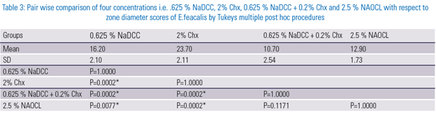
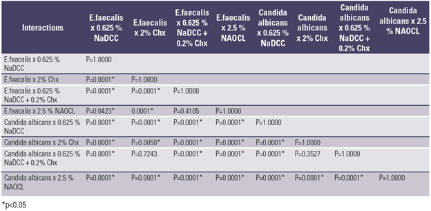
The present study used E.faecalis and C.albicans strains cultured on brain heart infusion agar and Sabouraud dextrose agar. The organisms were incubated under aerobic condition. The agar plates were prepared in sterile glass Petri dishes, punch wells of 6mm depth and 2mm diameter were made with a punch (punch well method). The testing solutions, sodium dichloro isocyanurate (NaDCC) (Ecolab, Garforth, LEEDS) available as tablet form, made in to solutions of 0.6%. Chlorhexidine gluconate (Dentochlor, Ammdent, INDIA) available as solution 2% form was tested. The combination of both 0.6% NaDCC and 0.2% Chlorhexidine and Sodium hypochlorite with 2% concentration were considered for the study. 50 microliters each was transferred into the wells. The E.faecalis and fungal growth inhibition was calculated by measuring the inhibition zones from the outer end of the well to the point where the zone ends with the help of vernier calipers. The point of abrupt diminution of growth, which corresponds to the point of complete inhibition of growth, was taken as zone edge. The results were statistically analyzed by using post hoc tukeys test and two way ANOVA test. (table.2 ) and (table.3)



STATISTICAL ANALYSIS:
Pair wise comparision between the groups reveal 2% Chlorhexidine was effective than 0.625% NADCC, combination of Chlorhexidine and 2% NaOCl for E.faecalis. This was statistically analyzed using post hoc tukeys test. For candida albicans, the mean zone diameter was greater for 0.625% NADCC when compared to the other solutions used in the study.
RESULTS
The results of NaDCC 0.6% (Fig 1) showed that it was effective against both the organisms with a substantial zone of inhibition better than the positive control normal saline and a combination used of both NaDCC 0.6% and Chlorhexidine 0.2%

Figure 1 : Interaction plot between organisms and concentrations on zone diameter in mm
NaDCC alone showed better antifungal efficacy against Candida albicans when compared to Chlorhexidine 2% and combination of NaDCC 0.6% & Chx0.2%.
Chlorhexidine 2% (Fig2) showed a better antimicrobial efficacy against E.faecalis when compared to other Chlorine releasing agents NaDCC and NaOCl at different concentrations.
DISCUSSION
NaDCC tablets are compact and stable (Hoffman, Death & Coates 1981) and dilutions can be prepared safely and accurately. The microbicidal activity of solutions prepared from NaDCC tablets possibly may be greater than that of NaOCl solutions containing same total available chlorine for two reasons. First the pH of solutions prepared from NaDCC tablets is much lower than that of NaOCl solutions and activity increases with acidity13. Secondly, in NaDCC solutions, only a proportion of the total available chlorine is free while the rest is combined; free available chlorine is used up so the rest part is released. Dichloro isocyanurate like sodium hypochlorite liberates free chlorine (OCl/HOCl) in water. Because of the neutral pH of NaDCC solutions, most of the free chlorine is in the form of hypochlorous acid (HOCl) which is more bactericidal than the hypochlorite ion (OCl) predominant in aqueous NaOCl solutions14. Stability of the antiseptic agent is also of importance. NaDCC tablets were stable for prolonged periods of time but solutions of NaDCC were unstable and decomposed much faster than NaOCl of the same strength. This can be overcome by dissolving tablets and preparing small amount of fresh solutions daily and are not prepared in bulk for distribution where they may be stored. NaDCC was found to be more effective in inhibiting E.faecalis than NaOCl which is known to be resistant to calcium hydroxide dressings15,16.
Chlorhexidine is a cationic biguanide with a broad spectrum of activity against bacteria and fungi13,17. Its efficacy is because of interaction of the positive charge of the molecule and the negatively charged phosphate groups on microbial cell walls, thereby altering the cells osmotic pressure. Membrane disruption is the lethal event in Chlorhexidine action. It is bactericidal because of precipitation and/or coagulation of intracellular constituents16.
In an in vitro study by Joshua et al Biopure MTAD showed significantly higher zones in inhibition when compared to NaOCl and Chlorhexidine but inhibition zones of Chlorhexidine and sodium hypochlorite was not so significant17,18. In a clinical study by Zamany et al19,20 showed that 2% CHX solution used as a final irrigant significantly decreased bacterial loads in root canals that had been irrigated with sodium hypochlorite during canal preparation.
CONCLUSIONS
- Sodium Dichloro isocyanurate is another Chlorine releasing agent same as sodium hypochlorite, so the combination of only sodium Dichloro isocyanurate and Chlorhexidine have been evaluated.
- Sodium Dichloro isocyanurate 0.6% showed better antibacterial and antifungal efficacy when compared to sodium hypochlorite 2%. Antifungal efficacy of Sodium Dichloro isocyanurate 0.6% was found to be better than Chlorhexidine 2%. Further in vivo studies should be done to know its biocompatibility and efficacy as alternative to sodium hypochlorite using as an antimicrobial root canal irrigant.
- Chlorhexidine 2% showed better antibacterial efficacy when compared to sodium Dichloro isocyanurate 0.6% and sodium hypochlorite 2%.
Acknowledgement
The author thanks to Dr. U. S. Krishna Nayak (Dean Academics, Senior Professor and Head Department of Orthodontics, A B Shetty Memorial Institute of Dental Sciences, Mangalore), Dr. Prathap M.S (Professor), and Dr. Harish K Shetty (Senior Professor and Head, Department of Conservative Dentistry and Endodontics, Yenepoya Dental College,Mangalore), For their highly valuable advice and support throughout this study.
REFERENCES
- Ingle J, Bakland L K, Baumgartner J C; Ingle’s Endodontics 6 ; 221-309; BC Decker ; 2008.
- . Porteneir S, Waltimo T & Haapasalo M, Enterococcus faecalis-the root canal survivor and star in post treatment disease. Endodontic topics 2004; 6: 135-159.
- Rocas IN, Siquera JF, Santos KRN .Association of Enterococcus faecalis with different forms of periradicular disease. J Endod 2004; 30: 315-20.
- love R M, Enterococcus faecalis- A mechanism for its role in endodontic failure, Int Endod J 2001; 39: 399-405
- Stuart Ch , Enterococcus faecalis: its role in root canal treatment failure and current concepts in retreatment. J Endod 2006 Feb;32(2):93-8
- Waltimo TMT,Siren EK,Torkko HLK, Olsen I ,Happasalo MPP.Fungi in therapy-resistant apical periodontitis. Int Endod J 1997; 30: 96-101
- Siquera JF Jr, Rocas IN, Lopes HP Elias CN,de Uzaeda M. Fungal infection of the radicular dentin. J Endod 2002; 28:(11): 770-3
- Siren EK, Haapasalo MP, Ranta K, Salmi P,Kerusuo EN. Microbiological findings and clinical treatment procedures in endodontic cases selected for microbiological investigation. Int Endod J 1997 ; 30: 91-5.
- Ohara PK, Torbinajad M, Ketting JD. Antibacterial effects of various endodontic irrigants on selected anaerobic bacteria. Endod Dent Traumatol 1993; 9: 95-100
- Leonardo MR,Tanontaru filho M,Silva LAB,Nelson Filho P, Bonifacio KC, In vivo antimicrobial activity of 2% chlorhexidine used as a root canal irrigation solution . Journal of endodontics 1999: 27; 616-9
- Mc Donnell G, Russell AD . Antiseptics and disenfectants : activity , action ,and resistance. Clin Microbiol Rev 1999: 12: 147-79
- Bloomfield SF , Miles GA. The antibacterial properties of Sodium dichloro isocyanurate and sodium hypochlorite formulations. J Appl Bacteriol 1978 ; 46: 65-73.
- Heling I ,Rotstein I ,Dinur.T , Szwec-Levine Y , Steinberg D. Bactericidal and Cytotoxic Effects of Sodium Hypochlorite and Sodium dichloroisocyanurate solutions in Vitro. J Endod 2001 ; 27, 4: 278-300
- Walton R E, Microbiologic analysis of teeth with failed endodontic treatment and the outcome of conservative re-treatment. Oral Surg, Oral Med,Oral Path, Oral Radiol Endod 1998; 85: 86-93.
- Bailey & Scott’s Diagnostic Microbiology, 11th edition, page no: 298- 315.
- D.Coates. A comparision of Sodium hypochlorite and sodium dichloro isocyanurate products. Journal of hosp Infection 1985;6:31-40
- . Russell AD Day MJ. Antibacterial activity of Chlorhexidine, J Hosp Infection 1993; 25: 229-38.
- Davis J, DDS, James M, James K. Bahcall D. An In Vitro Comparison of the Antimicrobial Effects of Various Endodontic Medicaments on Enterococcus faecalis. J Endod 2007; 33: 567-569
- . Zamany A, Safavi K, Spangberg L. The effect of Chlorhexidine as an endodontic disinfectant. Oral Surg Oral Med Oral Pathol 2003; 96:578-
- 81.
- . 20. Love R M, Bacterial adhesins--their role in tubule invasion and endodontic disease. Aust Endodontic J 2002 Apr; 28(1):25-8.
Disclaimer from Guident
The views and opinions expressed in articles published in Guident are solely those of the authors and do not necessarily reflect the views of the editors, the editorial board or the publisher, Ivory India. Guident does not endorse any product, technique or treatment described. Clinical content is intended for qualified dental and medical professionals and is not a substitute for individual clinical judgement. Guident, its editors and the publisher accept no liability for any consequence arising from the use of information published in the journal.
How to cite this article
Dr. Amarnath Shenoy, Dr. Shruti Nayak, Dr. Nagesh Bolla. An in-Vitro Comparison of the Antimicrobial Effects of Sodium Dichloroisocyanurate, Chlorhexidine and Sodium Hypochlorite Against Enterococcus Faecalis and Candida Albicans. Guident. 2013 Jul;6(9). Available from: https://ivoryindia.in/articles/research/an-in-vitro-comparison-of-the-antimicrobial-effects-of-sodium-dichloroisocyanurate-chlorhexidine-and-sodium.html