Clinical Management of MB2 Canal in Maxillary First Molar
Department of Conservative Dentistry & Endodontics, Pacific Dental College & Research Centre, Udaipur
ABSTRACT
Introduction: The mesiobuccal root of maxillary molar has generated more research and clinical investigation than any other root. Maxillary molar is the tooth with the largest volume and most complex root and root canal anatomy, also possibly the most treated and least understood posterior tooth. To identify and locate all root canal spaces along with thorough chemomechanical preparation and to achieve a fluid-impervious seal with an inert obturating material in all the portal of exits are the key requisites for successful endodontic therapy. Post treatment disease can be attributed to the presence of any undetected,unfilled anatomical spaces in the root canal system which can act as a nidus for infection leading to treatment failure.
Case Report: This case report describes endodontic treatment of maxillary first molar with two mesiobuccal canals. It also highlights the need for good anatomical knowledge of root canal morphology and its variations in order to achieve successful results.
Discussion: The incidence of MB2 canal in maxillary first molar is 86.36%, absent in 12.1% and untraceable in 1.5%. This case report on maxillary first molar in which MB2 in mesiobuccal root was located using visual, tactile and magnifying devices followed by endodontic treatment.
Conclusion: It is generally accepted that a major cause for failure of root canal therapy is an inability to recognize the presence of, and to adequately treat, all of the canals in the root canal system. Therefore, it is worthwhile to put the time and effort into properly locating and treating these canals in an attempt to increase the prognosis of endodontic therapy in maxillary molars.
Keywords: maxillary first molar, MB2 root canal, Udaipur District, magnifying loupes.
Introduction
The maxillary first molar is the largest tooth in volume and one of the complex in root and root canal anatomy. The pulp chamber of maxillary first molar is the largest in the dental arch, with four pulp horns: messiobuccal, distobuccal, mesiopalatal and distopalatal. The main mesiouccal canal orifice is buccal and mesial to the distobuccal orifice and is positioned within the acute angle of the pulp chamber. The mesiopalatal canal orifice (MB2) is located palatal and mesial to mesiobuccal orifice. The distobuccal angle is an obtuse angle, the palatal angle are basically right angle.1 It is important to identify and locate all root canal spaces along with thorough chemomechanical preparation in order to achieve a hermetic seal with an inert obturating material in all the portal of exit are the key requisites for successful endodontic therapy. Failure to do so can lead to post-treatment disease which can be attributed to the presence of any undetected and subsequently unfilled anatomical spaces in the root canal system which can act as a nidus for infection leading to treatment failure.2,3 Elusive “second mesiobuccal” (MB2) canal is one of the biggest mysteries in endodontics.4 The second mesiobuccal canal orifice in maxillary molars is usually located either mesial to or in the pulpal groove connecting the main mesiobuccal canal and palatal canals, within 3.5 mm palatally and 2 mm mesially from the main mesiobuccal canal.5,6,7 It has been found that endodontically retreated teeth contain more undetected MB2 canals than 1st time treated teeth suggesting inability to detect, debride and fill the MB2 canal.4I. Case Report
The patient reported to the Department Of Conservative Dentistry And Endodontics, Darshan Dental College and Hospital, Udaipur with a chief complaint of spontaneous pain on elaborating the chief complaint the pain was intermittent in nature, aggravated during mastication and during sleep. The patient’s medical history was non-contributory. On clinical examination it revealed the presence of deep carious lesion and there was no tenderness on percussion, no fistula and sinus tract was seen. On radiographic investigation (Fig 1), the tooth revealed the presence of radiolucency involving enamel, dentin and the pulp. Hence a diagnosis of symptomatic irreversible pulpitis with apical periodontitis was made. Access cavity is prepared using an endo access bur under rubber dam isolation. Once the pulp chamber was deroofed, a shaped access opening was obtained and the cavity was modified to a shamrock form (Fig 2). Glide path was achieved using stainless steel k file. 8,10 The canal was negotiated and the working length was determined with the help of an apex locator. Cleaning and shaping was done using hand protaper (SX, S1, S2, F1, F2) with crown down technique. Irrigation was performed using normal saline, 3% sodium hypochlorite solution and was activated with sonic activation with the use of endoactivator, 17% EDTA, 2% chlorhexidine digluconate was used as the final irrigant. The canals were dried with absorbent points and the master cone was taken (Fig 3) the canals were obturated using Endofloss sealer and single cone obturation with gutta-percha (Fig 4) and the teeth was subsequently restored with amalgam (Fig 5).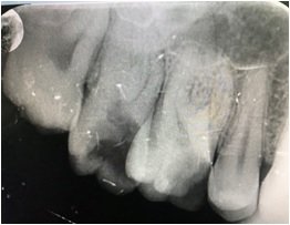
Figure 1: Pre-Operative

Figure 2: Access opening

Figure 3: Master Cone

Figure 4: Obturation IOPA

Figure 5: Post- Endo Restoration IOPA
II. Case Report
The patient reported to the Department Of Conservative Dentistry And Endodontics, Darshan Dental College and Hospital, Udaipur with a chief complaint of pain on elaborating the chief complaint the pain was intermittent in nature, aggravated during mastication and during sleep. The patient’s medical history was non-contributory. On clinical examination it revealed the presence of deep carious lesion and there was tenderness on percussion, no fistula and sinus tract was seen. On radiographic investigation (Fig 6), the tooth revealed the presence of radiolucency involving enamel, dentin and the pulp.Hence a diagnosis of symptomatic irreversible pulpitis with apical periodontitis was made. Access cavity is prepared using an endo access bur under rubber dam isolation. Once the pulp chamber was deroofed, a shaped access opening was obtained and the cavity was modified to a shamrock form. Glide path was achieved using stainless steel k file8,10.The canal was negotiated and the working length was determined with the help of an apex locator. Cleaning and shaping was done using hand protaper (SX, S1, S2, F1, F2) with crown down technique. Irrigation was performed using normal saline, 3% sodium hypochlorite solution and was activated with sonic activation with the use of endoactivator, 17% EDTA, 2% chlorhexidine digluconate was used as the final irrigant. The canals were dried with absorbent points and the master cone was taken (Fig 7) the canals were obturated using Endofloss sealer and single cone obturation with gutta-percha (Fig 9) and the teeth was subsequently restored with amalgam (Fig 10).
Figure 6. Pre-Operative IOPA

Figure 7. Access Opening

Figure 8 master cone

Figure 9. Obturation IOPA

Figure 10. Post- Endodontic Restoration
III. Case Report
The patient reported to the Department Of Conservative Dentistry And Endodontics, Darshan Dental College and Hospital, Udaipur with a chief complaint of spontaneous pain on elaborating the chief complaint the pain was intermittent in nature, aggravated during mastication and during sleep. The patient’s medical history was non-contributory. On clinical examination it revealed the presence of deep carious lesion and there was tenderness on percussion, no fistula and sinus tract was seen. On radiographic investigation (Fig 11), the tooth revealed the presence of radiolucency involving enamel, dentin and the pulp. Hence a diagnosis of symptomatic irreversible pulpitis with apical periodontitis was made. Access cavity is prepared using an endo access bur under rubber dam isolation. Once the pulp chamber was deroofed, a shaped access opening was obtained and the cavity was modified to a shamrock form. (Fig 12) Glide path was achieved using stainless steel k file. 8,10 The canal was negotiated and the working length was determined with the help of an apex locator. Cleaning and shaping was done using hand protaper (SX, S1, S2, F1, F2) with crown down technique. Irrigation was performed using normal saline, 3% sodium hypochlorite solution and was activated with sonic activation with the use of endoactivator, 17% EDTA, 2% chlorhexidine digluconate was used as the final irrigant. The canals were dried with absorbent points and the master cone was taken (Fig 13) the canals were obturated using Endofloss sealer and single cone obturation with gutta-percha (Fig14) and the teeth was subsequently restored with amalgam (Fig 15).
Figure 11. Pre- Operative IOPA

Figure 12. Access Opening

Figure 13. Master Cone

Figure 14. Obturation

Figure 15. Post- Endodontic Restoration
IV. Case Report
The patient reported to the Department Of Conservative Dentistry And Endodontics, Darshan Dental College and Hospital, Udaipur with a chief complaint of continuous pain on elaborating the chief complaint the pain was continuous in nature, aggravated during mastication and during sleep. The patient’s medical history was non-contributory. On clinical examination it revealed the presence of deep proximal carious lesion and there was tenderness on percussion, no fistula and sinus tract was seen. On radiographic investigation (Fig 16), the tooth revealed the presence of radiolucency involving enamel, dentin and the pulp. Hence a diagnosis of symptomatic irreversible pulpitis was made. Access cavity was prepared using an endo access bur under rubber dam isolation and pulp stone was removed (Fig 17). Once the pulp chamber was deroofed, a shaped access opening was obtained and the cavity was modified to a shamrock form. Glide path was achieved using stainless steel k file.8,10 The canal was negotiated and the working length was determined with the help of an apex locator. Cleaning and shaping was done using hand protaper (SX, S1, S2, F1, F2) with crown down technique. Irrigation was performed using normal saline, 3% sodium hypochlorite solution and was activated with sonic activation with the use of endoactivator, 17% EDTA, 2% chlorhexidine digluconate was used as the final irrigant. The canals were dried with absorbent points and the master cone was taken (Fig 18) the canals were obturated using Endofloss sealer and single cone obturation with gutta-percha (Fig 19) and the teeth was subsequently restored with amalgam (Fig 19).
Figure 16. Pre- Operative

Figure 17. Pulp Stone

Figure 18. Master Cone

Figure 19. Obturation

Figure 20. Post- Endodontic Restoration
Discussion
Lot of Variations are observed in root canal morphology, primarily in multi rooted teeth, are a persistent challenge for diagnosis and management. Existence of a second canal in the mesiobuccal (MB) root of maxillary molars has been a topic of numerous studies and its reported incidence is in the range of 33% and 96%.8,9 Weine’s classification has been used to describe four common configurations of the maxillary MB root. Type I is a single canal from orifice to apex, Type II has two orifices that converge to one, Type III has separate and distinct canals. Sempira, et al.10 Reported that only 33% of maxillary first molars had a negotiable MB2 canal, the most important causes of non-negotiable MB2 canal location are its narrowness, diffuse calcification, pulp stones debris and torturous pathways from orifice to apex, and Type IV begins as one canal and diverges into two separate canals.11 The age of the patients is also an important predictor for the detection of extra canals in maxillary molars, study have showed that with increase in age there is increase in calcification which makes detection of MB2 more difficult.12 As mentioned above keeping the age of the patient a factor which influence the detection and negotiation of MB2 canal the incidence of MB2 canal in people of Udaipur district was 70% when the patient of the age group 14-35 years was considered. As the age advances the detection and negotiation of MB2 canal is difficult because of diffuse calcifications and attached pulp stones. Use of magnification aids such as microscope and loupes helps in increasing the prevalence of the clinical detection of MB2. Other aids to enhance visualize the missed canals is by using sodium hypochloride, staining the chamber with methylene blue dye, transillumination, the use of sharp explorers, location of bleading point known as red test, white line test in cases of necrotiuc pulp and CBCT imaging.13 Stropko observed that by scheduling adequate clinical time and by use of recent magnification and detection instrumentation aids and by having thorough knowledge of how and where to search MB2, rate of location can approach 93% in maxillary first molar.14Conclusion
Conclusing that it is important for a dentist performing endodontic therapy on maxillary first molars to examine carefully the pulpal floor to locate the orifices of MB2 or any “extra” canals. It is worthwhile to put the time and effort into properly locating and treating these canals in an attempt to increase the prognosis of endodontic therapy in maxillary molars. More emphasis should be placed on the importance of using magnification to locate the MB2 canals. Their clinical use may improve the treatment prognosis of maxillary first molarsReferences
- Spagnuolo G, Ametrango G, D’ Anto V, et al. Micromcomputed tomography analysis of mesiobuccal orifices and major apical foramen in first maxillary molars, Open Dent J 6: 118, 2012.
- Schilder H. Cleaning and shaping the root canal. Dent Clin North Am. 1974; 18: 269–96.
- Hess W, Zurcher E. London: John Bane, Sons and Danielsson Ltd; 1925. The Anatomy of the Root Canals of the Teeth of the Permanent Dentition; pp. 4–49.
- Wolcott J, Ishley D, Kennedy W, Johnson S, Minnich S. Clinical investigation of second mesiobuccal canals in endodontically treated and retreated maxillary molars. J Endod. 2002;28: 477–9.
- Cantatore G, Berutti E, Castellucci A. Missed anatomy: Frequency and clinical impact. Endod Top. 2009;15: 3–31.
- Pomeranz HH, Fishelberg G. The secondary mesiobuccal canal of maxillary molars. J Am Dent Assoc. 1974;88: 119–24.
- Kulild JC, Peters DD. Incidence and configuration of canal systems in the mesiobuccal root of maxillary first and second molars. J Endod. 1990; 16:311– 317.
- Pattanshetti N, Gaidhene M, Al Kandari AM. Root canal morphology of the mesiobuccal and distal roots of permanent first molars in a Kuwait population. A clinical study. Int Endod J. 2008; 41:755–62.
- Weine FS, Healey HJ, Gerstein H, Evanson L. Canal configuration in the mesiobuccal root of the maxillary first molar and its endodontic significance. Oral Surg Oral Med Oral Pathol. 1969;28:419–25.
- Sempira HN, Hartwell GR. Frequency of second mesiobuccal canals in maxillary molars as determined by use of an operative microscope: A clinical study. J Endod. 2000; 26: 673–4.
- Ibarrola JL, Knowles KI, Ludlow MO, McKinley IB Jr. Factors affecting the negotiability of second mesiobuccal canals in maxillary molars. J Endod. 1997; 23: 236–8.
- Ahmad IA, Al-Jadaa A. Three root canals in the mesiobuccal root of maxillary molars: case reports and literature review. J Endod. 2014;40(12):2087–94
- Rudolf B, Michael AB. Color atlas of dental medicine. Endodontology 2000. p. 51-52.
- Wolcott J, Ishley D, Kennedy W, Johnson S, Minnich S, Meyers J. A 5 yr clinical investigation of second mesiobuccal canals in endodontically treated and re-treated maxillary molars. J Endod 2005;31: 262-4.
Disclaimer from Guident
The views and opinions expressed in articles published in Guident are solely those of the authors and do not necessarily reflect the views of the editors, the editorial board or the publisher, Ivory India. Guident does not endorse any product, technique or treatment described. Clinical content is intended for qualified dental and medical professionals and is not a substitute for individual clinical judgement. Guident, its editors and the publisher accept no liability for any consequence arising from the use of information published in the journal.
How to cite this article
Dr. Karishma Pathak, Dr. Jitendra Lohar, Dr. Tariq Mohammad Shekh. Clinical Management of MB2 Canal in Maxillary First Molar. Guident. 2020 Aug;13(9). Available from: https://ivoryindia.in/articles/endodontics/clinical-management-of-mb2-canal-in-maxillary-first-molar.html