Endocrown Restoration: A Conservative Approach to Restore Extensively Damaged Endodontically Treated Molar-a Case Report
Department of Conservative Dentistry & Endodontics, K.D. Dental College & Hospital, Mathura
ABSTRACT
The endocrown is a highly promising treatment option for endodontically treated molars. It permits tooth structure to be preserved and it is compatible with the objective of minimally invasive dentistry. This article discusses a case report of an endocrown-type restoration, fabricated from ZrO2-based ceramic in a mandibular 2nd molar with extensive coronal destruction.
Keywords-Endocrown, cervical butt joint.
INTRODUCTION
For restoring endodontically treated teeth, minimally invasive preparations with maximum tissue preservation are currently regarded as the ‘gold standard'.1 Pissis was the forerunner of endocrown process, which he referred to as the "mono-block porcelain technique." 2 Bindle and Mormann originally described the endocrown in 1999, referring to it as adhesive endodontic crowns and entire porcelain crowns.3
These crowns would be secured to the interior section of the pulp chamber and the cavity borders, resulting in macromechanical retention given by the pulpal walls and micromechanical retention gained via the application of adhesive cementation. This approach is especially useful when there is severe crown tissue loss, interproximal space is restricted, and typical rehabilitation method of post and core are ineffective because of insufficient ceramic thickness.4 These are also employed in cases of molars with short, obliterated, dilacerated, brittle roots, and highly mutilated tooth, as well as in cases involving considerable loss of coronal tooth structure with deficient interocclusal space.5 So the present article aims to discuss the case of grossly carious tooth managed by endocrown.
CASE REPORT
A 48-year-old female patient reported to the Department of Conservative Dentistry and Endodontics with a chief complaint of pain in the lower right back tooth region. Following radiographic and clinical examination, an extensive faulty composite restoration of a nonvital 47 tooth was identified. (Figures 1) Root canal therapy was advised in relation to 47.A conservative post -endodontic restoration with an endocrown was suggested, by accessing the amount of remaining tooth structure.
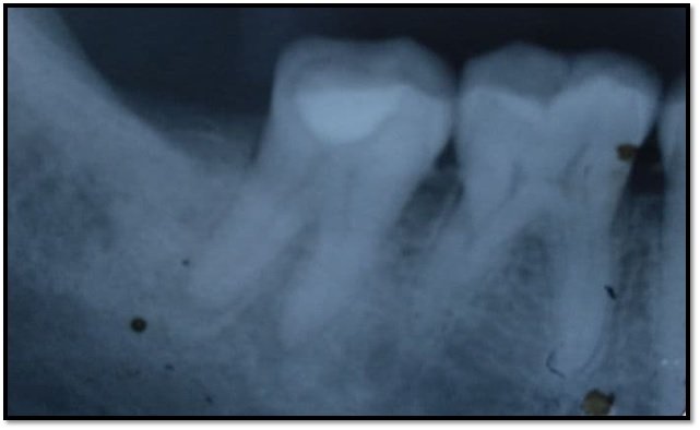
Figure 1: Preoperative radiograph of 47
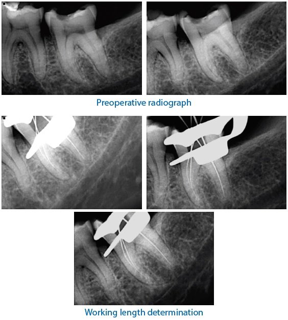
The patient was informed about the procedure and the treatment was executed following the patient consent. The tooth was treated endodontically. Figures (2,3,). Glass ionomer cement was used to block the orifices in the pulp chamber. The occlusal reduction was done using wheel diamond bur WR-13 (Diamond Burs FG, Mani) by placing it parallel to the long axis of the tooth. Then, the cervical margins were prepared in the shape of a chamfer using a diamond bur SO-21 (Diamond Burs, Mani) at high speed and under water coolant, creating a consistent thickness of 1.5 mm with the remaining coronal tooth structure. Axial preparation included removal of undercuts in the access cavity which was carried by cylindrical conical green diamond bur.

Figure 2: Working length radiograph of 47

Figure 3: Master cone radiograph of 47
The bur was oriented along the long axis of the tooth in such a way that it maintains an occlusal convergence of 7 to 10° thereby ensuring a continuous flow of the prepared coronal pulp chamber and the access cavity. Polishing of the cervical band was done. (Figure 4) Gingival retraction cord 0 (Coltene Roeko Stay Put Retraction Cord) was placed along the gingival crevice and an impression was made with poly vinyl siloxane (Dental-Avenue, Avue Gum Putty & Light Body) using putty wash technique. (Figure 5) Thereafter, shade selection was done. The impression was sent to the laboratory for further processing. The endocrown was fabricated in the laboratory (Figure 6,7) and was positioned on the master cast (Figure 8,). In the subsequent appointment, the evaluation of final fit of fabricated ZrO2-based ceramic endocrown in relation to 47 was done. Sandblasting of the fabricated endocrown was done to enhance the retention of the restoration and final cementation was done with glass ionomer cement. (GC Gold Label 1 Luting & Lining GIC) (Figure 9,10).


Figure 10-Radiographic view after cementation of endocrown
DISCUSSION
The endocrown is a monolithic (one-piece) ceramic bonded structure.6 Endocrown is made using feldsphatic and glass-ceramic materials, composite hybrid resin, and CAD/CAM ceramic and composite resin.7,8
The endocrown preparation consists of a circumferential 1.0-1.2 mm depth butt margin and a central retention cavity inside the pulp chamber, constructs both the crown and core as a single unit monoblock structure, and does not rely on root canal support.2,3
The objective is to create a broad, uniform, sturdy surface that can withstand the compressive pressures that are frequent on molars. To provide stress resistance along the primary axis of the tooth, the prepared surface is parallel to the occlusal plane. The pulp chamber cavity guarantees retention and stability. Its form is trapezoidal in mandibular molars and triangular in maxillary molars. There is no need to make any further preparations. The pulpal floor's saddle shape improves stability. This anatomy, together with the bonding material's adhesive properties, eliminates the need for any further post usage. The compressive stresses are dispersed throughout the cervical butt joint and the pulp chamber walls.9
Teeth with endocrown had lower stress levels than teeth with prosthetic crowns.10,11 The necessity for macroretentive crown preparation has diminished as adhesive cementation techniques have improved.12 Endocrown, according to Belleflamme et al., might be a viable technique to repair severely injured molars and premolars, especially in the presence of substantial coronal tissue loss or occlusal risk factors such as bruxism or unfavorable occlusal relationships.13 Endocrown are not recommended in the following situations: (1) when the pulp chamber depth is less than 3 mm or the cervical margin is less than 2 mm broad, (2) when adhesion cannot be guaranteed, and (3) when there is only a little amount of tooth structure remaining.14
CONCLUSION
Endocrowns are a relatively recent procedure that is simple and quick to do. The Endocrown is completely compatible with the notion of bio integration and might be the most conservative and cosmetic alternative for non-vital posterior tooth repair.
REFERENCES
- Dietschi D, Duc O, Krejci I, Sadan A. Biomechanical considerations for the restoration of endodontically treated teeth: A systematic review of the literature, part ii (evaluation of fatigue behaviour, interfaces, and in vivo studies). Quintessence Int 2008; 39:117-129.
- Pissis P. Fabrication of a metal-free ceramic restoration utilizing the monoblock technique. Pract Periodontics Aesthet Dent 1995; 7:83-94
- Bindl A, Mormann WH. Clinical evaluation of adhesively placed cerec endo-crowns after 2 years--preliminary results. J Adhes Dent 1999; 1:255-265.
- Chang CY KJ, Lin YS, Chang YH. Fracture resistance and failure modes of CEREC endo-crowns and conventional post and core-supported CEREC crowns. J Dent Sci 2009; 4:110-117.
- Amal S, Nair MG, Sreeja J, Babu A, Ajas A. Endocrown – an overlooked alternative. Arch Dent Med Res 2016;2: 34-38.
- Bernhart J, Brauning A, Altenburger MJ, Wrbas KT. Cerec3D endocrowns — two-year clinical examination of CAD/CAM crowns for restoring endodontically treated molars. Int J Comput Dent 2010; 13:141-54.
- Gohring TN, Peters OA. Restoration of endodontically treated teeth without posts. Am J Dent.2003;16:313–7.
- Dietschi D, Duc O, Krejci I, Sadan A. Biomechanical considerations for the restoration of endodontically treated teeth: A systematic review of the literature - part 1. Composition and micro- and macrostructure alterations. Quintessence Int 2007; 38:733–43.
- Fages M, Bennasar B. The endocrown: a different type of all-ceramic reconstruction for molars. J Can Dent Assoc. 2013;79: d140.
- Lin CL, Chang YH, Chang CY, Pai CA, Huang SF. Finite element and Weibull analyses to estimate failure risks in the ceramic endocrown and classical crown for endodontically treated maxillary premolar. Eur J Oral Sci. 2010; 118:87-93.
- Dejak B, MÅ‚otkowski A. 3D-Finite element analysis of molars restored with endocrowns and posts during masticatory simulation. Dent Mater 2013 ;29: e309-17.
- Lander E, Dietschi D. Endocrowns: a clinical report. Quintessence Int 2008; 39:99-106.
- Belleflamme MM, Geerts SO, Louwette MM, Grenade CF, Vanheusden AJ, Mainjot AK. No post-no core approach to restore severely damaged posterior teeth: An up to 10-year retrospective study of documented endocrown cases. J Dent. 2017; 63:1-7
- Gilboe DB, Teteruck WR. Fundamentals of extracoronal tooth preparation. Part I. Retention and resistance form. J Prosthet Dent 1974; 32:651-656
Disclaimer from Guident
The views and opinions expressed in articles published in Guident are solely those of the authors and do not necessarily reflect the views of the editors, the editorial board or the publisher, Ivory India. Guident does not endorse any product, technique or treatment described. Clinical content is intended for qualified dental and medical professionals and is not a substitute for individual clinical judgement. Guident, its editors and the publisher accept no liability for any consequence arising from the use of information published in the journal.
How to cite this article
Dr. Deepika, Dr. Ajay Nagpal. Endocrown Restoration: A Conservative Approach to Restore Extensively Damaged Endodontically Treated Molar-a Case Report. Guident. 2021 Mar;14(4). Available from: https://ivoryindia.in/articles/endodontics/endocrown-restoration-a-conservative-approach-to-restore-extensively-damaged-endodontically-treated-molar-a.html