The Tale of an Endodontic Non Surgical Retreatment of a Mandibular Molar: An Attempt to Salvage with One Year Follow Up
Department of Conservative Dentistry and Endodontics, Seema Dental College And Hospital, Rishikesh (UK)
Retreatment is common in endodontics. Endodontic failures must be evaluated so a decision can be made among nonsurgical retreatment, surgical retreatment, or extraction.1,2,3 Improvement in the diagnosis and treatment of lesions of endodontic origin (LEO) occurs with the recognition of the interrelationships between pulpal disease flow and the egress of irritants along these anatomical pathways.4
Endodontic failures can be attributable to inadequacies in shaping, cleaning and obturation, iatrogenic events, or re-infection of the root canal system when the coronal seal is lost after completion of root canal treatment. 9-12 Regardless of the etiology, the sum of all causes is leakage and bacterial contamination.5,6 Except in rare instances, lesions of endodontic origin will routinely heal following the extraction of pulpally involved teeth because the extraction not only removes the tooth, but more importantly serves to eliminate 100% of the contents of the root canal system. Endodontic treatment can approach 100% success discounting teeth that are nonrestorable, have hopeless periodontal disease or have radicular fractures.4
The goals of nonsurgical retreatment are to remove materials from the root canal space and if present, address deficiencies or repair defects that are pathologic or iatrogenic in origin.7 Nonsurgical endodontic retreatment procedures have enormous potential for success if the guidelines for case selection are respected and the most relevant technologies, best materials and precise techniques are utilized.8,9
CASE REPORT
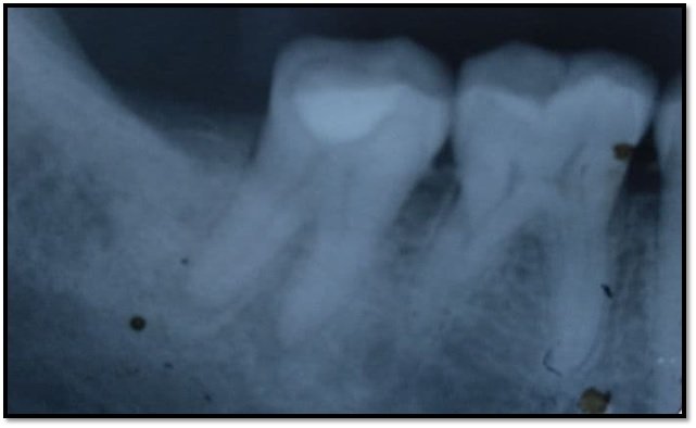
A 21-year-old female patient came with severe pain and swelling in lower left back tooth region. A thorough medical and dental history was taken. Medical history was noncontributory. The patient gave a history of dental treatment in mandibular posterior teeth 4 months back. She had undergone root canal treatment in 36. On examination revealed a diffuse swelling in buccal mucosa in relation to 36. Crown was placed in in the same. It was extremely tender to percussion. On radiographic examination, poorly obturated 36 was revealed. There was a very large radiolucent lesion involving apices of 36.
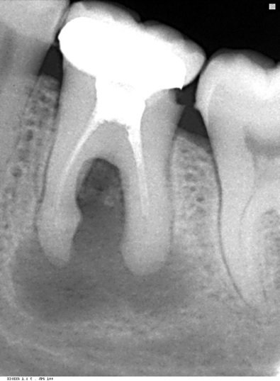
PREOPERATIVE
Following removal of crown Nonsurgical endodontic retreatment was planned to treat the involved tooth as the patient was unwilling to go for surgery. Under Local Anaesthesia access cavity was prepared and old gutta parcha was attempted to remove by using H files. While removing the instrument got separated in the one of the mesial canal at middle third. The gutta-percha was removed from other canals, there was drainage of large amount of straw-colored fluid into the pulp chamber providing immediate relief to patient from pain and swelling. When the drainage ceased, the root canal preparation was performed using rotary protaper files till No. F2. Apical patency was maintained throughout the procedure. During preparation, the canals were irrigated with 3% hypochlorite and saline.
The patient was recalled next day and access cavitie was sealed with provisional restoration. The patient was prescribed analgesics and antibiotics for 5 days .

H FILE SEPERATED
In the second visit patient reported of relief in pain and swelling. With the help of the loupes and ultrasonics the separated instrument was retrieved in the second visit. After retrieval, the canals was prepared irrigated and calcium hydroxide dressing was given for 15 days.

INSTRUMENT RETRIEVED
After 15 days the canals were obturated and acces cavity was sealed with provisional restoration. Patient recalled after 3 months.

FINAL OBTURATION
Patient reported after 1 year. The large periapical lesion was seen healing and patient was asymptomatic.

BEFORE & AFTER 1 YEAR
DISCUSSION
Retreatment ensures that the entire root canal system and communicating channels are thoroughly cleaned and a good quality root filling is placed under optimal asepsis in every canal. While assessing the outcomes of endodontic retreatment, damage to the root level walls should be considered.
The relative difficulty in removing gutta percha varies according to the obturation technique previously employed and further influenced by the canal’s length, cross-sectional dimension, curvature and internal configuration. Regardless of technique, gutta percha is best removed from a root canal in a progressive manner to prevent inadvertent displacement of irritants periapically. The removal techniques include rotary files, ultrasonic instruments, heat, hand files with heat or chemicals, and paper points with chemicals.10 Before commencing with the efforts to remove a broken instrument, the clinician should thoughtfully observe different horizontally angulated radiographs.11
Coronal access is the first step in the removal of broken instruments. Special attention should be directed towards flaring the axial wall that approximates the canal holding the broken instrument in efforts to subsequently improve the micro-sonic techniques below the orifice. Radicular access is the second step required in the successful removal of broken instrument. At times, when an ultrasonic instrument is introduced into a pre-enlarged canal, its activated tip does not have sufficient space lateral to the broken file segment to initiate trephining procedures. Ideally, radicular access should be performed in a way that the canal is pre-enlarged and shaped to the same diameter as that if there was no broken instrument obstructing the canal.11
Bioceramic-based root canal sealers show promising results as root canal sealers. MTA Fillapex (Angelus S/A, Londrina, PR, Brazil) is composed of salicylate resin, resin diluent, natural resin, bismuth oxide, silica nanoparticles, and calcium silicate. It was created combining a material with excellent biocompatibility and bioactive potential such as MTA with synthetic resins with good physical properties.
CONCLUSION
The root canal treatment proved successful in promoting the healing of a large periapical lesion. This confirms that even large periapical lesions can respond favorably to nonsurgical treatment and therefore, it must always be the first treatment of choice. The most important factors central to successful instrument removal are knowledge, training and competency in selecting the best developed and proven technologies and techniques available at present. However, the best antidote for a broken file is prevention.
REFERENCES
- Stabholz A, Friedman S: Endodontic retreatment- case selection and technique. Part 2: treatment planning for retreatment. J Endod 14:12, pp. 607-614, 1988
- Allen RK, Newton CW, Brown CE: A statistical analysis of surgical and non-surgical endodontic retreatment cases, J Endod 15:6, pp. 261-266, 1989
- Kvist T, Reit C: Results of endodontic retreatment: a randomized clinical study comparing surgical and nonsurgical procedures, J Endod 25:12, pp. 814-817, 1999.
- Schilder H: Filling root canals in three dimensions, Dent Clin North Am, pp. 723-744, November 1967.
- Ruddle CJ: Nonsurgical endodontic retreatment, J Calif Dent Assoc 25:11, pp. 765-800, 1997. 14.
- Ruddle CJ: Surgical endodontic retreatment, J Calif Dent Assoc 19:5, pp. 61-67, 1991.
- Ruddle CJ: Ch. 25, Nonsurgical endodontic retreatment. In Cohen S, Burns RC, editors: Pathways of the Pulp, pp. 875-929, 8th ed., Mosby, St. Louis, 2002.
- Blum JY, Machtou P, Ruddle CJ, Micallef JP: The analysis of mechanical preparations in extracted teeth using protaper rotary instruments: value of the safety quotient, J Endod 29:9, pp. 567-575, 2003.
- Ruddle CJ: Nickel-titanium rotary instruments: current concepts for preparing the root canal system, Australian Endodontic Journal 29:2, pp. 87-98, 2003.
- Wilcox LR, Krell KV, Madison S, Rittman B: Endodontic retreatment: evaluation of gutta-percha and sealer removal and canal reinstrumentation, J Endod 13:9, pp. 453-457, 1987.
- Gorni FG, Gagliani MM. The outcome of endodontic retreatment: A 2 year follow up. J Endod. 2004;30:1–4
Disclaimer from Guident
The views and opinions expressed in articles published in Guident are solely those of the authors and do not necessarily reflect the views of the editors, the editorial board or the publisher, Ivory India. Guident does not endorse any product, technique or treatment described. Clinical content is intended for qualified dental and medical professionals and is not a substitute for individual clinical judgement. Guident, its editors and the publisher accept no liability for any consequence arising from the use of information published in the journal.
How to cite this article
Dr. Deepika Rawat, Dr. Annil Dhingra. The Tale of an Endodontic Non Surgical Retreatment of a Mandibular Molar: An Attempt to Salvage with One Year Follow Up. Guident. 2021 Feb;14(3). Available from: https://ivoryindia.in/articles/endodontics/the-tale-of-an-endodontic-non-surgical-retreatment-of-a-mandibular-molar-an-attempt-to-salvage-with-one-year.html